Medical Image Segmentation

Inactive Publication Date: 2011-12-01
SAMSUNG ELECTRONICS CO LTD
View PDF7 Cites 17 Cited by
  • Summary
  • Abstract
  • Description
  • Claims
  • Application Information

AI Technical Summary

Benefits of technology

[0011]The present invention can be applied to different types of lesion, such as lung nodules, liver lesions, mammographic masses, and brain lesions. Mo

Problems solved by technology

However, the process can be tedious, time consuming and must be performed with great care to accurately delineate the object boundary.
Medical image segmentation is a difficult task because in most cases it is very hard to separate the object from the image background.
This is due to the nature of the image acquisition process in which noise is inherent for all medical

Method used


Image

  • Medical Image Segmentation
  • Medical Image Segmentation
  • Medical Image Segmentation

Examples


Embodiment Construction

Medical Imaging

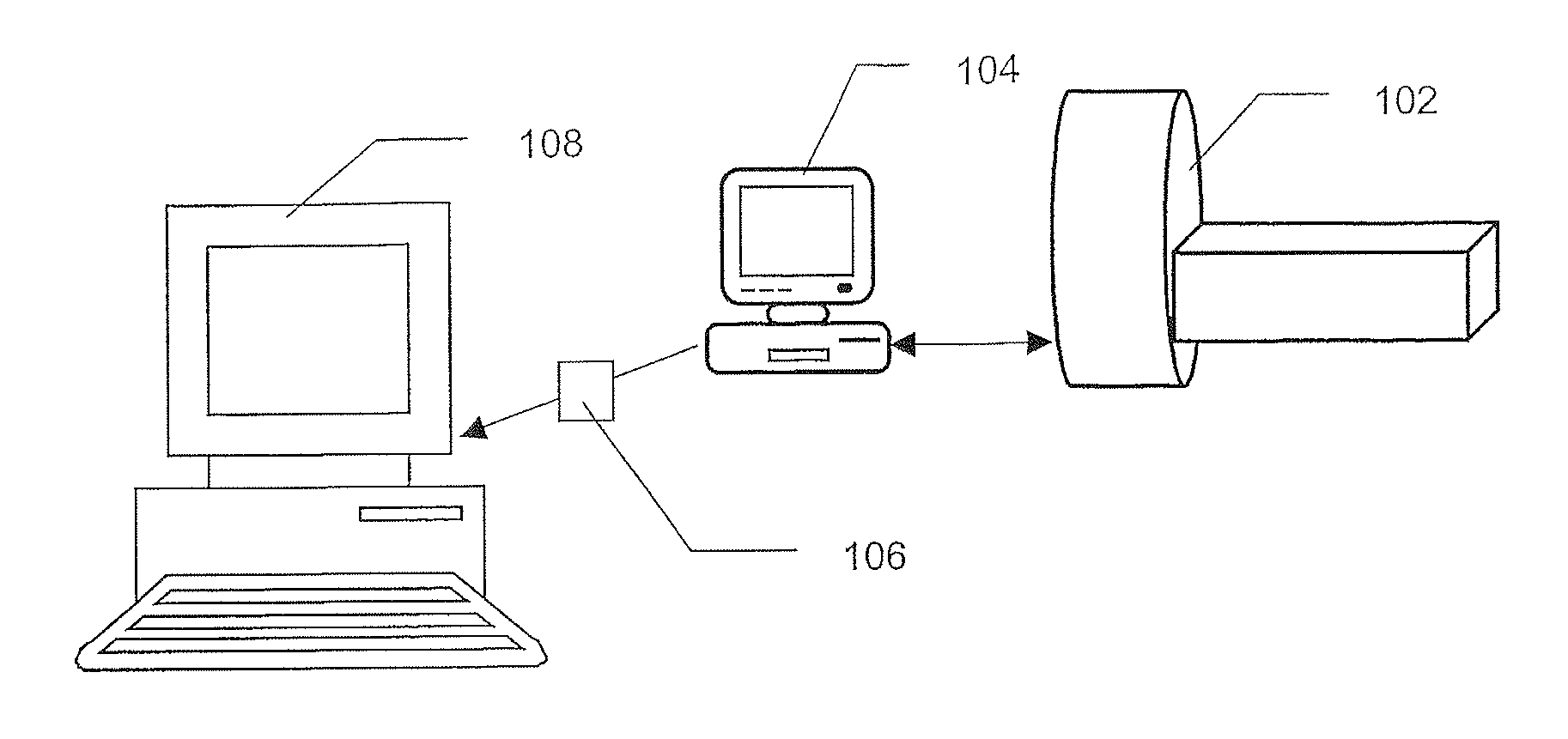
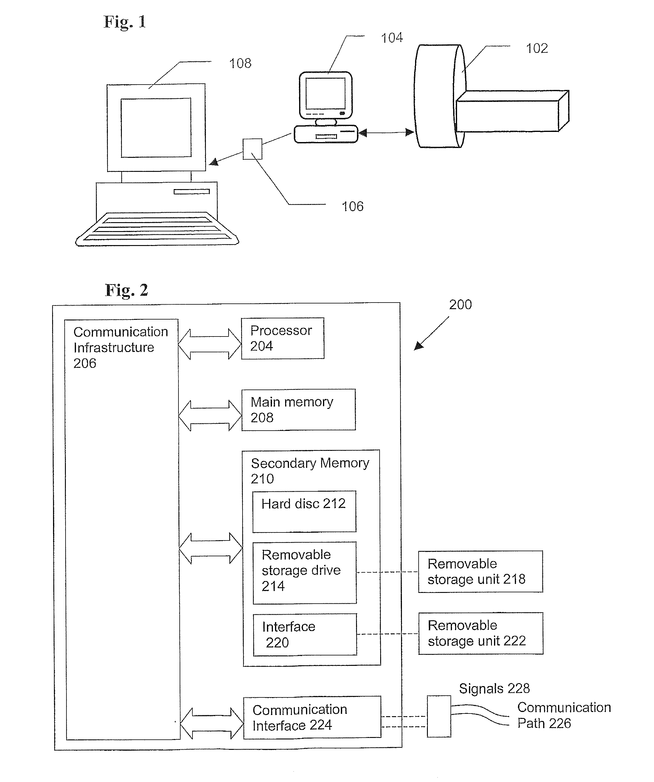
[0018]The present invention is applicable to digital medical images. One example of such an image is a CT scan image. A CT scan image is a digital image comprising one or a series of CT image slices obtained from a CT scan of an area of a human or animal patient. Each slice is a 2-dimensional digital grey-scale image of the x-ray absorption of the scanned area. The properties of the slice depend on the CT scanner used; for example, a high-resolution multi-slice CT scanner may produce images with a resolution of 0.5-1.0 mmG(pixel in the x and y directions (i.e. in the plane of the slice). Each pixel may have 32-bit greyscale resolution. The intensity value of each pixel may be expressed in Hounsfield units (HU). Sequential slices may be separated by a constant distance along the z direction (i.e. the scan separation axis); for example, by a distance of between 0.5-2.5 mm. Hence, the scan image may be a three-dimensional (3D) greyscale image, with an overall size depend...


PUM


Abstract

A segmentation method comprises clustering spatial, intensity and volumetric shape index to automatically segment a medical lesion. The algorithm has the following steps: (1) calculating volumetric shape index (SI) for each voxel in the image; (2) combining the SI features with the intensity range and the spatial position (x, y, z) to form a 5-dimensional feature vector set; (3) grouping the 5-dimensional feature vector set into clusters; (4) employing a modified expectation-maximization algorithm (EM) considering not only spatial but also shape features on an intensity mode map from the clustering algorithm to merge the neighbouring regions or modes. The joint spatial-intensity-shape feature provides rich information for the segmentation of the anatomic structures of interest, such as lesions or tumours.

Description

[0001]The present invention relates to the automated segmentation of medical images, and to the derivation of models for such automated segmentation.BACKGROUND TO THE INVENTION[0002]Medical imaging is generally recognized as key to better diagnosis and patient care. It has experienced explosive growth over the last few years due to imaging modalities such as X-ray, computed tomography (CT), ultrasound, and magnetic resonance imaging (MRI). Conventionally, medical images have been inspected visually and the anatomic structures of interest or some lesions (tumours) are manually delineated by trained radiologists. However, the process can be tedious, time consuming and must be performed with great care to accurately delineate the object boundary. To replace some or all of the work of the radiologists, segmentation of medical image data is a prerequisite in computer-assisted diagnosis.[0003]Medical image segmentation is a difficult task because in most cases it is very hard to separate ...

Claims


Application Information

Patent Timeline
IPC IPC(8): G06K9/00
CPCG06K9/6226G06T2207/30004G06T2207/10072G06T7/0081G06T7/11G06V10/763G06F18/2321G06T7/0012
Inventor YE, XUJIONGSLABAUGH, GREGORY GIBRANBEDDOE, GARETH RICHARDLIN, XINYUDOURI, ABDEL
Owner SAMSUNG ELECTRONICS CO LTD
Why Eureka
Try Eureka