Gene expression signature for classification of tissue of origin of tumor samples
a gene expression and tumor tissue technology, applied in the field of gene expression signatures, can solve the problems of undefined diagnostic accuracy of tumor markers, oncologists and pathologists are constantly faced with diagnostic dilemmas, etc., and achieve the effect of accurate identification of tumor tissue origin
- Summary
- Abstract
- Description
- Claims
- Application Information
AI Technical Summary
Benefits of technology
Problems solved by technology
Method used
Image
Examples
example 1
Samples and Profiling
[0192]A discovery process that profiled hundreds of samples on the array platforms was performed to identify candidate biomarkers. A training set of ˜400 FFPE samples was used. RNA was extracted from these samples and qRT-PCR was preformed. An assay was constructed using 48 microRNAs (Table 3; FIGS. 1-7), to differentiate between 26 classes representing 18 tissue origins. An alternative assay was constructed, which does not identify bladder as an origin, i.e., differentiates between 25 classes representing 17 tissue origins.
[0193]A validation set of 255 new FFPE tumor samples was used to assess the performance of the assay, representing 26 different tumor origins or “classes” (see Table 2 for a summary of samples). About half of the samples in the set were metastatic tumors to different sites (e.g., lung, bone, brain and liver). Tumor percentage was at least 50% for all samples in the set.
TABLE 2Cancer types, classes and histologyClassCancer types and histologic...
example 2
Decision-Tree Classification Algorithm
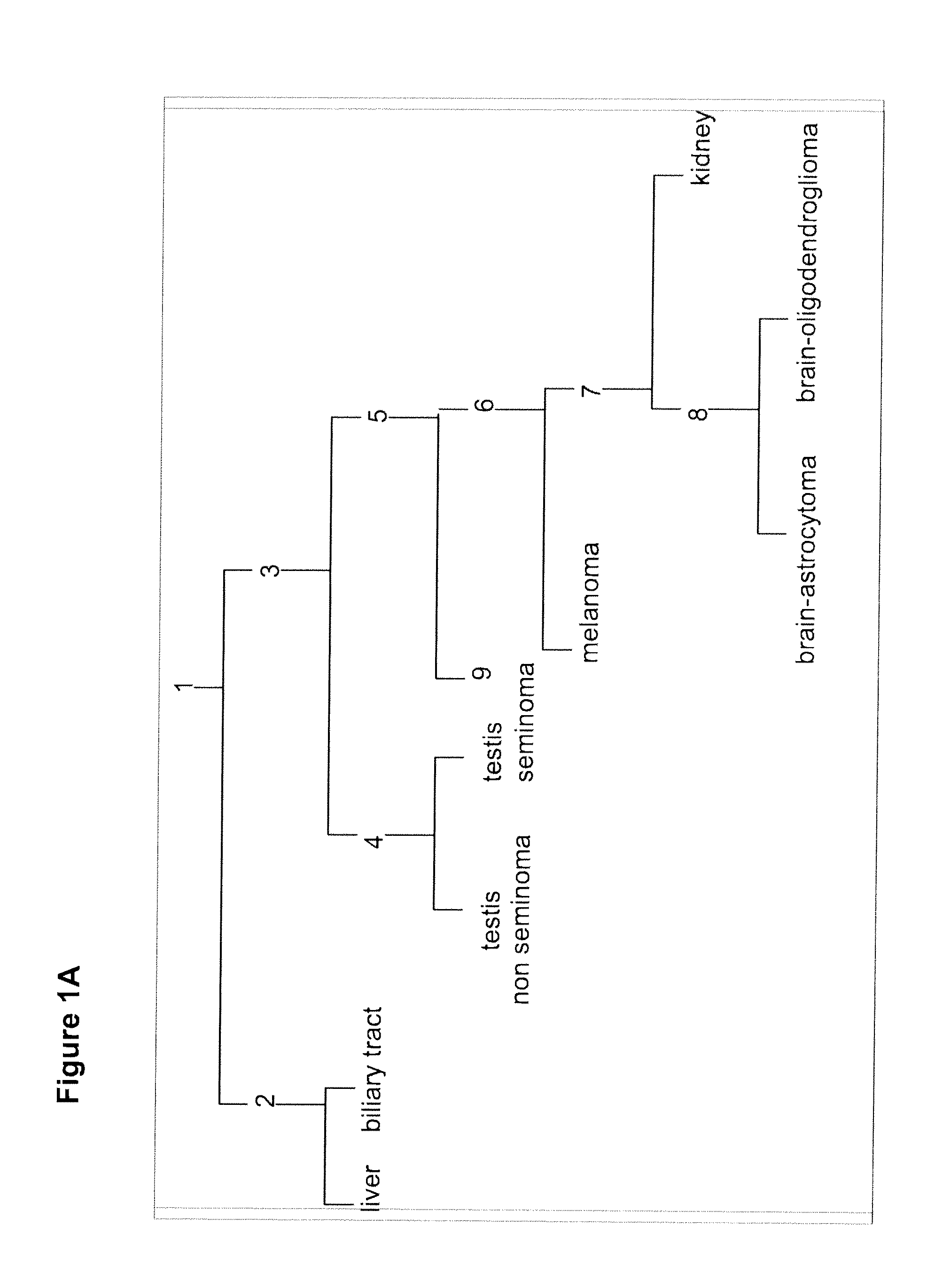
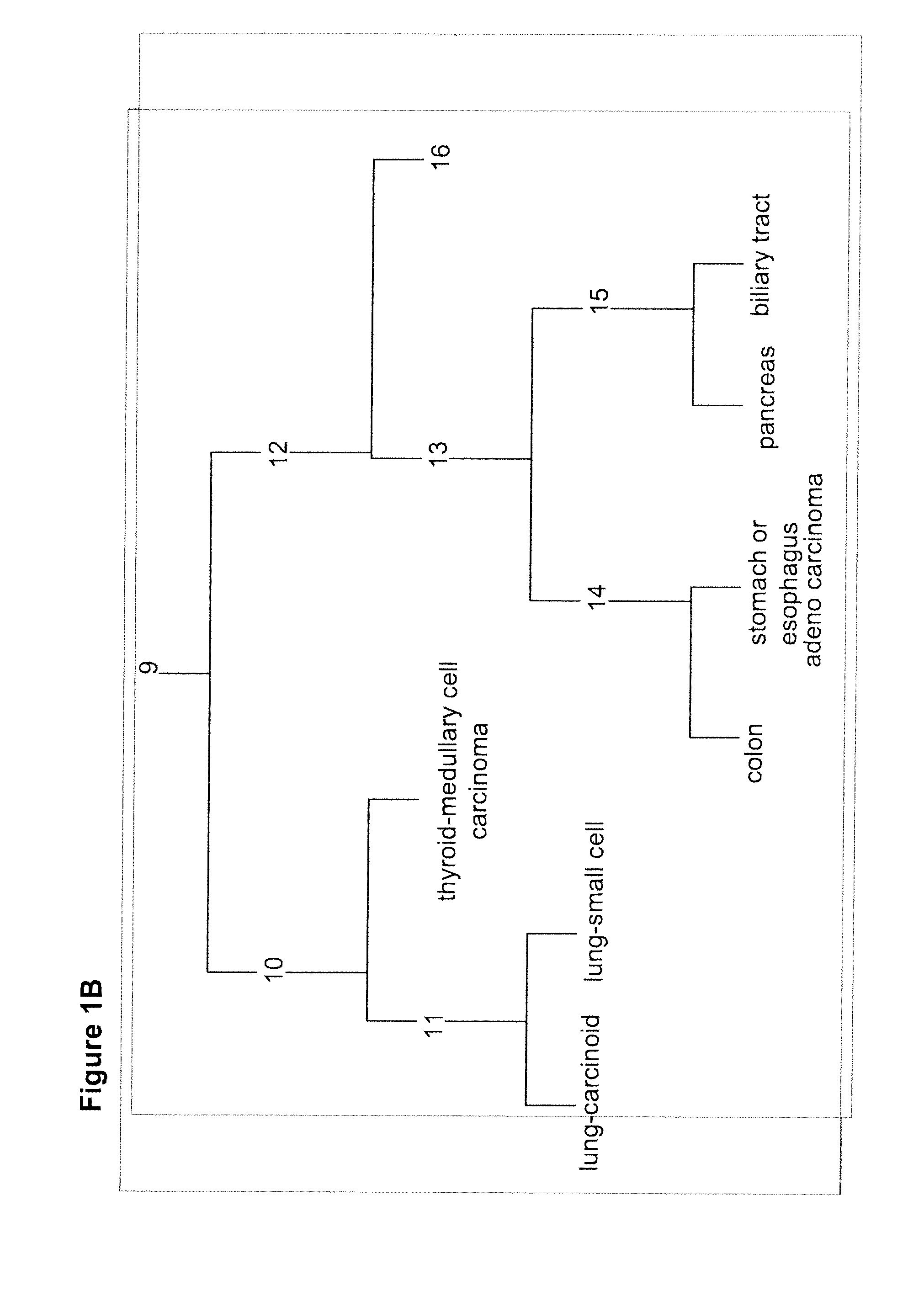
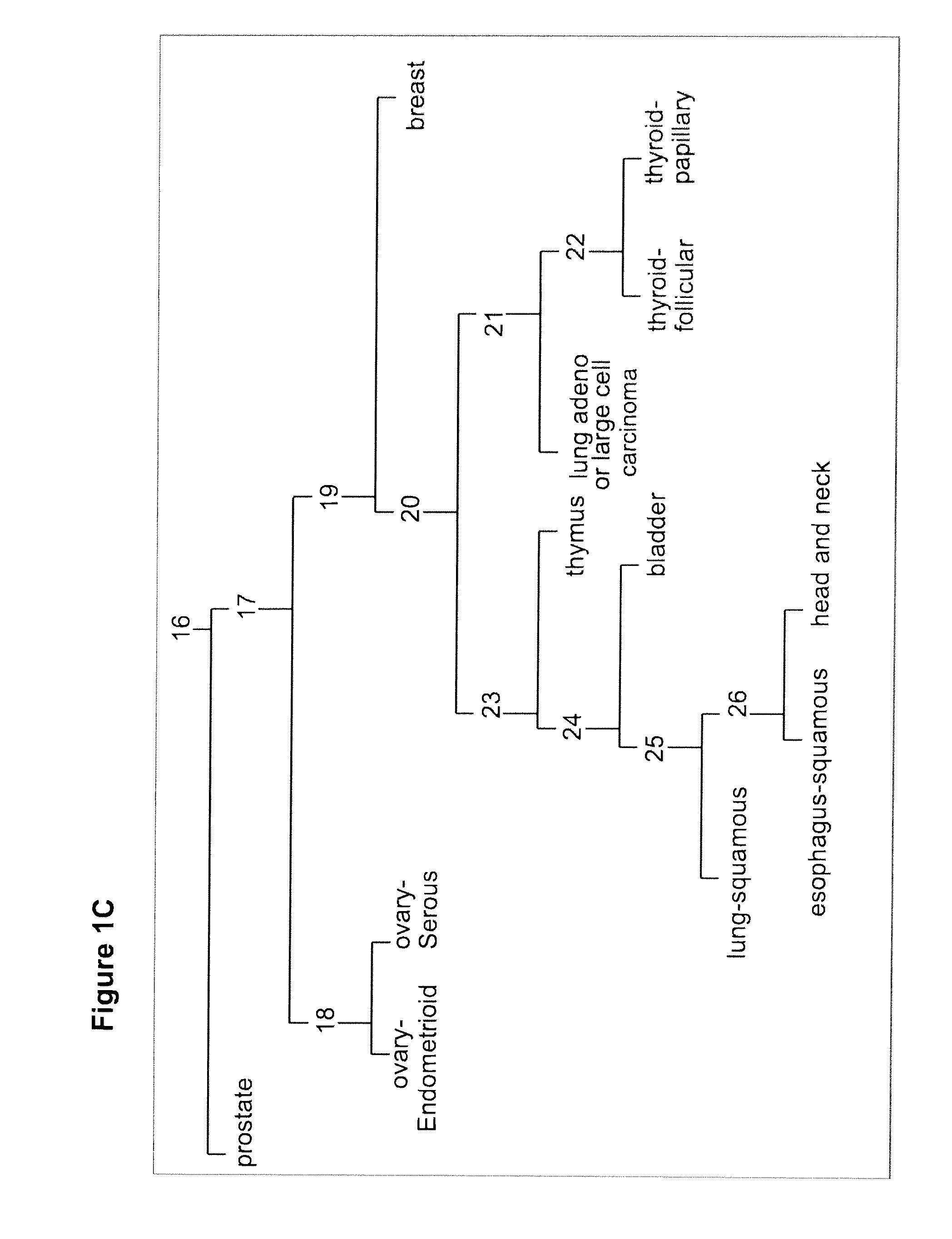
[0194]A tumor classifier was built using the microRNA expression levels by applying a binary tree classification scheme (FIG. 1). This framework is set up to utilize the specificity of microRNAs in tissue differentiation and embryogenesis: different microRNAs are involved in various stages of tissue specification, and are used by the algorithm at different decision points or “nodes.” The tree breaks up the complex multi-tissue classification problem into a set of simpler binary decisions. At each node, classes which branch out earlier in the tree are not considered, reducing interference from irrelevant samples and further simplifying the decision. The decision at each node can then be accomplished using only a small number of microRNA biomarkers, which have well-defined roles in the classification (Table 3). The structure of the binary tree was based on a hierarchy of tissue development and morphological similarityl8, which was modified by prom...
example 3
Defining High-Confidence Classifications
[0196]In clinical practice it is often useful to assess information of different degrees of confidence17,18. In the diagnosis of tumor origin in particular, a short list of highly probable possibilities is a practical option when no definite diagnosis can be made. Since the decision-tree and the KNN algorithms are designed differently and trained independently, improved accuracy and greater confidence can be obtained by combining and comparing their classifications. When the two classifiers agree, the diagnosis is considered high-confidence and a single origin is identified. When the two disagree, the classification is made with low-confidence and two origins are suggested. Sensitivity of the union refers to the percentage in which at least one of the classifiers (Tree and KLAN) was correct.
PUM
| Property | Measurement | Unit |
|---|---|---|
| Tm | aaaaa | aaaaa |
| temperature | aaaaa | aaaaa |
| temperature | aaaaa | aaaaa |
Abstract
Description
Claims
Application Information
 - R&D
- Intellectual Property
- Life Sciences
- Materials
- Tech Scout
- Unparalleled Data Quality
- Higher Quality Content
- 60% Fewer Hallucinations
Browse by: Latest US Patents, China's latest patents, Technical Efficacy Thesaurus, Application Domain, Technology Topic, Popular Technical Reports.
© 2025 PatSnap. All rights reserved.Legal|Privacy policy|Modern Slavery Act Transparency Statement|Sitemap|About US| Contact US: help@patsnap.com


