Image processing apparatus, image processing method, and image processing program

a technology of image processing apparatus and image processing program, which is applied in the direction of image enhancement, instruments, catheters, etc., can solve the problems of not focusing on the shift in the timing of contraction, unable to provide a solution to the above needs regarding analysis of cardiac motion, and unable to achieve the above needs, so as to achieve easy visual understanding, easy analysis and evaluation, and easy analysis and evaluation

Active Publication Date: 2012-03-15
FUJIFILM CORP
View PDF2 Cites 5 Cited by
  • Summary
  • Abstract
  • Description
  • Claims
  • Application Information

AI Technical Summary

Benefits of technology

[0035]The image processing apparatus, the image processing method, and the image processing program of the present invention obtains the myocardial thicknesses at a plurality of positions of the heart from a plurality of three dimensional images obtained by imaging the heart at a plurality of temporal phases within a single cardiac cycle. Representative values that represent the systolic phase of the myocardia are obtained for each of the plurality of positions, based on the myocardial thicknesses obtained for each temporal phase, then output. Thereby, users, such as physicians, can employ the representative values to easily analyze and evaluate the contraction timings of the myocardia at each of the plurality of positions of the heart.
[0036]In the image processing apparatus, the image processing method, and the image processing program of the present invention, the representative values may be output by generating a representative value image that represents the representative values such that visual recognition of differences in the sizes of the representative values of each of the plurality of positions is enabled, and displaying the generated representative value image with a display means. In this case, the display of the representative value image will enable users such as physicians to easily visually understand the contraction timings of the myocardia at each of the plurality of positions of the heart.
[0037]In addition, reference representative values that represent the systolic phase of myocardia may be obtained for positions of a healthy heart anatomically corresponding to the plurality of positions

Problems solved by technology

However, if changes occur in the contraction timings due to myocardial infarction or the like, efficient circulation of blood is precluded, which is a problem.
However, the aforementioned conventional te

Method used


Image

  • Image processing apparatus, image processing method, and image processing program
  • Image processing apparatus, image processing method, and image processing program
  • Image processing apparatus, image processing method, and image processing program

Examples


Embodiment Construction

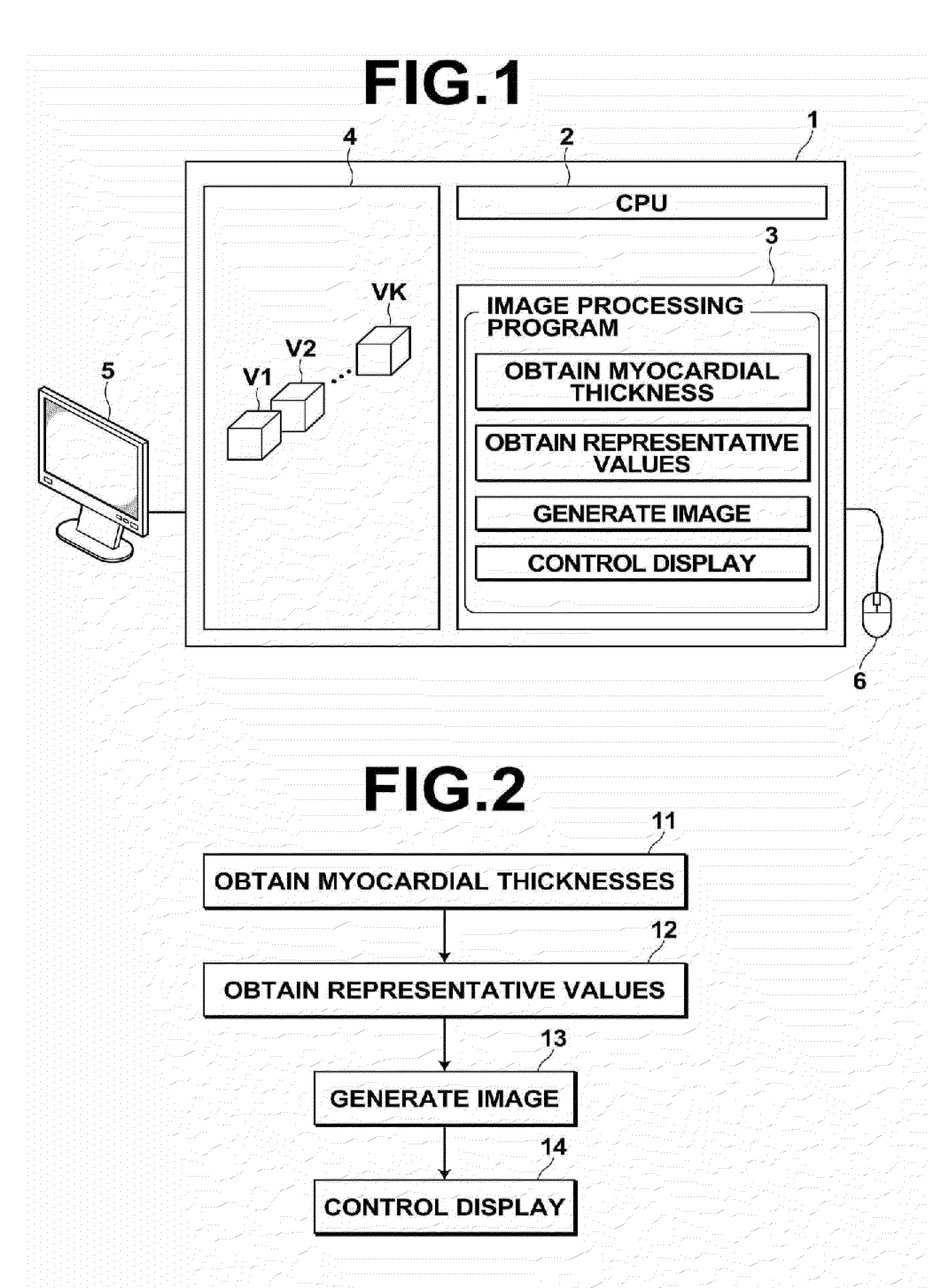
[0050]Hereinafter, embodiments of the image processing apparatus, the image processing method, and the image processing program of the present invention will be described in detail with reference to the attached drawings.

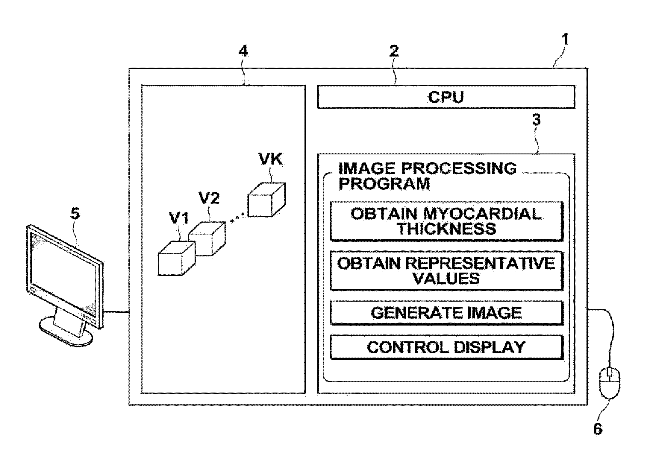
[0051]The image processing apparatus 1 of the present invention is a single computer, in which an image processing program is installed. The computer may be a workstation or a personal computer which is directly operated by a physician that performs diagnosis. Alternatively, the computer may be a server computer connected to such a workstation or a personal computer. The image processing program is distributed stored in a recording medium such as a DVD and a CD-ROM, and installed in the computer from the recording medium. Alternatively, the image processing program is recorded in a storage unit attached to the server computer or recorded in network storage such that it is accessible from the exterior, downloaded to the computer utilized by the physician upon request...


PUM


Abstract

The systolic timings of myocardia at various positions of a heart are analyzed and evaluated. An image processing apparatus 1 obtains myocardial thicknesses at various positions of a heart within a plurality of three dimensional images V1 through VK, obtained by imaging a heart at a plurality of temporal phases within a single cardiac cycle, at each of the temporal phases. The image processing apparatus 1 obtains representative values that represent the systolic phase of the myocardia at each position, based on the obtained myocardial thicknesses at each temporal phase, and outputs the obtained representative values.

Description

BACKGROUND OF THE INVENTION[0001]1. Field of the Invention[0002]The present invention is related to an image processing apparatus, an image processing method, and an image processing program for investigating states of cardiac motion.[0003]2. Description of the Related Art[0004]The heart pumps out blood, which is temporarily stored therein, to circulate the blood throughout the body by periodic contracting and expanding motions of myocardia. If circulation of blood deteriorates due to incomplete cardiac motion, immunity decreases, which is a factor that may cause various diseases.[0005]In this respect, various methods for analyzing and evaluating states of cardiac motion have been proposed. For example, there is a known technique in which amounts of myocardial wall motion and changes in myocardial wall thickness are measured from a plurality of three dimensional images of a heart by imaging in temporal series, then the state of cardiac motion is evaluated employing the results of me...

Claims


Application Information

Patent Timeline
IPC IPC(8): A61B5/02A61B5/0402
CPCG06T7/0012G06T7/602G06T2207/10072G06T2207/30048A61B6/541A61B6/503A61B6/5217A61B6/5288A61B6/03G06T7/62G16H50/30
Inventor MASUMOTO, JUN
Owner FUJIFILM CORP
Why Eureka
Try Eureka