Eureka AIR delivers breakthrough ideas for toughest innovation challenges, trusted by R&D personnel around the world.

Magnetic resonance method and system to generate an optimized mr image of an examination subject

a magnetic resonance and optimized technology, applied in the field of magnetic resonance method and system to generate an optimized mr image of an examination subject, can solve the problems of not being able to directly affect the homogeneity of bb>1, the ability to recognize the desired details of the imaged examination subject, and the use of such examinations. the effect of simple and quick manner

Inactive Publication Date: 2012-03-29
SIEMENS HEALTHCARE GMBH
View PDF12 Cites 4 Cited by
  • Summary
  • Abstract
  • Description
  • Claims
  • Application Information

AI Technical Summary

Benefits of technology

[0010]An object of the present invention is to provide a method, a non-transitory electronically readable data medium, and an imaging system with which an optimized MR image can be generated in a simple and quick manner.

Problems solved by technology

However, the B1 field distribution in the examination volume typically is non-uniform in real MR systems, which leads to image inhomogeneities (image artifacts in the MR images reconstructed from the signals, and therefore to a poorer ability to recognize the desired details in the imaged examination subject.
Due to the resulting poor image quality, this has prevented more extensive use of such examinations in the clinical field.
In conventional MR imaging with a transmission channel, it is not possible to directly affect the homogeneity of B1 fields.
However, these have only limited applicability with regard to achievable flip angle and phase response in use with slice selection, as well as with regard to pulse times and SAR intensity (SAR: “specific absorption rate”).
They are therefore typically used only for magnetization preparation and moreover have found no broad application in typical MR imaging sequences.
Such magnetization preparations can in turn reduce the sensitivity of a subsequent imaging sequence, but they naturally lengthen the required transmission time of the complete pulse sequence.
2D and 3D modulations, however, lead to very long pulse times and an inefficient use of the RF pulses since the average flip angle per radiated power is reduced.
Most notably, the length of the required pulse sequences has previously prevented an establishment of this method in MR imaging.
The achievable homogeneity is limited by the hardware, i.e. the number of available parallel transmission channels, for example.
However, the achievable pulse times are still longer than could be used to replace the previously typical slice-selective or non-selective pulses in the prevalent imaging sequences.
Furthermore, it is unclear how robustly (i.e. with regard to B0 field variations, chemical shifts, movements of the examination subject and / or relaxation times), and with what SAR efficiency, these complex pulses can be used for in vivo imaging.

Method used


Image

  • Magnetic resonance method and system to generate an optimized mr image of an examination subject
  • Magnetic resonance method and system to generate an optimized mr image of an examination subject
  • Magnetic resonance method and system to generate an optimized mr image of an examination subject

Examples


Embodiment Construction

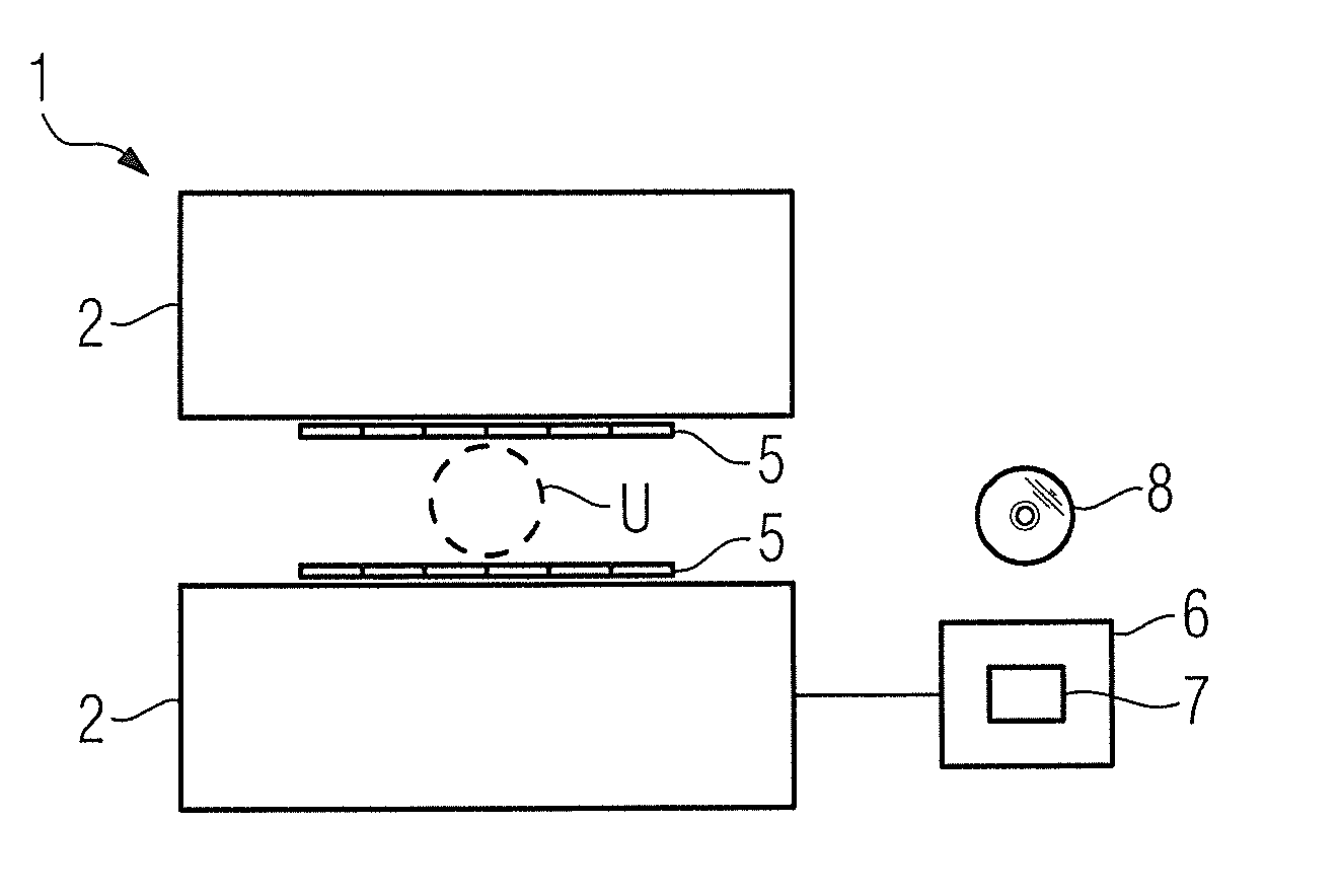
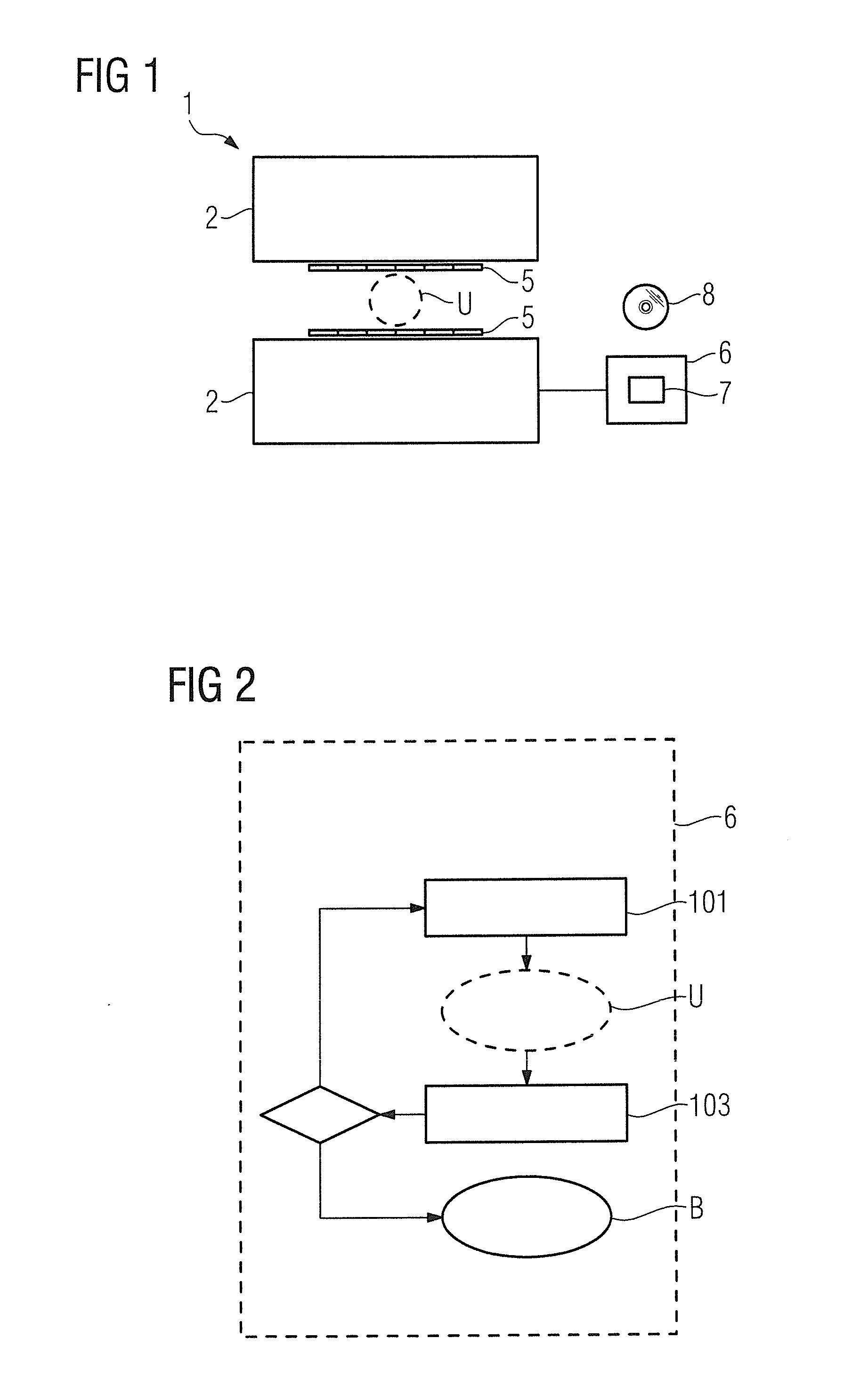
[0023]In FIG. 1 a magnetic resonance apparatus 1 is schematically represented by a magnet unit 2 and parallel RF transmission and / or reception coils 5. The basic design of a magnetic resonance apparatus composed of a magnet unit, radio-frequency coils and gradient coil unit as well as the associated control units of such a magnetic resonance apparatus are known and therefore need not be explained in detail herein.

[0024]During an MR examination (data acquisition) an examination subject—for example a region of a patient that is to be examined—is located in an examination volume U of the magnetic resonance apparatus 1. As described above, RF pulses are radiated into the examination subject located in the examination volume U in the course of the MR examination. The signals resulting in the examination subject due to the radiation of the RF pulses are acquired.

[0025]The magnetic resonance apparatus 1 is connected with a processing unit 6 which can receive data from the magnetic resonanc...


PUM


Abstract

A magnetic resonance method and system for generation of an optimized MR image of an examination subject operate as follows. A pulse sequence including a series of at least two RF pulses is radiated into the examination subject to generate at least one optimized signal, wherein the second and possibly every additional RF pulse is radiated before the effect of the first or a preceding RF pulse on the spin system in the examination subject has decayed. The radiated RF pulses are generated by parallel transmission coils. At least the signal resulting after the last radiated RF pulse of the pulse sequence is acquired. The pulse sequence is repeated with modified spatial coding until signals have been generated and acquired in a desired positional space. The optimized MR image per pulse sequence is calculated from at least one of the acquired signals.

Description

BACKGROUND OF THE INVENTION[0001]1. Field of the Invention[0002]The present invention concerns a method to generate an optimized MR image of an examination subject, a corresponding electronically readable data storage medium, and a corresponding magnetic resonance apparatus.[0003]2. Description of the Prior Art[0004]Magnetic resonance (MR) imaging is a known modality with which images of the inside of an examination subject can be generated. Expressed in simple terms, for this purpose the examination subject is positioned in a strong, static, homogeneous basic magnetic field B0 (field strengths from 0.2 to 7 Tesla or more) in a magnetic resonance apparatus so that nuclear spins in the subject orient along the basic magnetic field. For spatial coding of the measurement data, rapidly switched gradient fields are superimposed on the basic magnetic field. To trigger nuclear magnetic resonances, radio-frequency excitation pulses (RF pulses) are radiated into the examination subject by at...

Claims


Application Information

Patent Timeline
IPC IPC(8): G01R33/48G01R33/28
CPCA61B5/055G01R33/5659G01R33/5612
Inventor FAUTZ, HANS-PETER
Owner SIEMENS HEALTHCARE GMBH
Why Eureka
Eureka Blog
Learn More