Medical imaging apparatus
a medical imaging and apparatus technology, applied in the field of medical imaging apparatus, can solve the problem of difficulty in adjusting the same slice position for the operator, and achieve the effect of prolonging the examination tim
- Summary
- Abstract
- Description
- Claims
- Application Information
AI Technical Summary
Benefits of technology
Problems solved by technology
Method used
Image
Examples
first embodiment
[0044]Hereafter, the first embodiment of the present invention will be explained. In all of the drawings for explaining the embodiments of the present invention, elements having the same function are indicated with the same numerals or symbols, and repetitive explanations thereof are omitted.
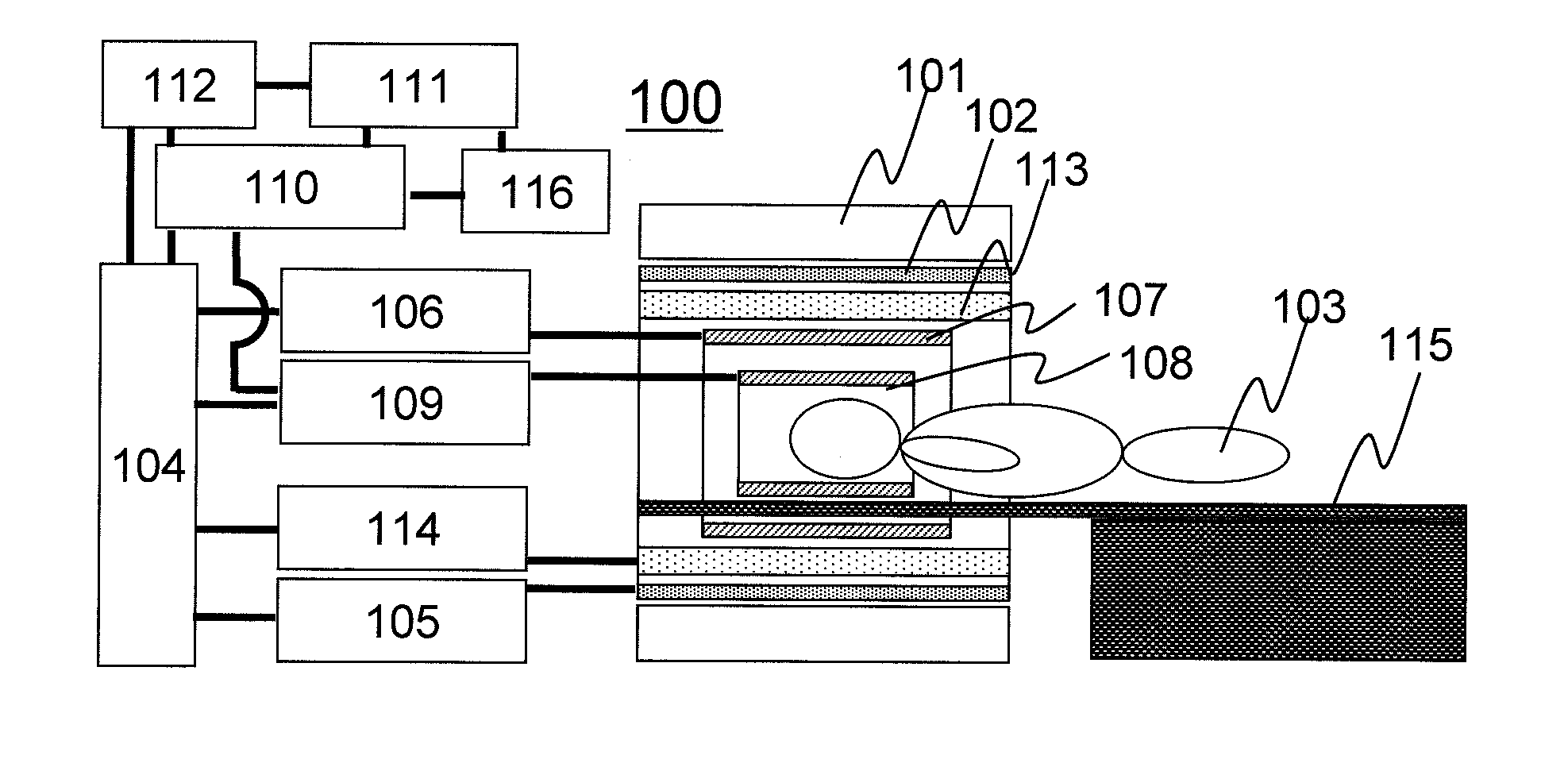
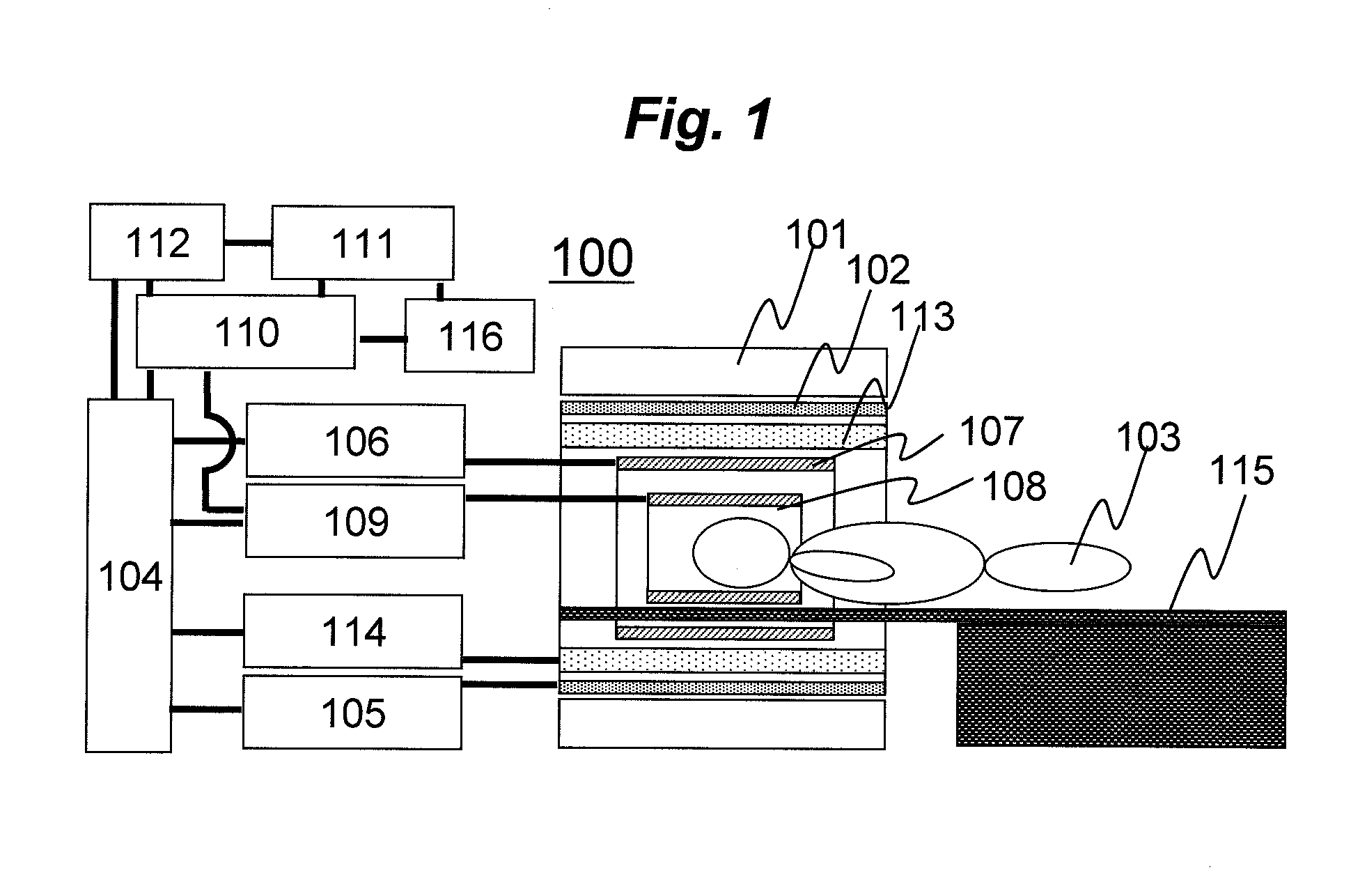
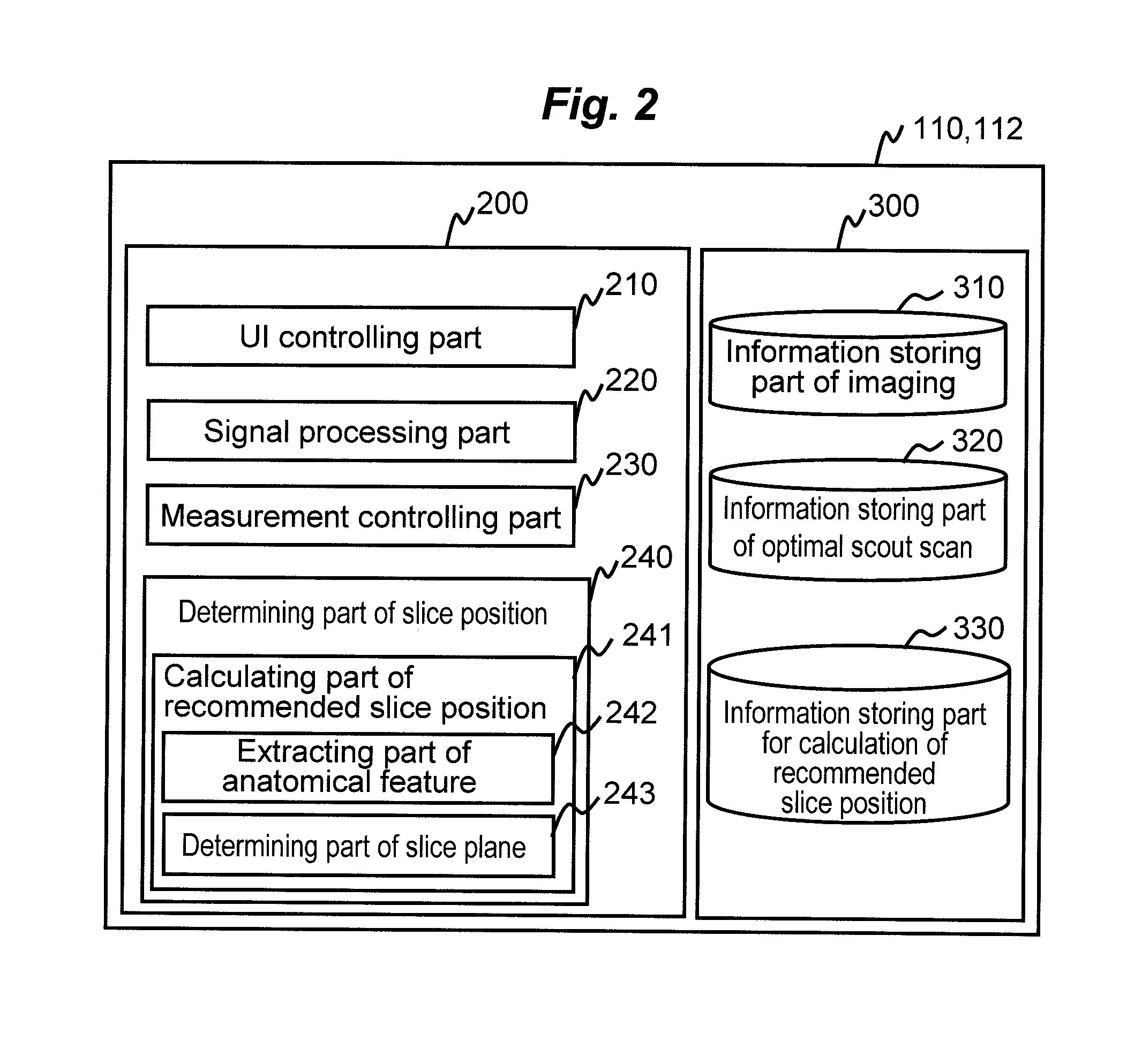
[0045]First, a magnetic resonance imaging (MRI) apparatus of this embodiment will be explained. An MRI apparatus 100 of this embodiment applies a radio frequency magnetic field to a subject 103 placed in a static magnetic field to excite nuclear magnetization in the subject 103, and measures generated magnetic resonance signals (echo signals), as described above. In this operation, a gradient magnetic field is applied to add positional information to the magnetic resonance signals to be measured for obtaining an image (imaging). FIG. 1 is a block diagram showing a typical configuration of the MRI apparatus 100 according to this embodiment, which implements the above operation. The MRI apparatus ...
second embodiment
[0158]Hereafter, the second embodiment of the present invention will be explained. The MRI apparatus according to this embodiment has basically the same configurations as those of the first embodiment. However, in this embodiment, there is provided a function of memorizing amount of adjustment received from the operator as learning data after a recommended slice position or a recommended slice for scout scan is displayed, and reflecting it in the subsequent processings. Hereafter, explanation will be made with focusing on the configurations different from those of the first embodiment.
[0159]FIG. 14 is a functional block diagram of an information processor constituted by a computer 110A and a storage device 112A according to this embodiment. In the information processor according to this embodiment, in addition to the same configurations as those of the first embodiment, the determining part of slice position 240 of a control part 200A is provided with a learning function part 244, a...
third embodiment
[0170]Hereafter, the third embodiment of the present invention will be explained. The MRI apparatus according to this embodiment has basically the same configurations as those of either one of the aforementioned embodiments. However, in this embodiment, the calculating part of recommended slice position used in the first embodiment and the second embodiment is used for MPR processing. Hereafter, this embodiment will be explained with focusing on the configurations different from those of the embodiments described above.
[0171]FIG. 15 is a functional block diagram of an information processor constituted by a computer 110B and a storage device 112B according to this embodiment. The information processor according to this embodiment is basically the same as that of either one of the information processors of the aforementioned embodiments, but in the information processor of this embodiment, in addition to the same configurations as those of the aforementioned embodiments, the control p...
PUM
 Abstract
Description
Claims
Application Information
 - R&D
- Intellectual Property
- Life Sciences
- Materials
- Tech Scout
- Unparalleled Data Quality
- Higher Quality Content
- 60% Fewer Hallucinations
Browse by: Latest US Patents, China's latest patents, Technical Efficacy Thesaurus, Application Domain, Technology Topic, Popular Technical Reports.
© 2025 PatSnap. All rights reserved.Legal|Privacy policy|Modern Slavery Act Transparency Statement|Sitemap|About US| Contact US: help@patsnap.com


