Method and System for Propagation of Myocardial Infarction from Delayed Enhanced Cardiac Imaging to Cine Magnetic Resonance Imaging Using Hybrid Image Registration

a technology of enhanced cardiac imaging and hybrid image registration, applied in image enhancement, image analysis, instruments, etc., can solve the problem that the regional myocardial changes in the motion pattern caused by suspicious scars cannot be visually assessed, and achieve accurate alignment and deformation correction

Active Publication Date: 2012-05-17
SIEMENS HEALTHCARE GMBH
View PDF0 Cites 219 Cited by
  • Summary
  • Abstract
  • Description
  • Claims
  • Application Information

AI Technical Summary

Benefits of technology

[0005]Without an accurate mapping of the infarction zone to the cine images, regional myocardial changes in motion pattern caused by suspicious scars can only be visually assessed. Accordingly, accurate alignment and deformation correction between cine an...

Problems solved by technology

Without an accurate mapping of the infarction zone to the cine images, regional myoc...

Method used


Image

  • Method and System for Propagation of Myocardial Infarction from Delayed Enhanced Cardiac Imaging to Cine Magnetic Resonance Imaging Using Hybrid Image Registration
  • Method and System for Propagation of Myocardial Infarction from Delayed Enhanced Cardiac Imaging to Cine Magnetic Resonance Imaging Using Hybrid Image Registration
  • Method and System for Propagation of Myocardial Infarction from Delayed Enhanced Cardiac Imaging to Cine Magnetic Resonance Imaging Using Hybrid Image Registration

Examples


Embodiment Construction

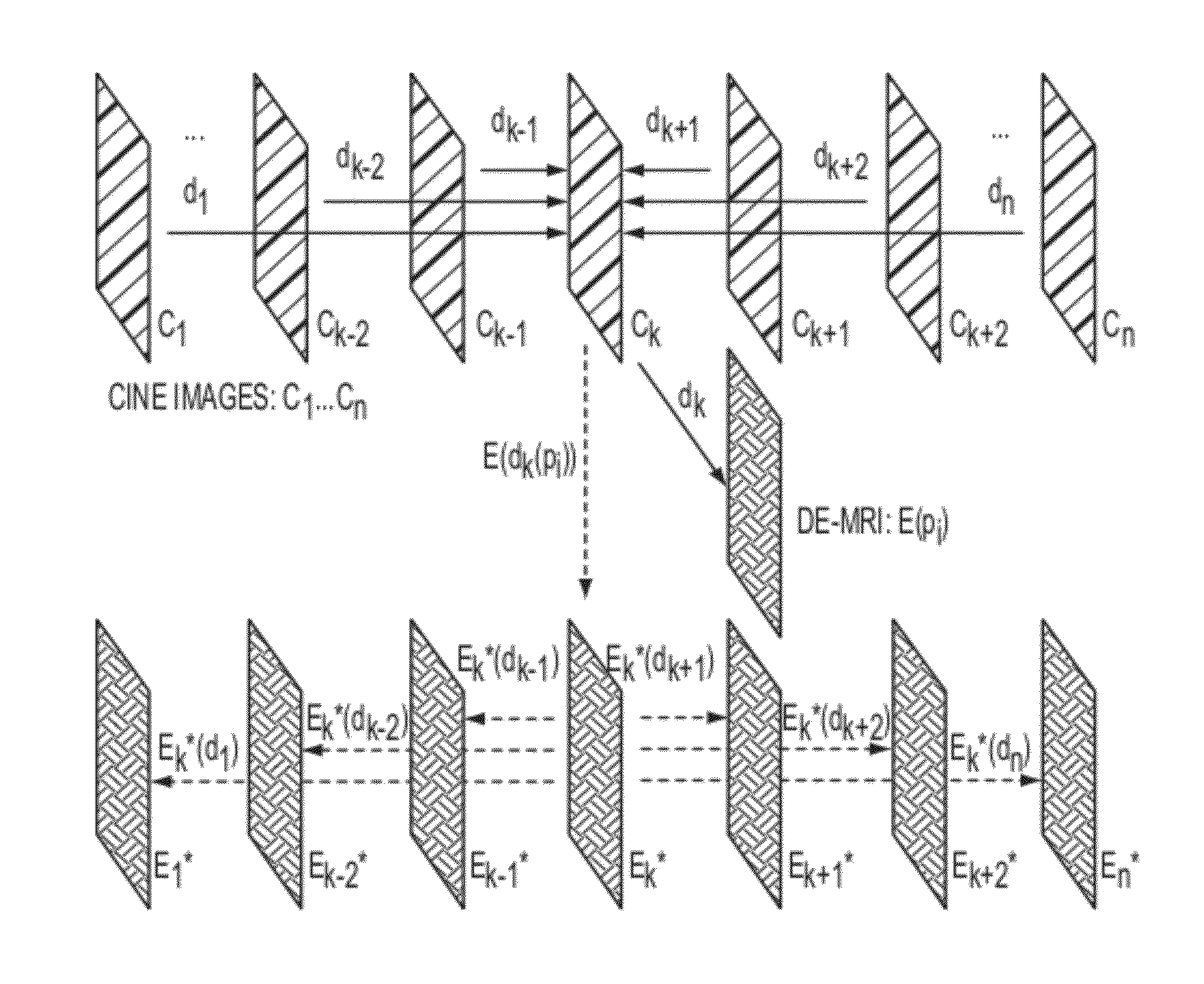
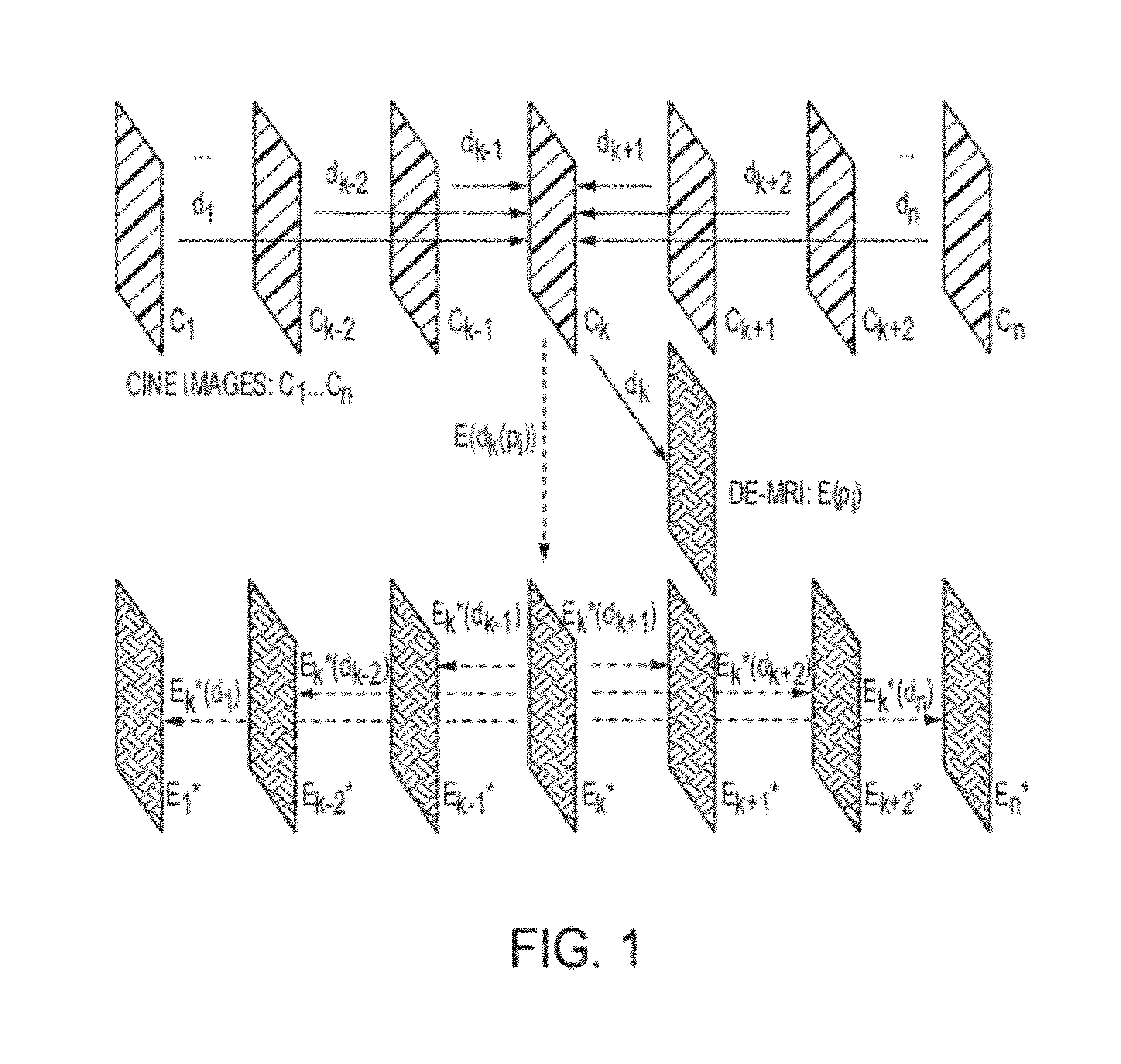
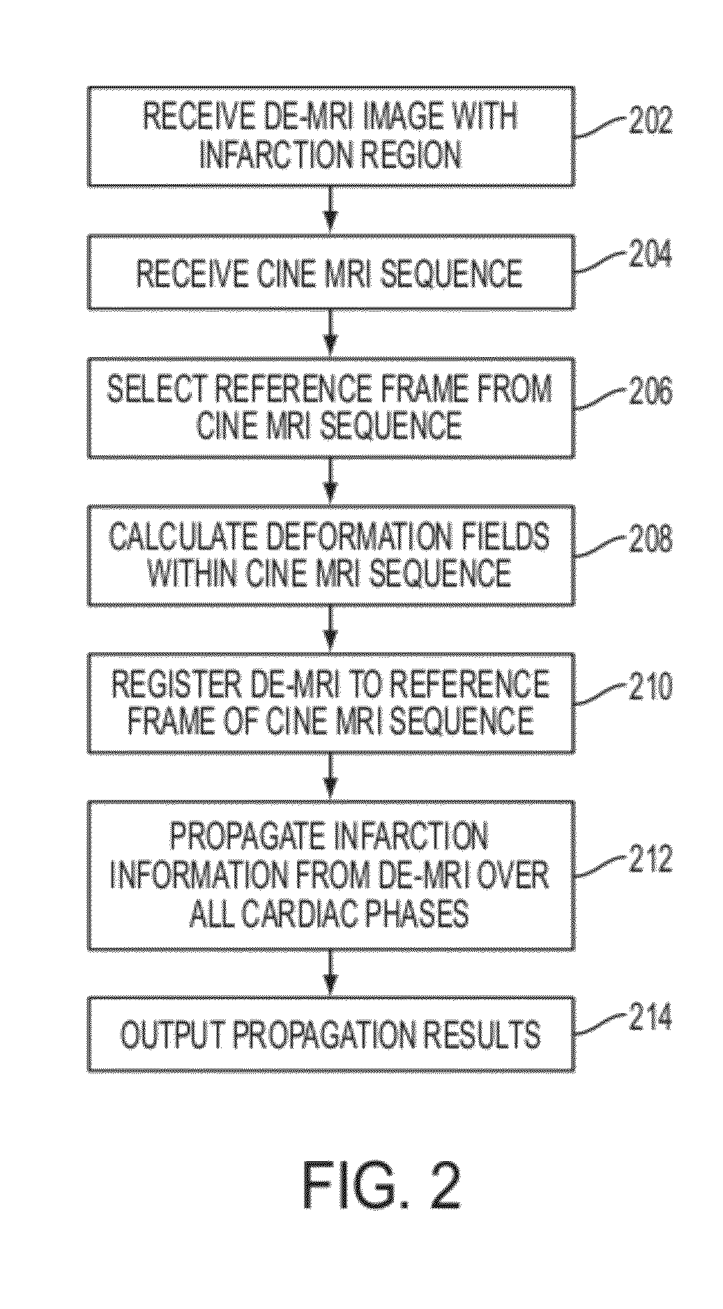
[0017]The present invention is directed to a method and system for propagation of myocardial infarction from delayed enhanced magnetic resonance imaging (DE-MRI) over a cardiac cycle using cine MRI. Embodiments of the present invention are described herein to give a visual understanding of the DE-MRI infarction propagation method. A digital image is often composed of digital representations of one or more objects (or shapes). The digital representation of an object is often described herein in terms of identifying and manipulating the objects. Such manipulations are virtual manipulations accomplished in the memory or other circuitry / hardware of a computer system. Accordingly, is to be understood that embodiments of the present invention may be performed within a computer system using data stored within the computer system.

[0018]Embodiments of the present invention provide align a DE-MRI image with a corresponding cine image and propagating a suspicious infarction zone from the DE-MR...


PUM


Abstract

A method and system for propagation of myocardial infarction from delayed enhanced magnetic resonance imaging (DE-MRI) to cine MRI is disclosed. A reference frame is selected in a cine MRI sequence. Deformation fields are calculated within the cine MRI sequence to register the frames of the cine MRI sequence to the reference frame. A DE-MRI image having an infarction region is registered to the reference frame of the cine MRI sequence. The DE-MRI image may be registered to the infarction region using a hybrid registration algorithm that unifies both intensity and feature points into a single cost function. Infarction information in the DE-MRI image is then propagated cardiac phases of the frames in the cine MRI sequence based on the registration of the DE-MRI image to the reference frame and the plurality of deformation fields calculated within the cine MRI sequence.

Description

[0001]This application claims the benefit of U.S. Provisional Application No. 61 / 413,606, filed Nov. 15, 2010, the disclosure of which is herein incorporated by reference.BACKGROUND OF THE INVENTION[0002]The present invention relates to cardiac imaging, and more particularly, to propagation of myocardial infarction from delayed enhanced cardiac imaging to cine magnetic resonance imaging.[0003]Cardiac magnetic resonance imaging (MRI) has been proven effective for determining patient-specific myocardial motion and functional information using cine imaging, as well as for detection of myocardial infarction using delayed enhanced MRI (DE-MRI). Recent studies have compared myocardial tissue viability revealed in the DE-MRI to the functional deficits measured with cine MRI, showing the so-called “peri-infarction zone” defined in DE-MRI is correlated well with the dysfunctional myocardial region defined in cine. This information is potentially valuable for reperfusion therapy, as regional ...

Claims


Application Information

Patent Timeline
IPC IPC(8): G06K9/00
CPCG06T7/0028G06T2207/30048G06T2207/10088G06T7/33
Inventor XUE, HUILIU, YIXUNGUETTER, CHRISTOPHJOLLY, MARIE-PIERREGUHRING, JENS
Owner SIEMENS HEALTHCARE GMBH
Why Eureka
Try Eureka