Phosphopeptides as melanoma vaccines

a technology of phosphopeptides and melanoma, applied in the field of cancer immunotherapy and immunodiagnostics, can solve problems such as limited clinical success, and achieve the effect of reducing the risk of primary cancer

Active Publication Date: 2012-07-12
THE JOHN HOPKINS UNIV SCHOOL OF MEDICINE
View PDF2 Cites 8 Cited by
  • Summary
  • Abstract
  • Description
  • Claims
  • Application Information

AI Technical Summary

Benefits of technology

[0014]These and other embodiments which will be apparent to those of skill in the art upon reading the specification as a whole provide the a...

Problems solved by technology

Immunotherapies directed against currently defined tumor-associated or tumor-specific antigens can enhance anti-...

Method used


Image

  • Phosphopeptides as melanoma vaccines
  • Phosphopeptides as melanoma vaccines
  • Phosphopeptides as melanoma vaccines

Examples


example 1

T Cell Recognition of the Candidate Phosphopeptide B-Raf mutpT599

[0034]To assess the potential for human CD4+ T cells to specifically discriminate phosphoresidues, we first explored the candidate mutant melanoma antigen B-RafV600E (B-Raf mut) shared by 60% of melanomas (20). The somatic V600E mutation constitutively activates the B-Raf serine-threonine kinase and hence the MAPK cascade, presumably by mimicking a phosphorylation event. V600E is juxtaposed to the known dominant phosphosite T599. We previously reported that in vitro stimulation of melanoma patient peripheral blood mononuclear cells (PBMC) with a non-phosphorylated 29-mer candidate B-Raf mut peptide generated B-Raf mut-specific HLA-DRβ1*0404-restricted CD4+ T cells that did not cross-react with the wild type peptide (B-Raf wt) and that specifically recognized HLA compatible melanoma cells expressing B-Raf mut(21). To investigate the fine specificity of MHC II-restricted CD4+ T cells for phosphate moieties, T cells rais...

example 2

Identification and Characterization of HLA-DR-Associated Phosphopeptides

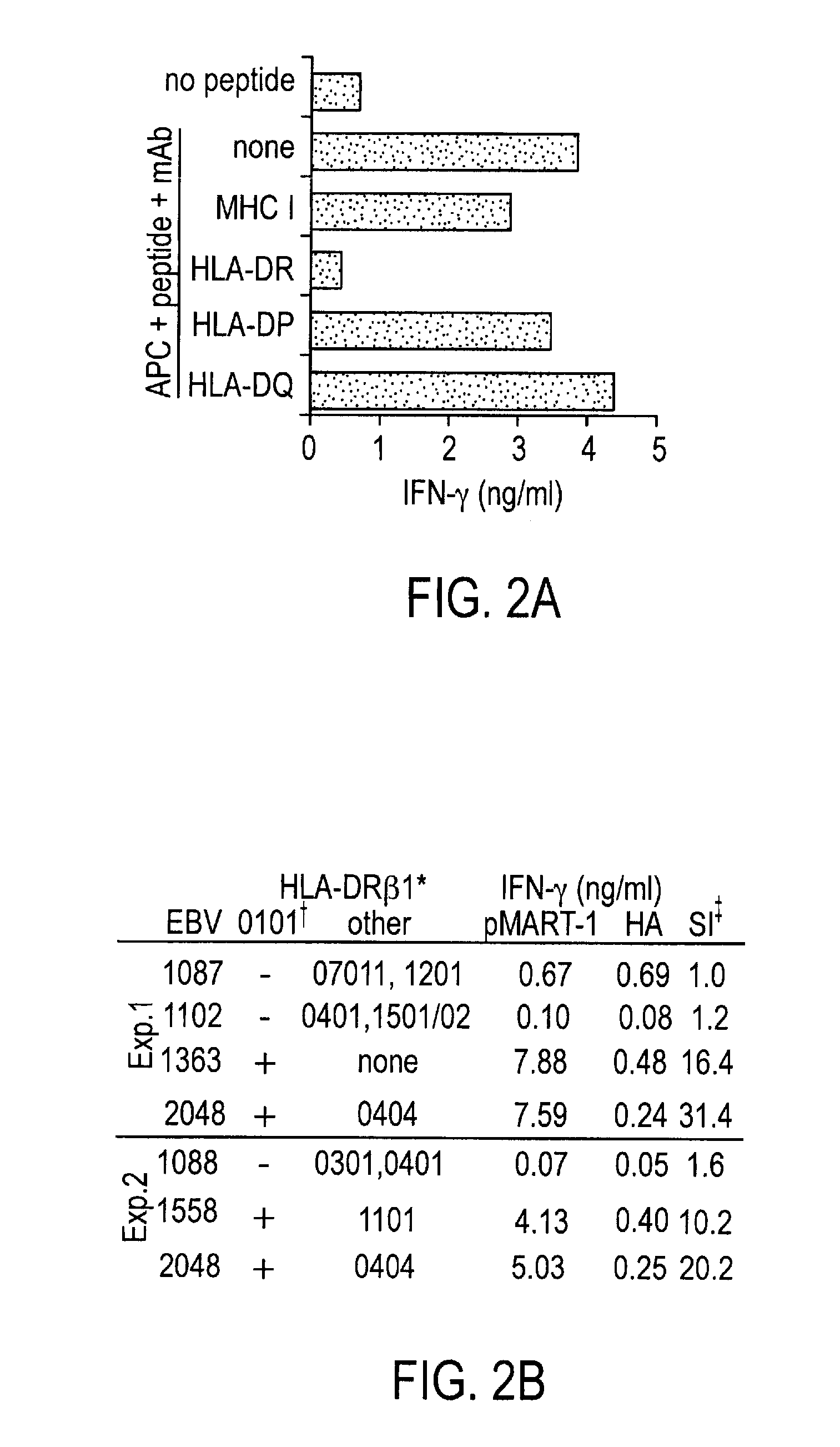
[0035]To identify naturally processed tumor-associated MHC II-restricted phosphopeptides as potential targets for immune recognition, we affinity isolated HLA-DR-peptide complexes from 2 cultured melanoma lines (1363-mel and 2048-mel) and their autologous EBV-B cell counterparts (1363-EBV and 2048-EBV). These cell lines were selected because they constitutively express significant levels of common HLA-DR molecules, as assessed with flow cytometric analysis using the pan-DR mAb L243 and HLA allele-specific mAbs (not shown). By HLA genotyping, 2048-mel and 2048-EBV contain HLA-DRβ1*0101, which is found in 31% of melanoma patients (23); DRβ1*0404, found in 6.5% of patients (23); and DRβ4*0103. Notably, 1363-mel and -EBV contain a single DR molecule, HLA-DR DRβ1*0101, affording an opportunity to isolate phosphopeptides with unambiguous HLA allele restriction. Patients 2048 and 1363 share HLA-DR DRβ1*0101, enabling t...

example 3

Specific CD4+ T Cell Recognition of Phospho-MART-1

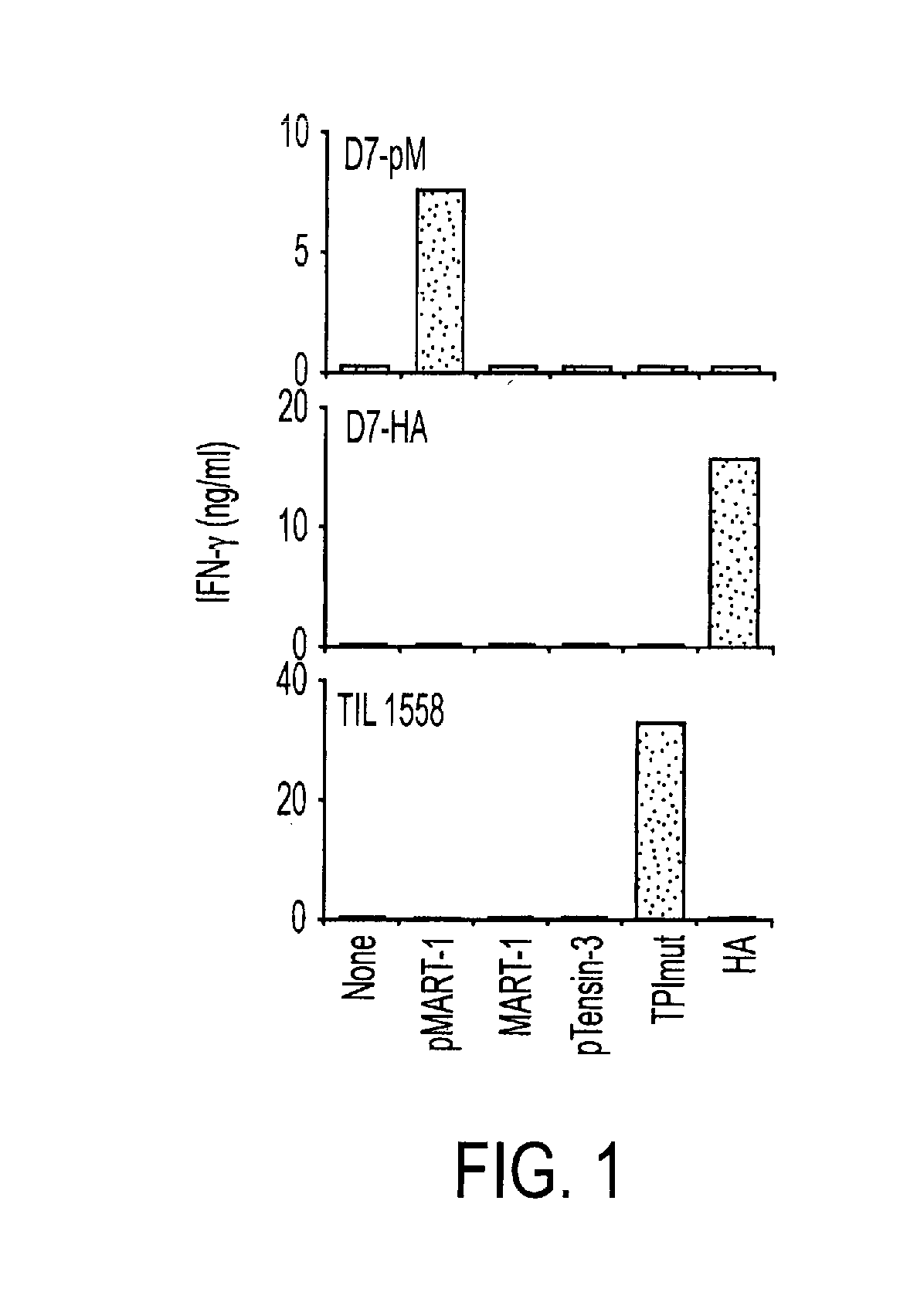
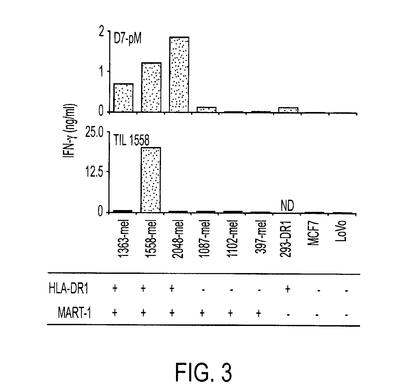
[0040]To assess the ability of human CD4+ T cells to specifically recognize tumor-associated phosphopeptides, we selected the MART-1100-111 phosphopeptide (pMART-1, containing pS108) for further study. As shown in FIG. 4, a nested set of phosphopeptides derived from the C-terminus of MART-1 was eluted from both 1363- and 2048-mel which share HLA-DR DRβ1*0101, but not from the autologous EBV-B cell lines. Because of its selective expression pattern in cells of the melanocytic lineage, including normal melanocytes and melanoma cells, MART-1 (also termed Melan-A) is an important target of immunotherapeutic approaches for the treatment of melanoma including vaccines and adoptive T cell transfer (31, 32) This transmembrane protein, which is localized to melanosomes and functions to regulate mammalian pigmentation (33), was not known to be phosphorylated prior to the current report. However, several non-phosphorylated MHC class I- and II-r...


PUM

PropertyMeasurementUnit
Densityaaaaaaaaaa
Densityaaaaaaaaaa
Densityaaaaaaaaaa

Abstract

We characterized a total of 175 HLA-DR-associated phosphopeptides using sequential affinity isolation, biochemical enrichment, mass spectrometric sequencing and comparative analysis. Many were derived from source proteins which may have roles in cancer development, growth and metastasis. Most were expressed exclusively by either melanomas or transformed B cells, suggesting the potential to define cell type-specific phosphatome “fingerprints”. We generated HLA-DRβ1*0101-restricted CD4+ T cells specific for a phospho-MART-1 peptide identified in two melanoma cell lines. These T cells showed specificity for phosphopeptide-pulsed antigen presenting cells as well as for intact melanoma cells. MHC II-restricted phosphopeptides recognizable by human CD4+ T cells are potential targets for cancer immunotherapy.

Description

[0001]The invention was made using funding from the U.S. government. The U.S. retains certain rights in the invention according to the terms of a grant from the National Institute of Allergies and Infectious Diseases 2 R37 AI 33993-15.TECHNICAL FIELD OF THE INVENTION[0002]This invention is related to the area of immunotherapy and immunodiagnostics. In particular, it relates to cancer immunotherapy and immunodiagnostics.BACKGROUND OF THE INVENTION[0003]Immunotherapies directed against currently defined tumor-associated or tumor-specific antigens can enhance anti-tumor immunity in patients, as detected with in vitro immune monitoring, and yet have had limited clinical success (1). One reason may be the nature of the targeted antigens, the majority of which are proteins overexpressed in tumor cells but not essential to maintaining their malignant phenotype.[0004]Phosphorylation is the most common and ubiquitous form of enzyme-mediated post-translational protein modification, and transi...

Claims


Application Information

Patent Timeline
IPC IPC(8): A61K39/00A61K35/12C07K16/30A61P35/00A61P35/04A61P37/04C12N5/0784C07K7/08C07K14/74C07K14/47G01N33/566C07K7/06C12N5/0783
CPCA61K39/0011A61K2039/5154G01N33/5743C07K14/4748A61K2039/5158A61P35/00A61P35/04A61P37/04A61K39/001191A61K39/39
Inventor TOPALIAN, SUZANNE L.DEPONTIEU, FLORENCEHUNT, DONALDSHABANOWITZ, JEFFREYQIAN, JIEENGELHARD, VICTORZARING, ANGELA
Owner THE JOHN HOPKINS UNIV SCHOOL OF MEDICINE
Why Eureka
Try Eureka