Method, an optical probe and a confocal microscopy system for inspecting a solid organ

a confocal microscopy and optical probe technology, applied in the direction of dianostics using fluorescence emission, sensors, catheters, etc., can solve the problems of inability to accurately diagnose, and inability to perform a diagnosis

Active Publication Date: 2012-07-19
MAUNA KEA TECHNOLOGIES
View PDF18 Cites 13 Cited by
  • Summary
  • Abstract
  • Description
  • Claims
  • Application Information

AI Technical Summary

Benefits of technology

[0033]Preferably, the optical fiber bundle, the ferule and the sheath each have an external diameter of preferably less than 0.9 mm, preferably less than 0.8 mm, preferably less than 0.7 mm.
[0034]Preferably, the length of the ferule is less than 8 mm, preferably less than 7 mm, preferably less than 6 mm. Preferably, the axial length of the ferule is less than 8 mm, preferably less than 7 mm, preferably less than 6 mm.

Problems solved by technology

Histological examination of suspected lesions and structural alterations in solid organs, e.g. in liver cirrhosis or staging of pancreatic malignant disease, is a continuous challenge.
Radiology only offers a partial answer to this issue because Magnetic Resonance Imaging (MRI), helical Computed Tomography (CT) scan, endoscopic ultrasonography and Positron Emission Tomography (PET) only allow a low accuracy diagnosis with significant over and understaging status.
However, cytology has also major limitations including: incremental cost, risk, time needed to perform a diagnosis, lack of in vivo information such as blood flow, and limited ability to predict disease course.
Fine needle aspirations are particularly limited by sampling errors due to the limited number of aspirations and delayed diagnosis because of time consuming sampling processing.

Method used


Image

  • Method, an optical probe and a confocal microscopy system for inspecting a solid organ
  • Method, an optical probe and a confocal microscopy system for inspecting a solid organ
  • Method, an optical probe and a confocal microscopy system for inspecting a solid organ

Examples


Embodiment Construction

[0050]Specific embodiments of the present disclosure will now be described in detail with reference to the accompanying Figures. Like elements in the various Figures may be denoted by like numerals.

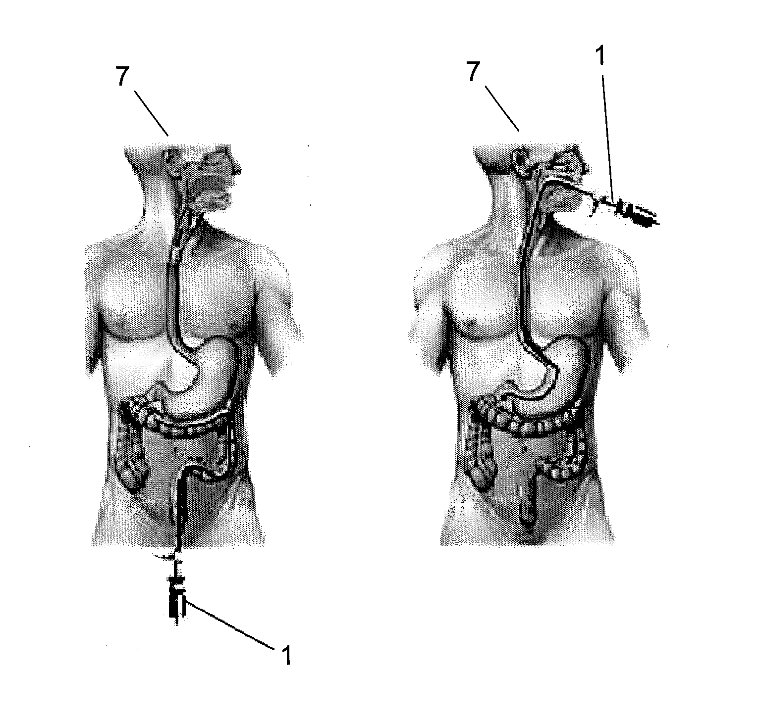
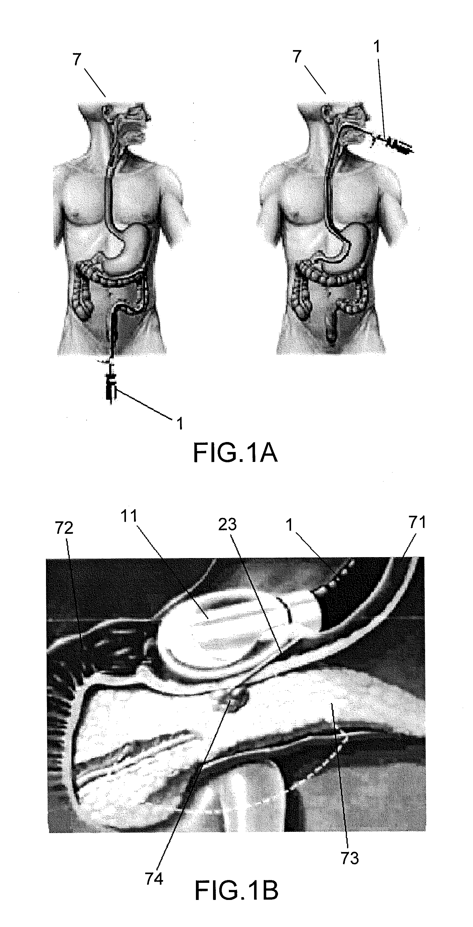
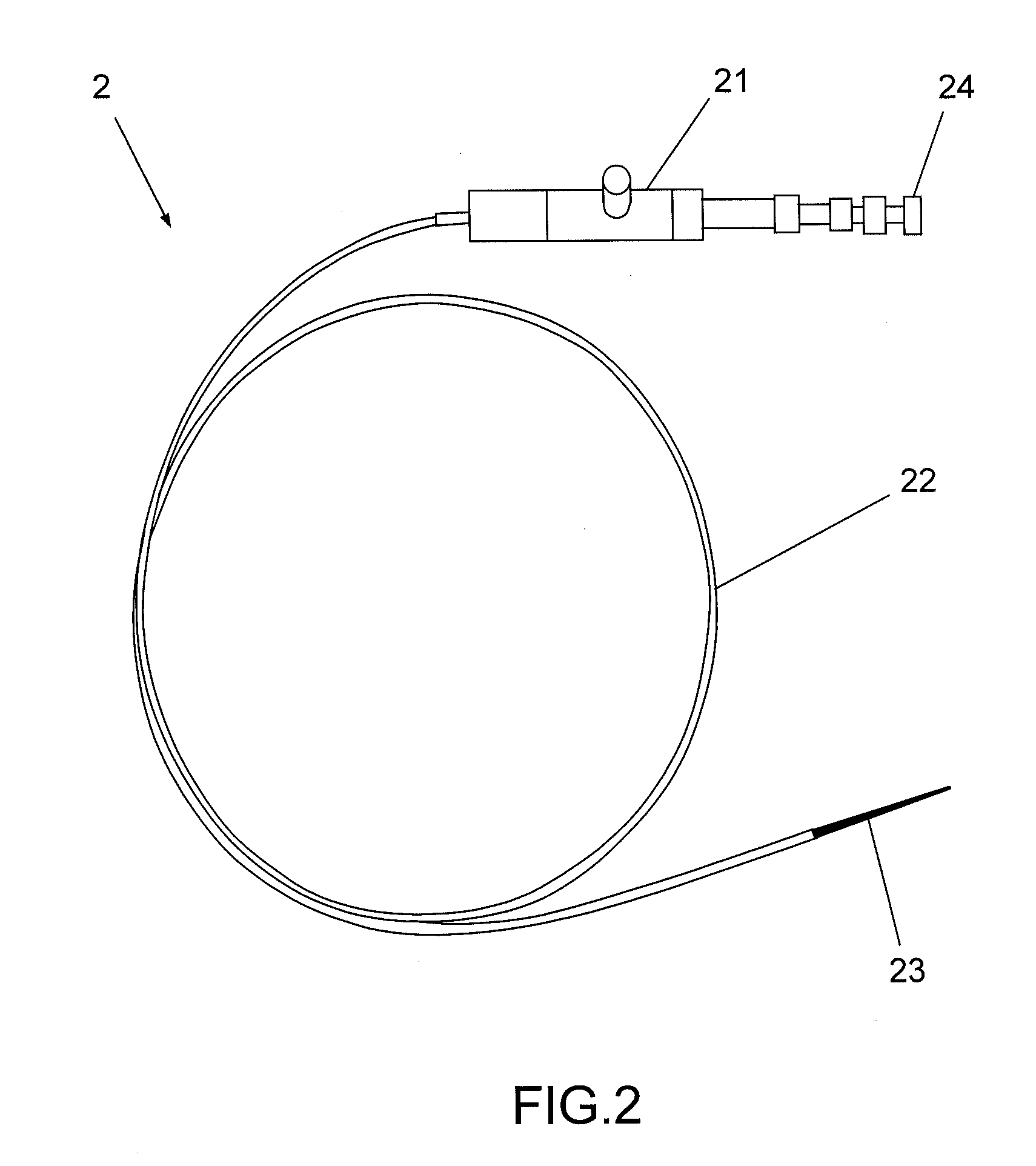
[0051]In a method for inspecting solid organs according to the present disclosure, a needle may be introduced in a solid organ of a subject. An optical probe inserted in a lumen of the needle may be brought in contact of a predetermined area of the organ to image the predetermined area. The optical probe may be used together with a confocal microscopy system. Imaging the organ according to this method may enable to obtain microscopic pictures of the predetermined area and may help establishing a diagnosis in real time. The solid organ may be one selected from the group consisting of a pancreas, a liver, a spleen, a lymph node, breast, ovaries, a kidney or a prostate.

[0052]The step of introducing the needle in the solid organ may comprise puncturing the solid organ, preferably with a tip o...


PUM


Abstract

A method to inspect a solid organ in a subject includes introducing a needle in a predetermined area of the solid organ, inserting an optical probe through a lumen of the needle, and imaging the predetermined area using the optical probe. An optical probe to inspect a solid organ in a subject, the optical probe being intended to be positioned in the solid organ through a needle, the optical probe includes an optical fiber bundle, a ferule to protect the distal tip of the optical fiber bundle, the ferule comprising a shank and a head, and a sheath wrapping the fiber bundle and the shank, wherein the head of the ferule has a length adapted for the optical probe to image the solid organ while keeping the sheath inside the needle.

Description

BACKGROUND[0001]1. Field of the Present Disclosure[0002]The disclosure generally relates to organ inspection. More specifically, it relates to organ inspection of living subjects for diagnosing purpose and / or therapeutic applications.[0003]2. Background Art[0004]In the following description, a solid organ is defined as an organ that does not contain a cavity or lumen and that is not gaseous. A solid organ may for example consist of parenchyma and stroma, the latter often arranged as trabeculae or surrounding groups of parenchymatous cells to provide support (e.g. liver, kidney). A solid organ may also comprise cysts. Histological examination of suspected lesions and structural alterations in solid organs, e.g. in liver cirrhosis or staging of pancreatic malignant disease, is a continuous challenge. Radiology only offers a partial answer to this issue because Magnetic Resonance Imaging (MRI), helical Computed Tomography (CT) scan, endoscopic ultrasonography and Positron Emission Tomo...

Claims


Application Information

Patent Timeline
Patent Type & Authority Applications(United States)
IPC IPC(8): A61B1/06A61B5/055A61B6/03A61B8/00
CPCA61B5/0068A61B5/0071A61B5/0084A61B5/6848A61B5/416A61B5/418A61B5/415
Inventor BOULAROT, NICOLASGENET, MAGALIESCHWARZ, FRANCE
Owner MAUNA KEA TECHNOLOGIES
Why Eureka
Try Eureka