Pneumatically driven ophthalmic scanning endoprobe

a scanning endoscope and pneumatic drive technology, applied in the field of ophthalmic microsurgical endoscopes, can solve the problems of difficult to discard after one or only a few surgical procedures, complex operation of endoscopes, and cost of most prior art devices

Active Publication Date: 2012-09-27
ALCON INC
View PDF11 Cites 88 Cited by
  • Summary
  • Abstract
  • Description
  • Claims
  • Application Information

AI Technical Summary

Problems solved by technology

Current state of the art provides endoprobes that are quite complex in operation, often times requiring moving parts that are operated using complex mechanical systems.
Most of the prior art devices have a cost and that makes them difficult to discard after one or only a few surgical procedures.
Furthermore, prior art devices gener

Method used


Image

  • Pneumatically driven ophthalmic scanning endoprobe
  • Pneumatically driven ophthalmic scanning endoprobe
  • Pneumatically driven ophthalmic scanning endoprobe

Examples


Embodiment Construction

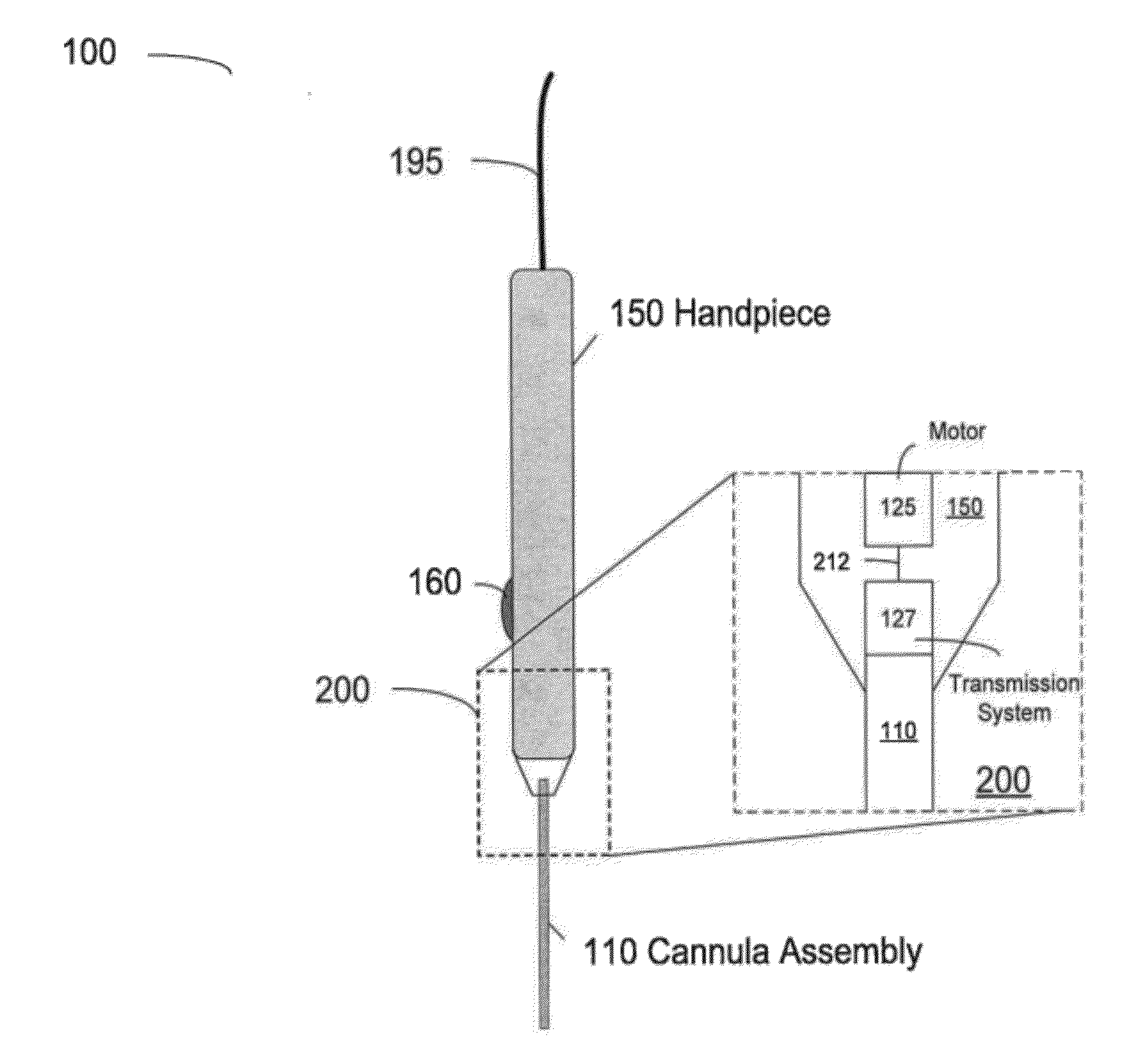
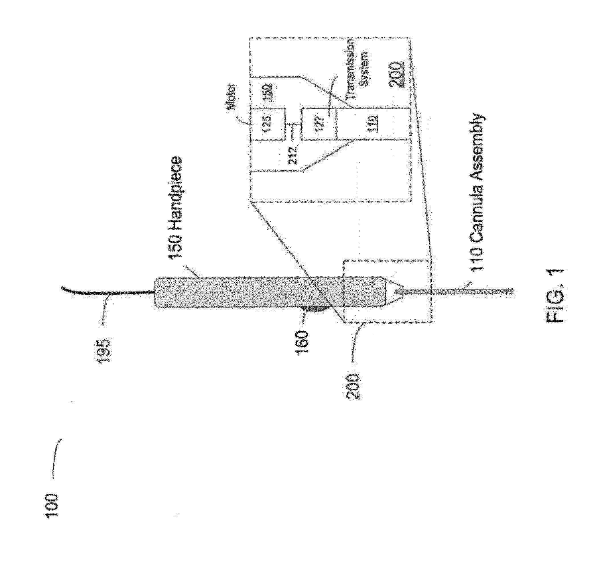
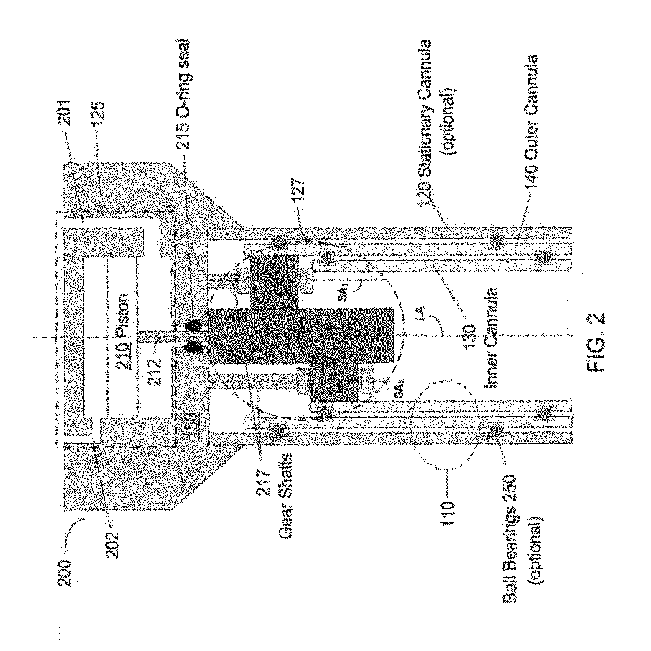
[0026]Microsurgical procedures using endoscopic instruments may include an endoprobe having a simple and cost-effective drive coupling system. The endoprobe may be a hand-held endoprobe for direct manipulation by specialized personnel. In some embodiments, the endoprobe may be controlled by a robotic arm or a computer-controlled device. Endoprobes have a proximal end close to the operation controller (be it a specialist or a device), and a distal end, close to or in contact with the tissue. Endoprobes according to embodiments disclosed herein may have small dimensions, be easy to manipulate from a proximal end, and be minimally invasive to the surrounding tissue. In the distal portion, the endoprobe ends with a tip, from where the endoprobe performs certain action on a target tissue located in the vicinity of the tip. For example, the endoprobe may deliver light from its tip, and receive light reflected or scattered from the tissue, coupled through the tip. The tip of the endoprobe ...


PUM


Abstract

An ophthalmic endoprobe including a hand-piece and a cannula assembly having a longitudinal axis is provided. The cannula assembly including an inner tube concentric with an outer tube; wherein the hand-piece may further include a motor powered by a pneumatic energy source, the motor providing motion to a transmission shaft; and a transmission system to couple the shaft motion to the cannula assembly; wherein the transmission system provides a counter-rotating motion to the inner tube and the outer tube about the longitudinal axis of the cannula. A fluid console for use in endoscopic ophthalmic microsurgery including a pneumatics module to obtain a pneumatic force from an external source and provide an adjustable pneumatic force; a scanning module coupled to the pneumatics module; and an endoprobe coupled to the scanning module is also provided.

Description

CROSS-REFERENCE TO RELATED APPLICATION[0001]This application claims priority to and benefit of U.S. provisional application Ser. No. 61 / 466,364 filed Mar. 22, 2011, which is herein incorporated in its entirety by reference.BACKGROUND[0002]1. Field of the Invention[0003]Embodiments described herein relate to the field of ophthalmic microsurgical endoprobes. More particularly, embodiments described herein are related to the field of endoscopic Optical Coherence Tomography (OCT) and to the field of ophthalmic microsurgical techniques.[0004]2. Description of Related Art[0005]The field of ophthalmic microsurgical procedures is evolving rapidly. Typically, these procedures involve the use of endoprobes that are capable of reaching the tissue that is being operated or diagnosed. Such procedures make use of endoscopic surgical instruments having an endoprobe coupled to a control device in a remote console. Current state of the art provides endoprobes that are quite complex in operation, oft...

Claims


Application Information

Patent Timeline
IPC IPC(8): A61B17/00
CPCA61B1/00172A61B5/0066A61B2017/00539A61B2017/00544A61F9/00763A61F9/00802A61B2017/320028A61B3/102A61F9/007A61B17/00A61B5/00
Inventor PAPAC, MICHAEL JAMESYADLOWSKY, MICHAEL J.HUCULAK, JOHN CHRISTOPHER
Owner ALCON INC
Why Eureka
Try Eureka