Laser Video Endoscope

a laser endoscope and endoscope technology, applied in the field of laser video endoscopes, can solve the problems of small diameter of the probe, fragile and tend to break, and problems such as the risk of breaking, and achieve the effect of reducing the risk of breaking

Inactive Publication Date: 2012-10-18
BEAVER VISITEC INT US
View PDF18 Cites 12 Cited by
  • Summary
  • Abstract
  • Description
  • Claims
  • Application Information

AI Technical Summary

Benefits of technology

[0006]One embodiment of the surgical instrument of this invention employs a stainless steel probe having a distal portion and a proximal portion. The distal portion has an OD that is less than 25 mils (thousandths of an inch) with a two mil wall thickness. Thus it can be inserted through a 23 gauge sleeve. The proximal portion of the probe, exiting from the hand piece, has a 31 mil OD and a wall thickness of five mils. The distal 25 mils diameter portion has a length of 710 mils. This combination of three design features provides a probe that can fit through a 25 mil (23 gauge) sleeve yet be robust enough to minimize the risk of breaking. Most breakage occurs at the juncture between the hand piece and the probe.

Problems solved by technology

Reuse is important because of the expense of the endoscope.
One problem is that a 23 gauge probe is so small in diameter (25 mils) that it is fragile and tends to break.
This breakage problem becomes a major concern when using a laser video endoscope because of the cost of these endoscopes.

Method used


Image

  • Laser Video Endoscope
  • Laser Video Endoscope
  • Laser Video Endoscope

Examples


Embodiment Construction

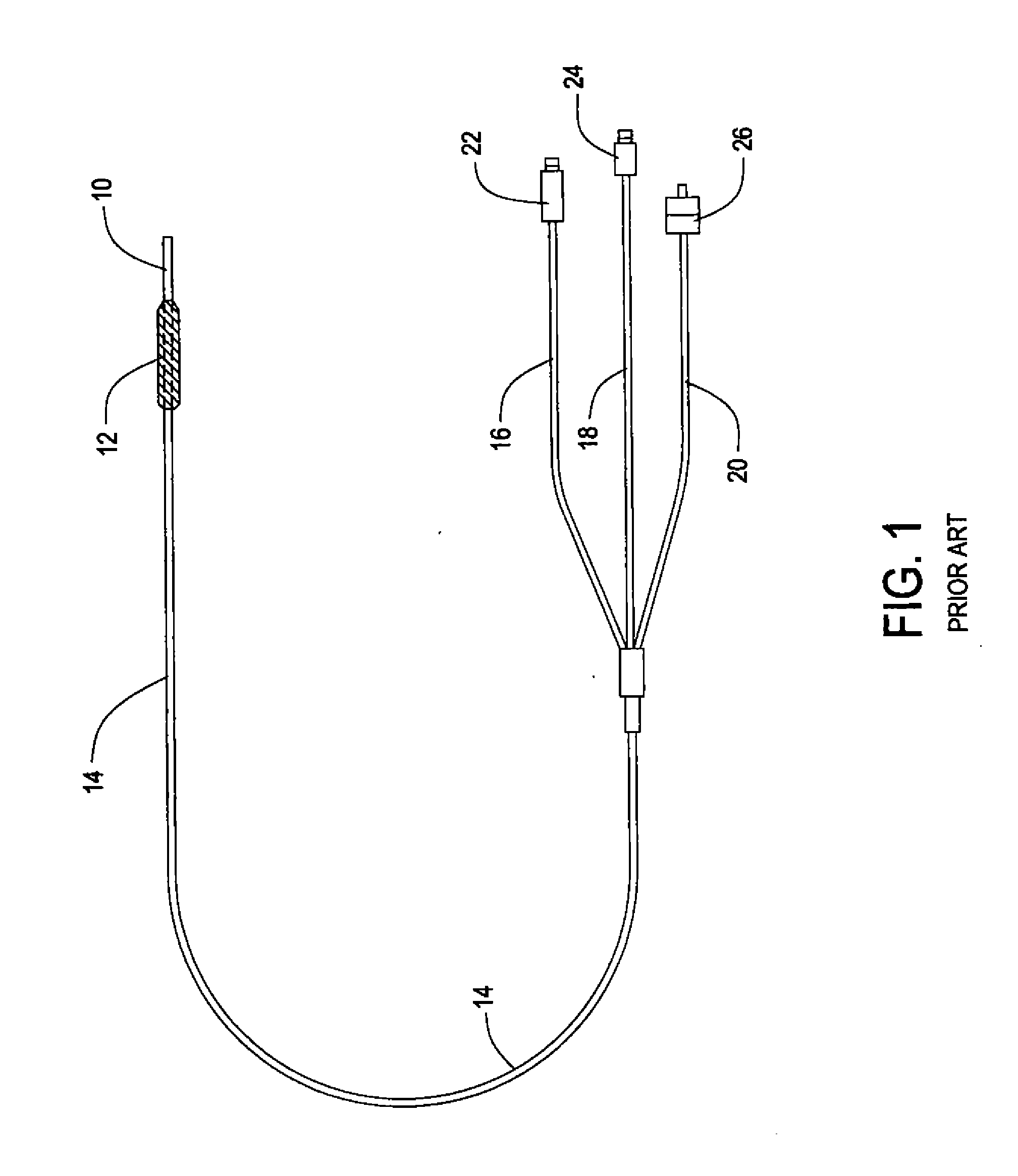
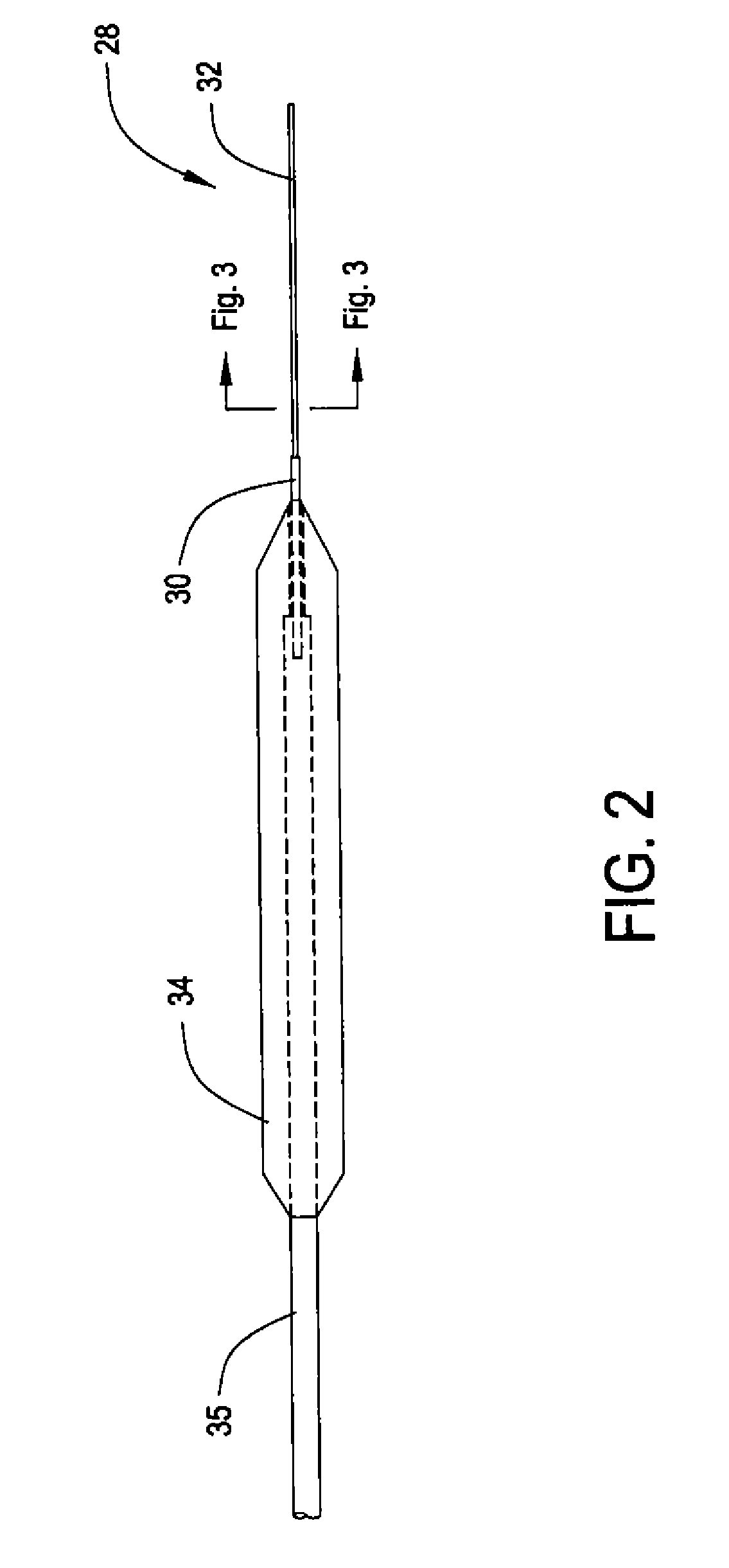
[0014]FIG. 1 illustrates one prior art device. The rest of the figures are all to a single embodiment of the device of this invention.

[0015]As shown in FIG. 1, the known video endoscope has an operating probe 10, a hand piece 12 and a cable 14. Extending through the probe, the hand piece and cable are a laser guide 16, illumination guide 18, and an image guide 20. These are all fiber optic guides which extend from the distal end of the probe 10 to the terminals 22, 24 and 26.

[0016]FIGS. 2 through 4 illustrate an embodiment of this invention showing probe 28, hand piece 34 and cable 35. The probe 28 has a proximal portion 30 and a distal portion 32. The proximal portion 30 has a 20 gauge (35 mil) outer diameter and a five mil wall thickness. The probe is stainless steel. The proximal portion extends into the hand piece 34. Thus, at the juncture of the end of the hand piece 34 and the probe 28, there is a diameter having sufficient robustness to contribute to minimizing the likelihood...


PUM


Abstract

A laser video endoscope provides a small diameter (25 mils) probe. This size probe requires a minimum access lesion. The tradeoff that produces such a probe includes reducing the laser guide fiber to 100 microns in diameter, employing an image bundle having approximately 6,000 optical fibers and an illumination bundle having only about 210 optical fibers. The probe where it extends into the handle has a 45 mil outer diameter and a 5 mil thick sidewall to provide resistance to breaking at the juncture with the handle. The probe is rigid, preferably metal. The probe has a larger diameter proximal portion and a smaller diameter distal portion. The distal portion of the probe has a length limited to about 710 mils. A green laser of 532 nanometers wavelength provides a collimated laser beam that causes minimal loss in the 100 micron laser optical fiber.

Description

BACKGROUND OF THE INVENTION[0001]This invention relates in general to a laser video endoscope for use in ophthalmology operations and more particularly to one in which the operating probe has a small diameter so that, for example, it can be passed through a 23 gauge sleeve such as a trocar sleeve.[0002]Laser video endoscopes are known and examples are described in Applicant's issued U.S. Pat. No. 5,121,740 issued on Jun. 16, 1992 and U.S. Pat. No. 6,997,868 issued on Feb. 14, 2006. Disclosures of these two patents are incorporated herein by reference. These endoscopes used in ophthalmology operations are either disposable or reused after autoclaving or sterilization. Reuse is important because of the expense of the endoscope. These prior art endoscopes are employed with the probe passing through a 20 gauge tissue incision during ophthalmological surgery. A 20 gauge incision has been a standard in the art and is used for entry by instruments employed during an ophthalmological surgic...

Claims


Application Information

Patent Timeline
Patent Type & Authority Applications(United States)
IPC IPC(8): A61F9/008
CPCA61F9/008A61B1/042A61B1/00167A61B3/156A61B1/07A61B1/04A61B17/00
Inventor URAM, MARTIN
Owner BEAVER VISITEC INT US
Why Eureka
Try Eureka