
Method and Device for Perfusing Tissue by ExVivo Attachment to a Living Organism

Inactive Publication Date: 2012-11-01
GILLIS JOHN ARCHIE
View PDF0 Cites 17 Cited by
  • Summary
  • Abstract
  • Description
  • Claims
  • Application Information

AI Technical Summary

Benefits of technology

[0039]When the holding vessel is in use it will contain a tissue selection that will be attached to the circulatory system of a living organism by connecting the existing vasculature of the organism to engineered or grafted vascular cables. The other ends of the vascular cables are then connected to the vasculature of the tissue selection. A tubular construct containing a protective solution will protect the vascular cables.
[0040]The holding vessel will provide support, oxygen and nutrient delivery to the tissue selection. The present methods will provide a novel and superior means of supplying a tissue selection with nutrient delivery along with biochemical and mechanical signals that are superior to known methods.

Problems solved by technology

One of the major challenges facing tissue engineering today is the requirement for more complex functionality.
A major dilemma with most current tissue engineering technologies is that most tissue engineered structures and organs require a means of providing vascularization and perfusion to survive.
Creating this vascular supply and more viable methods of perfusion to a thick-engineered tissue construct remains one of the great challenges in the field today.
These methods have their place in medical procedures, but immunosuppressant drugs are usually always required when introducing foreign tissues and if the tissue selection contains tissues that are not a good match rejection can occur.
Current methods of perfusing a tissue structure are limited, due to time constraints.
Even with our advanced technologies, helicopters and database matching systems organs are often lost due to injuries during brain death, ischemia, cell death and other causes.
These methods of perfusion are great advances in medical technologies, but still have their limitations.
This damage often occurs due to the formation of ice.
These and similar methods are great for protecting certain portions of existing tissues for a limited amount, but are not often successful at penetrating deep into thicker tissue constructs.
It can be very difficult to position the protective solutions deep within these already existing structures.
Lab grown tissue engineered structures are also limited by these same problems.
Preserving the tissue selection or construct after it has been fabricated makes it extremely difficult to reach all the desired areas.
Preservation of organs and tissues are commonplace in medicine, but again because organs are most often donated rather that fabricated it can be difficult to place these solutions in areas that can deeply penetrate the structure, especially if the tissue or organ is a thick structure.
These methods are aimed at constructing functional organ modules however at present there has been limited success and the printing of entire organs layer-by-layer has not yet been realized.
These methods of tissue engineering still suffer from some of the limitations of traditional scaffolding methods.
There have been some great successes with these methods, but the issue of nutrient delivery is still a major concern.
A common problem with thick tissue structures is that cells deep inside the structure are damaged due to a lack of nutrient delivery.
One can delay this problem for a short by preserving the tissue with a cryoprotectant solution, but unless the tissue is prepared as described in the present invention the problems of getting cryoprotectant solutions into all the desired locations, including cells deep within the structure remains a large and limiting problem.

Method used


Image

  • Method and Device for Perfusing Tissue by ExVivo Attachment to a Living Organism
  • Method and Device for Perfusing Tissue by ExVivo Attachment to a Living Organism
  • Method and Device for Perfusing Tissue by ExVivo Attachment to a Living Organism

Examples


Embodiment Construction

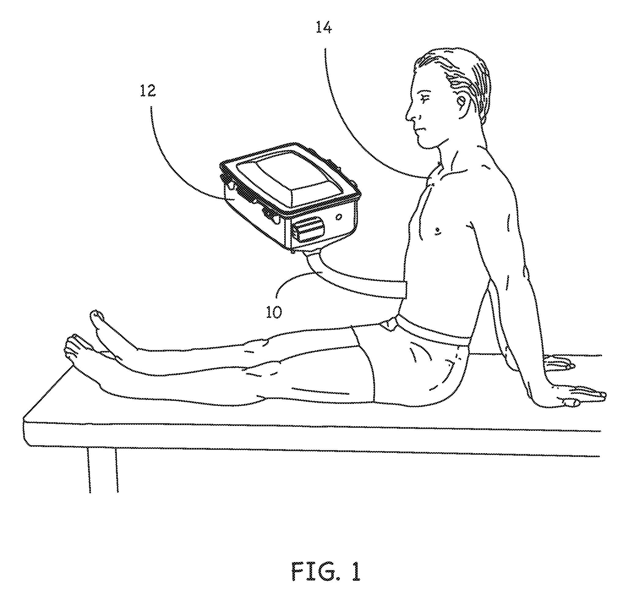
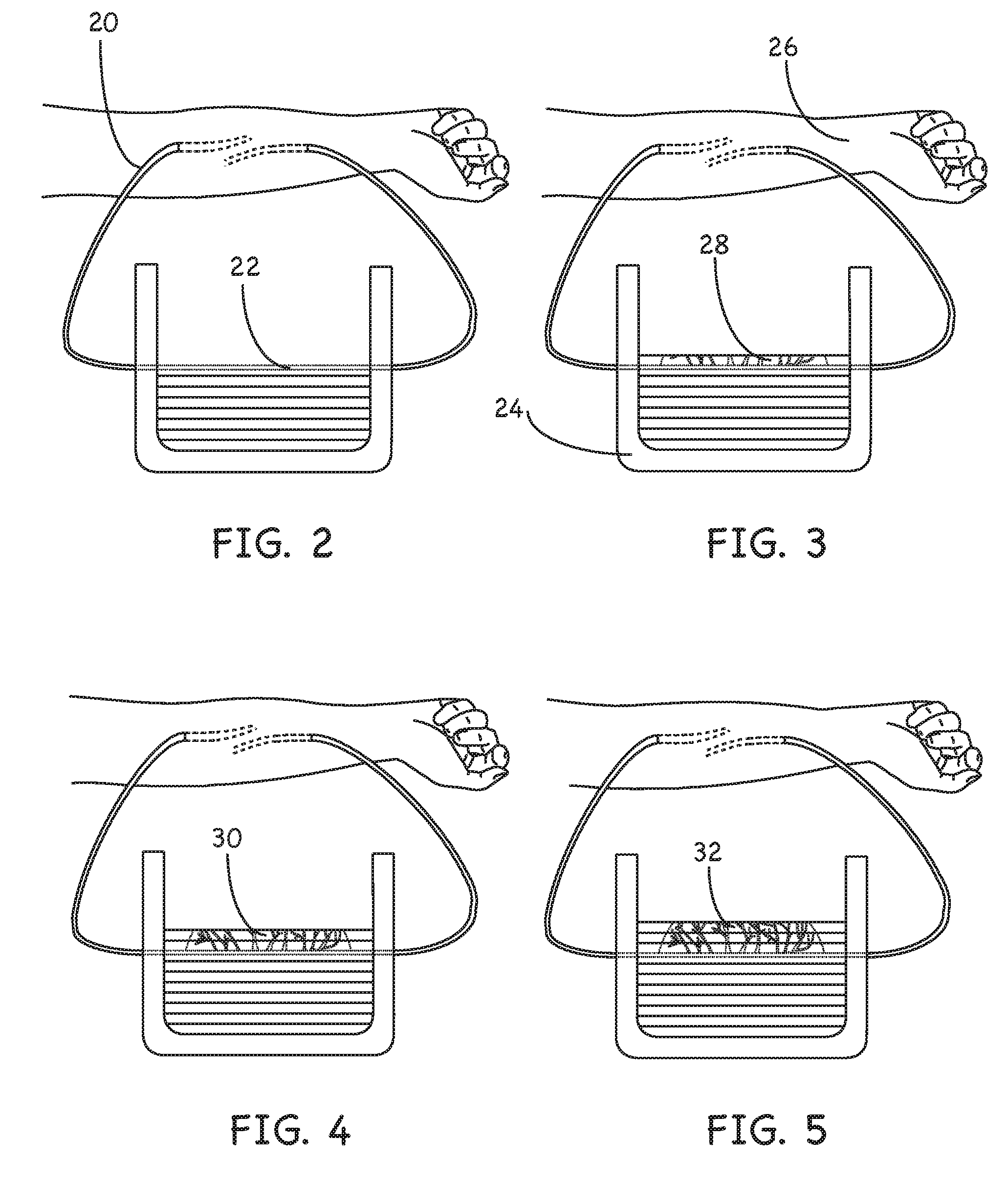
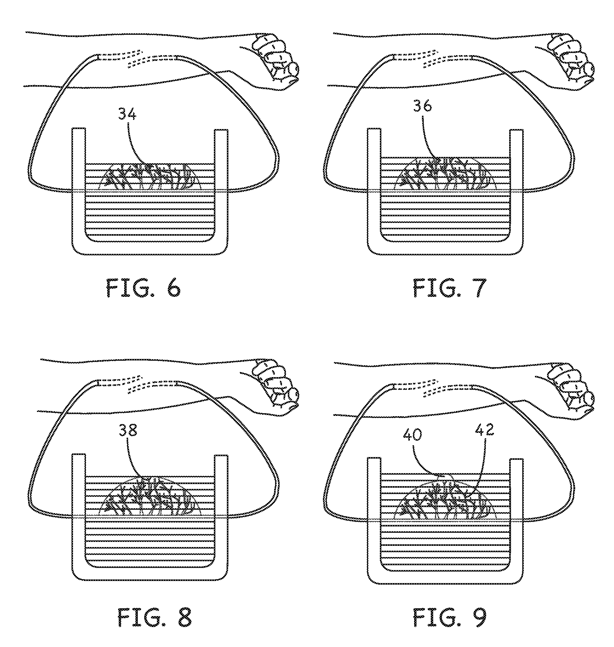
[0092]Reference will now be made in detail to various embodiments of the invention, one or more examples of which are set forth below. Each embodiment is provided by way of explanation of the invention, not limitation of the invention. In fact, it will be apparent to those skilled in the art that various modifications and variations may be made in the present invention without departing from the scope or spirit of the invention. For instance, features illustrated or described as part of one embodiment, may be used in another embodiment to yield a still further embodiment. Thus, it is intended that the present invention cover such modifications and variations as come within the scope of the appended claims and their equivalents.

[0093]In the preferred embodiments the present invention describes a novel method for the perfusion and vascularization of a tissue selection. In the preferred embodiment the selection consists of tissue-engineered constructs, but may also be very useful in pe...


PUM


Abstract

The present invention is a holding vessel that has bioreactor and perfusion bioreactor components, a temperature specific environment and holes for transporting substances from a living organism.When the holding vessel is in use it will contain a tissue selection that will be attached to the circulatory system of a living organism by connecting existing vasculature of the organism to engineered or grafted umbilical / vascular cables and then connecting the other end of the umbilical cables to the vasculature of the tissue selection. A tubular construct containing a protective solution will protect the vascular cables.The tissue selections used will be selected from existing or fabricated tissues, but preference is given to cryogenically prepared tissues electronically dispensed from a three-dimensional printing device.

Description

CROSS-REFERENCE TO RELATED APPLICATIONS[0001]U.S. Provisional Application No. 61 / 479,341FEDERALLY SPONSORED RESEARCH[0002]Not ApplicableSEQUENCE LISTING OR PROGRAM[0003]Not ApplicableBACKGROUND OF THE INVENTION[0004]1. Field of Invention[0005]The present invention relates to methods of perfusing tissue constructs.[0006]2. Prior Art[0007]One of the major challenges facing tissue engineering today is the requirement for more complex functionality. For a greater number of tissue engineered structures to be considered useful in areas such as transplantation, more biomechanical stability is required along with an advanced means of supplying these structures with nutrients and removal of waste products, especially when discussing thick tissue structures.[0008]Bioreactors and perfused bio-reactors have had some success with delivering some of the required nutrients to a construct or existing tissue selection, but designing or discovering better systems for nutrient delivery for tissue cons...

Claims


Application Information

Patent Timeline
IPC IPC(8): A01N1/02C12M3/00
CPCA01N1/0221B33Y80/00A01N1/02
Inventor GILLIS, JOHN ARCHIE
Owner GILLIS JOHN ARCHIE
Learn More