Method for Manufacturing a Medical Implant With a Radiopaque Marker

a radiopaque marker and medical implant technology, applied in the field of medical implants, can solve the problems of easy visible markers, b>5/b>% or more of the medical implants manufactured have to be rejected, and the product of peek composites is not easily visible on x-rays and other imaging techniques

Inactive Publication Date: 2012-11-22
MEDOS INT SARL
View PDF16 Cites 39 Cited by
  • Summary
  • Abstract
  • Description
  • Claims
  • Application Information

AI Technical Summary

Benefits of technology

[0009]Accordingly, one aspect of the present invention provides a method for manufacturing a medical implant comprising a radiopaque marker, in which the radiopaque marker is formed from a curable mixture of a biocompatible polymer and a radiopaque material. This allows the radiopaque mixture to be inserted into the medical implant in liquid form, removing the difficulties with press-fitting in production and allowing more complex shapes of radiopaque marker to be formed.

Problems solved by technology

However, PEEK has a disadvantage because it is radio translucent, so that PEEK and PEEK composite based products are not easily visible on X-rays and other imaging techniques.
Difficulties in achieving these tolerances mean that sometimes 5% or more of the medical implants manufactured have to be rejected due to difficulties with the press-fit connection of the radiopaque marker.
The use of wires and beads to form the radiopaque marker also has a disadvantage that when viewed on an X-ray or other imaging technique, only the markers are easily visible.
This results in a relatively poor representation of the product when imaged.
This technique is not appropriate for manufacture of medical implants.

Method used


Image

  • Method for Manufacturing a Medical Implant With a Radiopaque Marker
  • Method for Manufacturing a Medical Implant With a Radiopaque Marker
  • Method for Manufacturing a Medical Implant With a Radiopaque Marker

Examples


Embodiment Construction

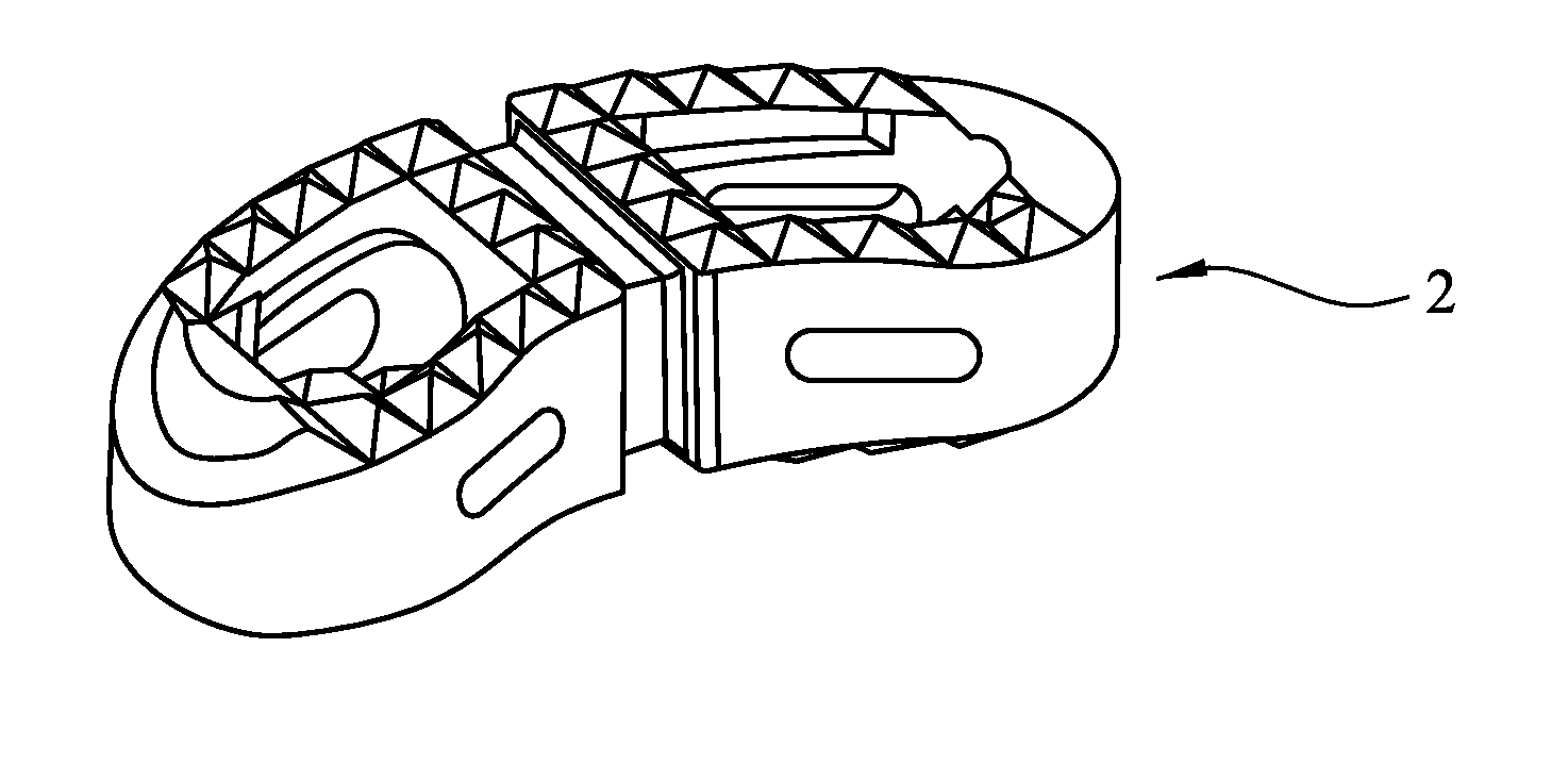
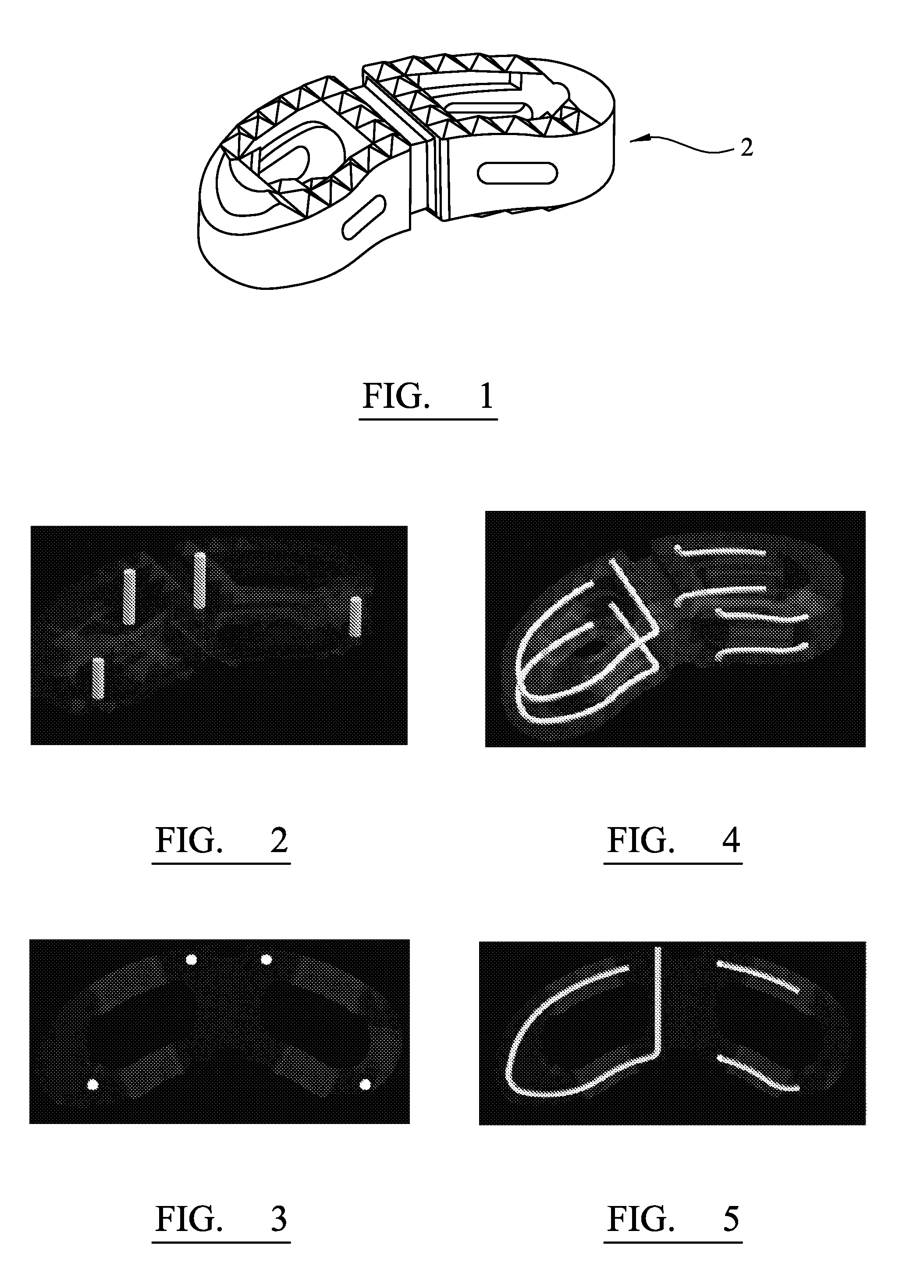
[0050]FIG. 1 depicts an example of a spinal implant 2, which can be provided with radiopaque markers according to the present invention.

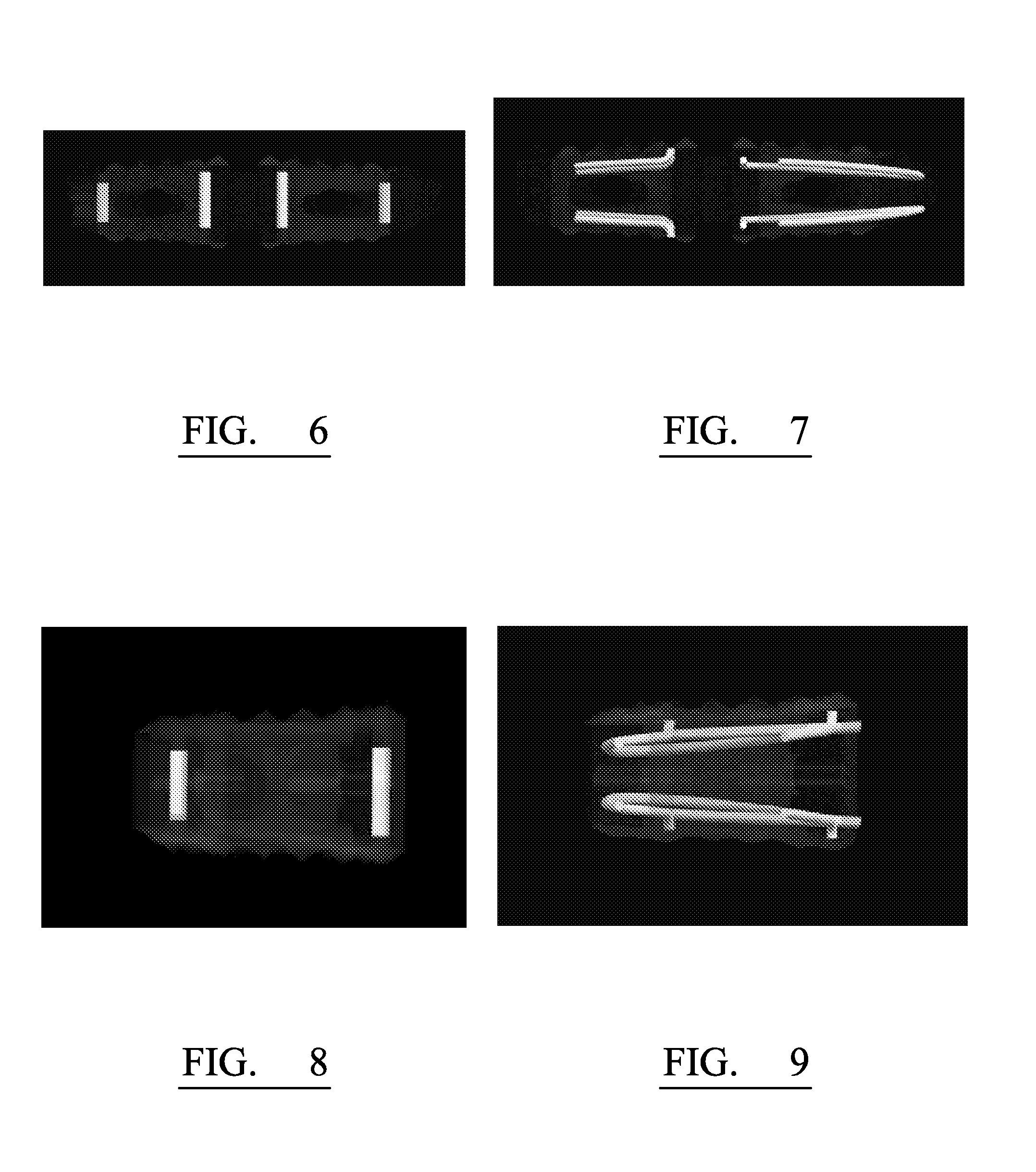
[0051]FIGS. 2, 4, 6 and 8 depict simulated X-rays of the spinal implant 2 using prior art radiopaque wire based markers which are press-fit into openings in the implant. As can be seen, these markers are limited to straight lines, resulting in a poor representation of the medical implant 2 under X-ray.

[0052]FIGS. 3, 5, 7 and 9 depict simulated X-rays of the medical implant 2 incorporating radiopaque markers according to the present invention. As can be seen, these markers include curved portions and / or turns defining complex paths that enable the shape of the implant to be determined more readily under X-ray.

[0053]In order to manufacture the complex channels within the medical implant 2, the medical implant 2 is preferably manufactured by an additive manufacturing technique, for example, stereo lithography. In stereo lithography, the shape of the im...


PUM

PropertyMeasurementUnit
weight %aaaaaaaaaa
weight %aaaaaaaaaa
diameteraaaaaaaaaa

Abstract

A method of manufacturing a medical implant comprising a radiopaque marker is described. The method comprises manufacturing a medical implant using stereolithography, wherein the medical implant has an external surface that delimits an opening to a channel. A curable mixture of a biocompatible polymer and a radiopaque material is then inserted into the channel while in a liquid state and cured in the channel so that it solidifies in the channel.
The combination of an implant manufactured by stereolithography and a radiopaque marker which is inserted into the channel in a liquid state enables complex internal structures to be formed in the implant for the radiopaque marker and for the marker to take on those complex forms more easily. This allows a better visualisation of the marker under X-Ray or other medical imaging technique.

Description

FIELD OF THE INVENTION[0001]The present application relates to a medical implant comprising at least one radiopaque marker, a method for manufacturing a medical implant including at least one radiopaque marker and a curable mixture for forming a radiopaque marker in a medical implant.DESCRIPTION OF THE PRIOR ART[0002]Medical implants are used in many surgical procedures. Examples of such procedures include orthopaedic surgery, such as spinal surgery.[0003]Recently, the use of polyetheretherketone (PEEK) has been proposed for use in medical implants. PEEK and PEEK composites are biocompatible and have good mechanical properties. However, PEEK has a disadvantage because it is radio translucent, so that PEEK and PEEK composite based products are not easily visible on X-rays and other imaging techniques.[0004]To improve visibility of an implant under X-ray, it is known to provide a medical implant with radiopaque markers. The radiopaque markers increase visibility under X-ray and other ...

Claims


Application Information

Patent Timeline
Patent Type & Authority Applications(United States)
IPC IPC(8): B29C70/68
CPCA61F2/30942A61F2002/3008A61F2002/30583A61F2002/30962A61F2310/00131A61F2002/30056A61F2002/30619A61F2002/3071A61F2/4465A61F2002/30617B33Y80/00
Inventor SPRATT, FRANKBETTS, DUNCANMAGLIANO, GIANCARLO
Owner MEDOS INT SARL
Why Eureka
Try Eureka