Method for providing a 3D image data record with suppressed aliasing artifacts overlapping the field of view and computed tomograph

Inactive Publication Date: 2012-11-22
SIEMENS HEALTHCARE GMBH
View PDF5 Cites 4 Cited by
  • Summary
  • Abstract
  • Description
  • Claims
  • Application Information

AI Technical Summary

Benefits of technology

[0016]If the second 3D image data record obtained on the biological object is to be incomplete with respect to the geometric capture of the biological object, this is in particular a cause of aliasing artifacts overlapping the field of view occurring in the resulting computed tomography images. By providing the at least one first 3D image data record, incomplete data can then in particular be extended in the second 3D image data record such that the aliasing artifacts overlapping the field of view are suppressed or even completely prevented. By means of the first 3D image data record, the appearance of aliasing artifacts overlapping the field of view is already prevented so that if necessary, a subsequent modification and / or processing of a resulting computed tomography image can be omitted with respect to the retouching of aliasing artifacts overlapping the field of view. The method therefore does not suppress aliasing artifacts overlapping the field of view by processing the image of a resulting image but instead assesses in advance, by extending the data structure underlying the image so that the artifacts do not appear in the first place. The provision of a concrete first 3D image data record dispenses with imprecise and rough assumptions for the modification of the second 3D image data record. A qualitatively improved and modified second 3D image data record is thus available, from which meaningful computed tomography images can be obtained. Aliasing artifacts overlapping the field of view are prevented very efficiently. Assessing the resulting computed tomography images is thus easier for a person.
[0018]The method preferably includes the further step of obtaining a 2D image data record from the modified second 3D image data record and herefrom a computed tomography image, in particular a forward projection image and / or an x-ray sectional image is generated. Forward projection images and x-ray sectional images allow a meaningful interpretation of the measuring data by means of an operating person. Since, within the scope of the method, aliasing artifacts overlapping the field of view in such 2D images are prevented particularly effectively, a very realistic image of the actual characteristics of the measuring object is obtained.
[0024]The approximation of the first 3D image data record to the second 3D image data record preferably therefore takes place iteratively. In this way, deviations in respect of the shape and design of a mirroring image object in the first 3D image data record can be adjusted such that the image registration in the second 3D image data record is optimized. The extension and / or amendment of the second 3D image data record then take place in an even more realistic fashion.
[0031]First data is preferably selected in the at least one first 3D image data record provided, in particular by way of a binary segmentation, which can be assigned to a specific biological tissue type of the biological object and in which obtained second 3D image data record, second data is selected, in particular by way of a binary segmentation, which can be assigned to the same biological tissue type. In step c), the image registration preferably then takes place with the aid of the first and second data. For instance, provision can be made for the first and second data to be selected such that it corresponds to bone material of the biological object if the biological object includes bone material and soft tissue. Then, in step c), the image registration can be implemented in two stages, wherein in a first stage, an image registration takes place with the aid of the first and second data assigned to the bone material and in a second stage downstream of the first stage, a refined image registration takes place with the aid of the data of the first and second image data record, which are assigned to the soft tissue. A particularly precise and reliable image registration is ensured in this way, thereby rendering possible a correct extension and amendment of the data of the second 3D image data record.

Problems solved by technology

If the second 3D image data record obtained on the biological object is to be incomplete with respect to the geometric capture of the biological object, this is in particular a cause of aliasing artifacts overlapping the field of view occurring in the resulting computed tomography images.

Method used


Image

  • Method for providing a 3D image data record with suppressed aliasing artifacts overlapping the field of view and computed tomograph
  • Method for providing a 3D image data record with suppressed aliasing artifacts overlapping the field of view and computed tomograph
  • Method for providing a 3D image data record with suppressed aliasing artifacts overlapping the field of view and computed tomograph

Examples


Embodiment Construction

[0037]Identical or functionally identical elements are provided with the same reference characters in the figures.

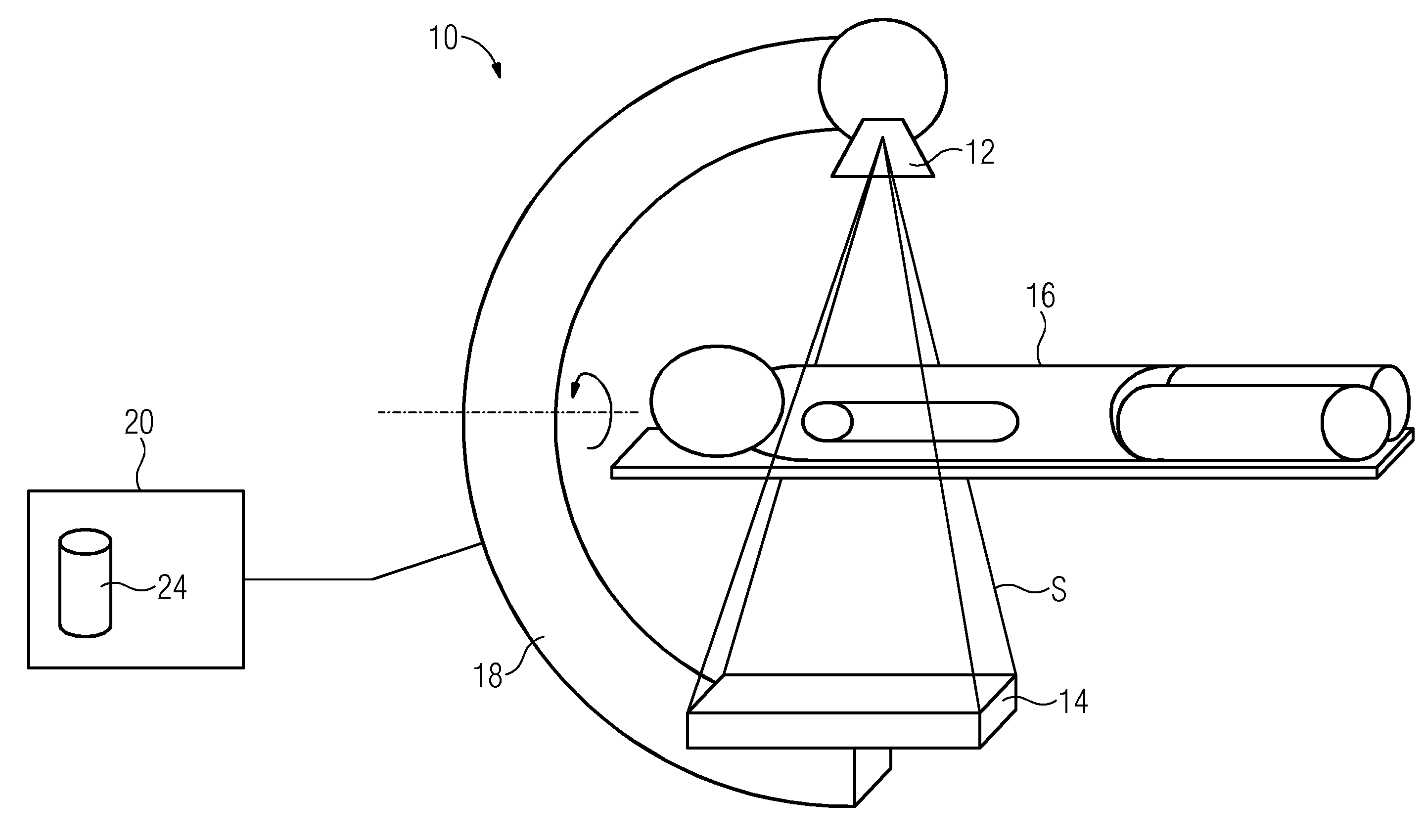
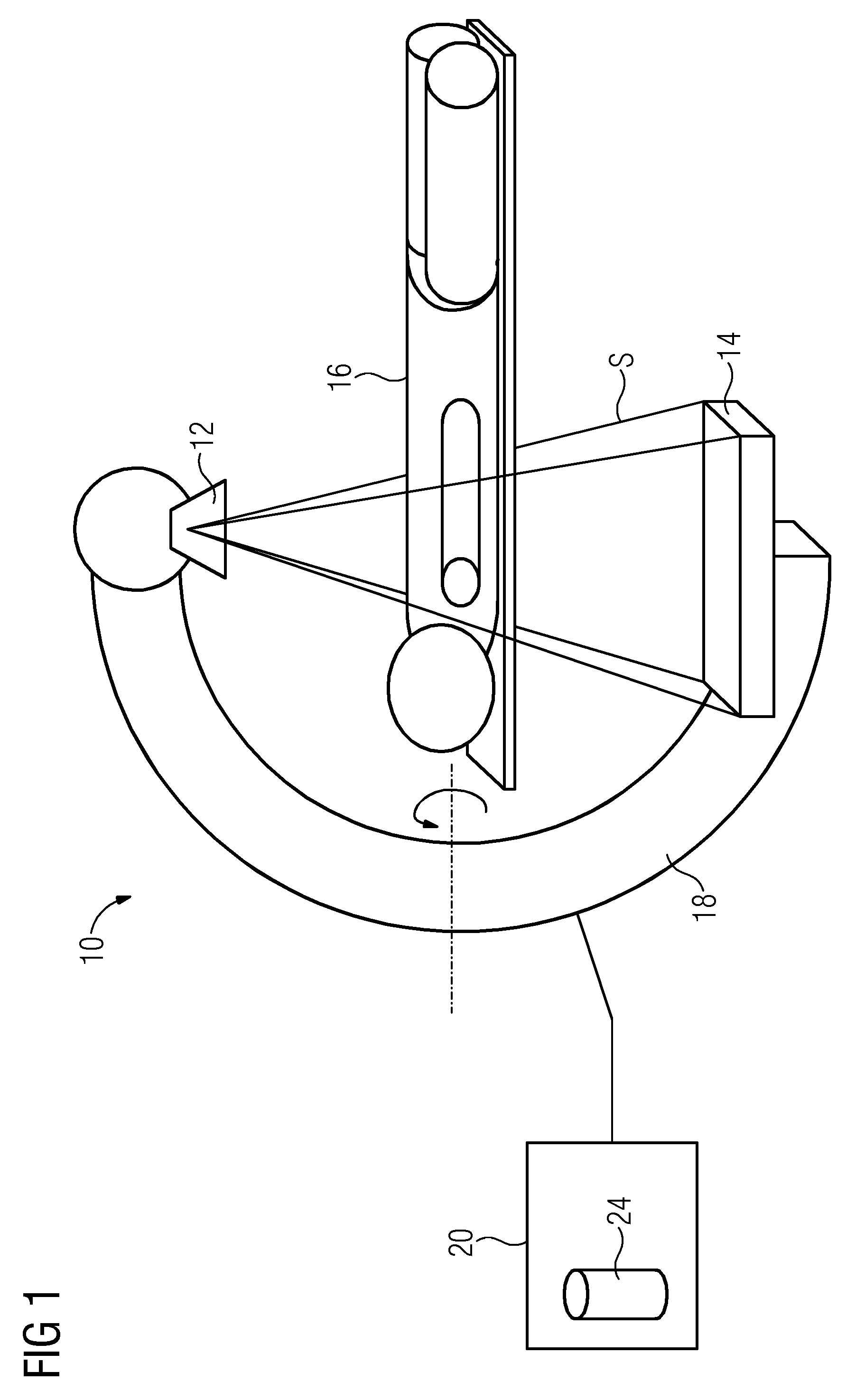
[0038]FIG. 1 shows a computed tomograph 10 with an x-ray C-arm 18, to one end of which an x-ray source 12 is fastened which emits x-ray radiation S in the direction of an x-ray detector 14. A patient 16 is arranged on a couch between the x-ray source 12 and the x-ray detector 14, wherein a body part of the patient is irradiated by the x-ray radiation S.

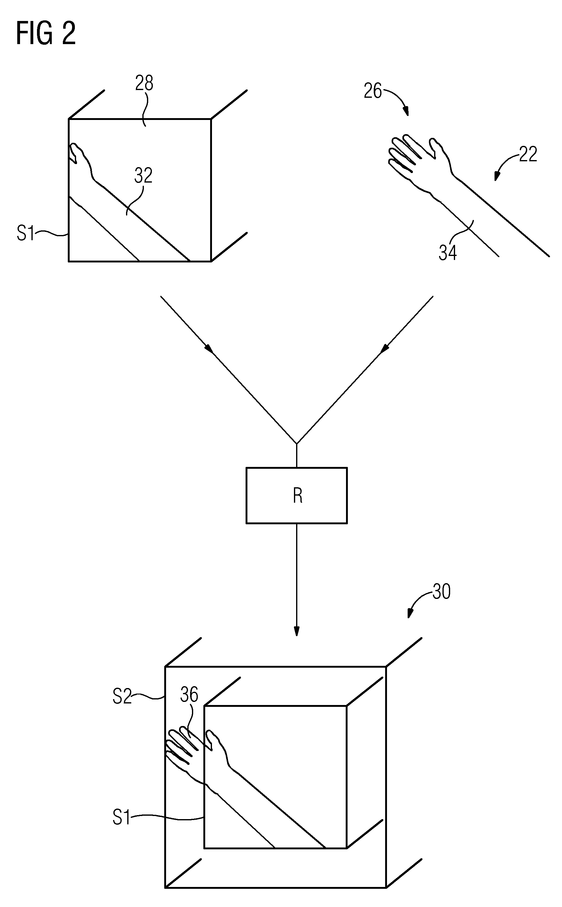
[0039]The x-ray C-arm 18 is embodied to be rotatable and can in this way detect the body part of the patient 16 from different perspectives or at different angles. In this way, different x-ray projection images can be detected by way of the x-ray detector 14, which are transferred by way of a computer 20. A 3D image data record 28 can be reconstructed from the projection images by way of a method for back projection. The 3D image data record 28 of the body part is shown schematically in FIG. 2. In the exemplary embodiment,...


PUM


Abstract

A method is provided for providing a 3D image data record relating to a biological object with suppressed aliasing artifacts overlapping the field of view caused by an incomplete geometric capture of the object by a computed tomography. A first 3D image data record is provided to describe a subarea of the object. A second 3D image data record is obtained by the computed tomography including data relating to the subarea of the object and is registered with the first 3D image data record. Data of the second 3D image data record is extended and / or amended according to data of the first 3D image data record. A part of such data of the second 3D image data record can be assigned to an aliasing artifact overlapping the field of view and thus generates a modified second 3D image data record with suppressed aliasing artifacts overlapping the field of view.

Description

CROSS REFERENCE TO RELATED APPLICATION[0001]This application claims priority of German application No. 10 2011 075 917.4 filed May 16, 2011, which is incorporated by reference herein in its entirety.FIELD OF INVENTION[0002]The invention relates to a method for providing a 3D image data record relating to a biological object with suppressed aliasing artifacts overlapping the field of view, which are caused by an incomplete geometric capture of the biological object by means of a computed tomograph. The invention also relates to a computed tomograph having an x-ray source, a detector and an image evaluation apparatus, which is embodied to execute such a method.BACKGROUND OF INVENTION[0003]The x-ray and / or tomography images obtained by x-ray image recording apparatuses, in particular computed tomographs, may comprise various image artifacts. One type of image artifact is used such that the measured object is not completely captured during the measuring process in terms of its geometric...

Claims


Application Information

Patent Timeline
IPC IPC(8): G06T5/00A61B6/03
CPCA61B6/5258A61B6/5241
Inventor KYRIAKOU, YIANNIS
Owner SIEMENS HEALTHCARE GMBH
Why Eureka
Try Eureka