Targeting and in vivo imaging of tumor-associated macrophages
a macrophage and tumor technology, applied in the field of tumor growth and biology, can solve the problems of poor penetration of solid tumors and high fc-mediated aspecific binding, limited conventional antibodies, and inability to detect tumors in time,
- Summary
- Abstract
- Description
- Claims
- Application Information
AI Technical Summary
Benefits of technology
Problems solved by technology
Method used
Image

Examples
example 1
TS / A Tumors are Highly Infiltrated with a Heterogeneous Population of Myeloid Cells Containing Distinct Granulocyte and Monocyte / Macrophage Subsets
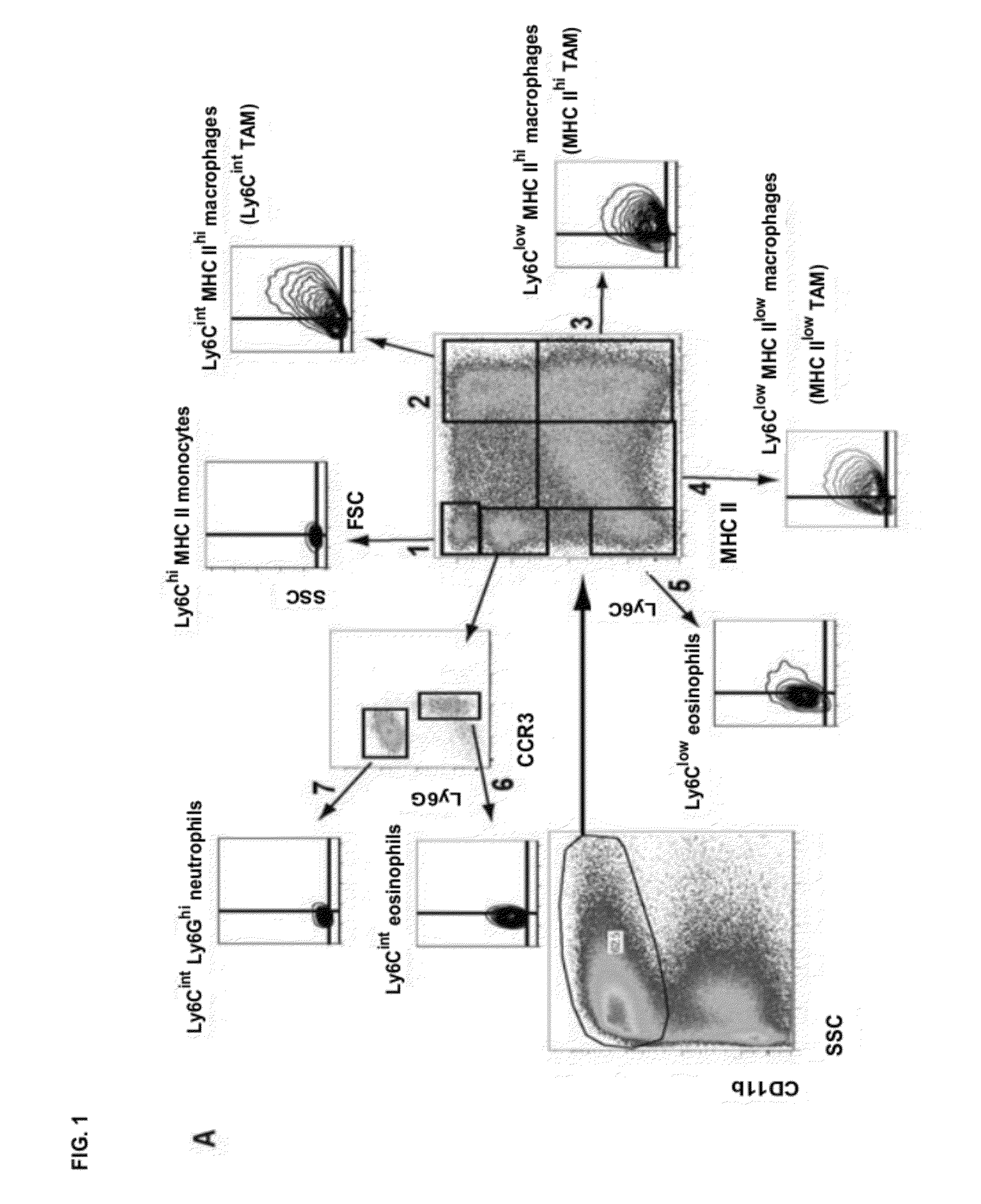
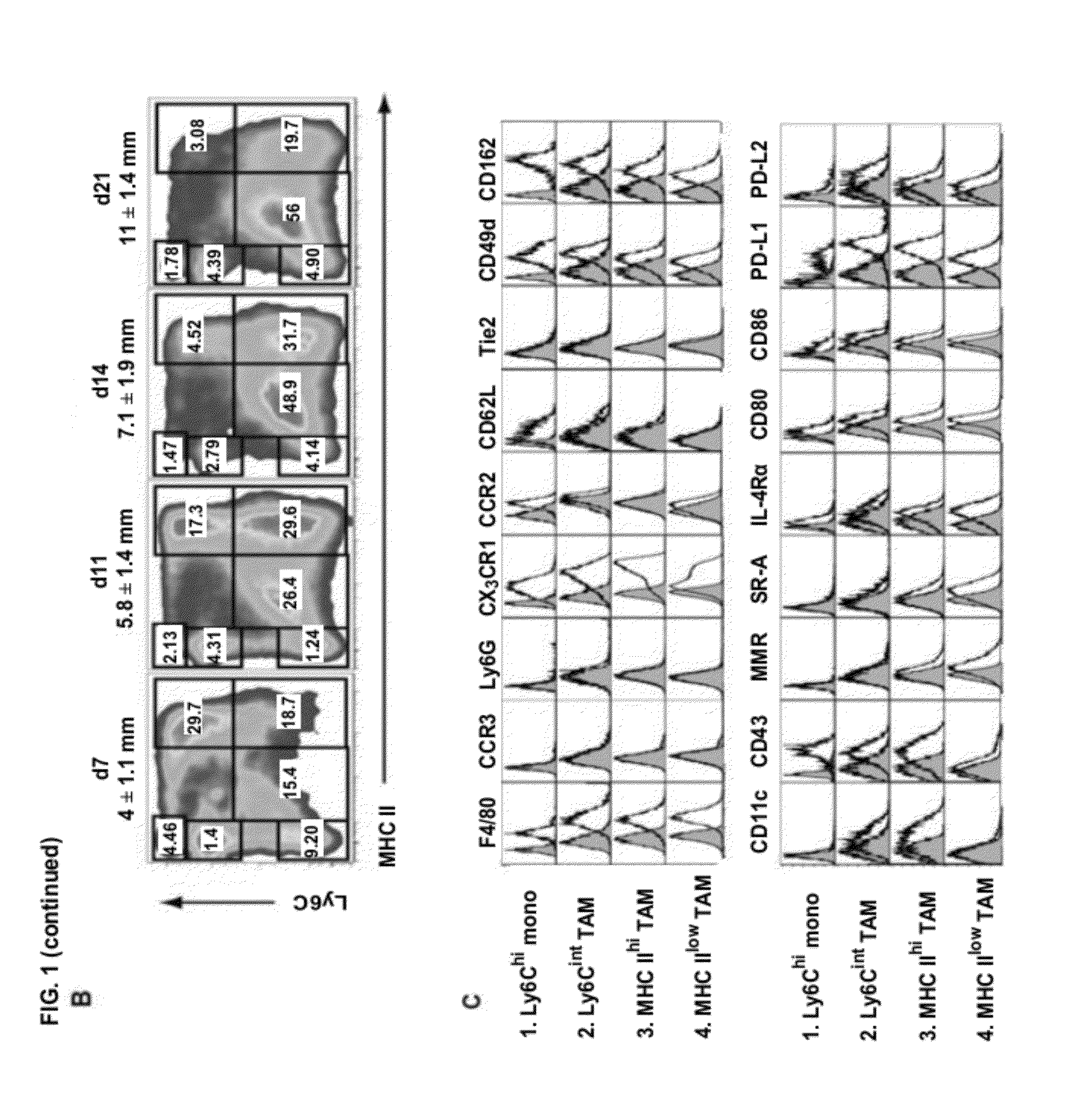
[0157]To study the tumor-infiltrating myeloid compartment, we employed the Balb / c mammary adenocarcinoma model TS / A. Subcutaneous tumors contained a large CD11b+ fraction, indicating a high infiltration of myeloid cells (FIG. 1A). Interestingly, this CD11b+population was heterogeneous and encompassed at least 7 subsets, which could be readily distinguished based on their differential expression of MHC class II and Ly6C (FIG. 1A). Ly6ChiMHC II− cells (Gate 1: FIG. 1A) were F4 / 80+CX3CR1lowCCR2hiCD62L+, did not express the granulocyte markers Ly6G or CCR3 and had a small size and granularity (FSClowSSClow), indicating that they were Ly6Chi monocytes (FIGS. 1A, 1C and FIG. 6). The CD11b+MHC II+ cells in Gates 2-4 were reminiscent of macrophages, having an enlarged macrophage-like scatter and expressing high levels of F4 / 80 (FIGS. 1A, 1C). Rem...
example 2
Ly6Chi Monocytes are the Precursors of all TAM Subsets in TS / A Tumors
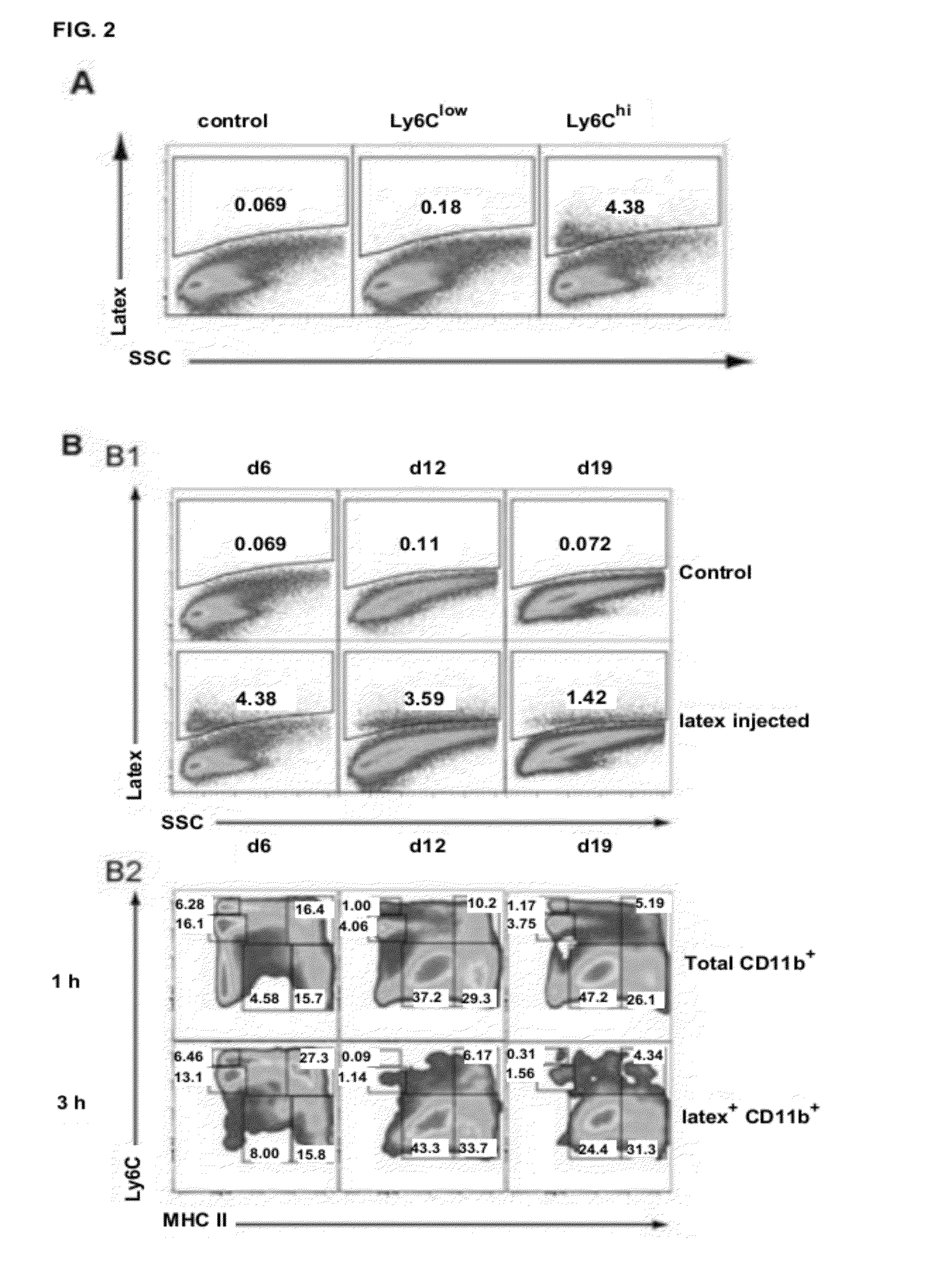
[0159]Macrophages typically derive from circulating blood-borne precursors such as monocytes. The presence of Ly6Chi, but not Ly6Clow, monocytes in TS / A tumors suggested that the former could be more efficiently recruited to tumors and function as the TAM precursor. To investigate this, we selectively labeled Ly6Chi or Ly6Clow monocyte subsets in vivo with fluorescent latex beads, using a previously described procedure.(11, 12). This method has been validated to stably label the respective monocyte subsets for 5 to 6 days in naïve mice. Hence, TS / A was injected after Ly6Clow or Ly6Chi monocyte labeling and tumors were collected 6 days pi. No appreciable numbers of tumor-infiltrating latex+monocytes were observed when applying the Ly6Clow labeling strategy (FIG. 2A). In contrast, Ly6Chi labeling resulted in the detection of a significant fraction of CD11b+latex+ monocytes, illustrating that Ly6Chi monocytes are the ...
example 3
Ly6Cint, MHC IIhi and MHC TAMs have Distinct Differentiation Kinetics and Turnover Rates
[0160]To determine the turnover rate and differentiation kinetics of the monocyte / TAM subsets, BrdU was administered continuously to tumor-bearing animals and its incorporation was measured at consecutive time points. Tumor-infiltrating Ly6Chi monocytes quickly became BrdU+, reaching plateau values after 48 hours of BrdU administration (FIG. 2D). This indicates a rapid monocyte turnover rate and / or proliferation of monocytes inside tumors. Remarkably, intratumoral Ly6Chi monocytes were Ki67+, suggesting a proliferative potential (FIG. 2C). In contrast, TAMs were non-proliferating (Ki6T) and hence unable to directly incorporate BrdU. Therefore, BrdU+ TAMs must differentiate from BrdU+ monocytes, resulting in a lag phase of BrdU positivity. Indeed, only a minor fraction of MHC IIhi and MHC IIlow TAMs were BrdU+ upon 24 hours BrdU administration (FIG. 2D). However, compared with these subsets, Ly6Ci...
PUM
| Property | Measurement | Unit |
|---|---|---|
| volume | aaaaa | aaaaa |
| dissociation constant | aaaaa | aaaaa |
| dissociation constant | aaaaa | aaaaa |
Abstract
Description
Claims
Application Information
 - R&D
- Intellectual Property
- Life Sciences
- Materials
- Tech Scout
- Unparalleled Data Quality
- Higher Quality Content
- 60% Fewer Hallucinations
Browse by: Latest US Patents, China's latest patents, Technical Efficacy Thesaurus, Application Domain, Technology Topic, Popular Technical Reports.
© 2025 PatSnap. All rights reserved.Legal|Privacy policy|Modern Slavery Act Transparency Statement|Sitemap|About US| Contact US: help@patsnap.com