Reporting organ volume for a medical digital image

a medical digital image and organ volume technology, applied in image analysis, image enhancement, instruments, etc., can solve the problems of liver segmentation map produced being over-segmented, type of approach unusable, and proves to be particularly challenging for conventional segmentation approaches

Inactive Publication Date: 2013-02-07
CARESTREAM HEALTH INC
View PDF0 Cites 16 Cited by
  • Summary
  • Abstract
  • Description
  • Claims
  • Application Information

AI Technical Summary

Benefits of technology

[0010]It is an aspect of the present invention to address the need for an improved volume computation method for the liver and other anatomical organs for diagnostic imaging applications.

Problems solved by technology

The liver, because of the relatively complex tissue structure of this organ, proves to be particularly challenging for conventional segmentation approaches.
Applicants have noted that noise levels in the CT image make this type of approach unusable.
However, the resulting liver segmentation maps produced can be over-segmented, often including substantial regions of non-liver tissue.
However, computation of the liver volume requires accurate segmentation and it has proved difficult to obtain repeatable results.
One problem that affects volume computation relates to image contrast and the contrast-to-noise ratio (CNR).
Even where a contrast agent is used, volume computation can be difficult and results less accurate, since the most useful series of images of the liver, in a particular case, may not be those for which contrast is enhanced using the contrast agent.

Method used


Image

  • Reporting organ volume for a medical digital image
  • Reporting organ volume for a medical digital image
  • Reporting organ volume for a medical digital image

Examples


Embodiment Construction

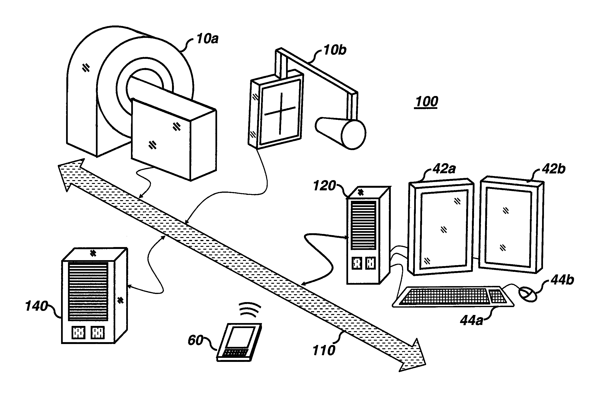
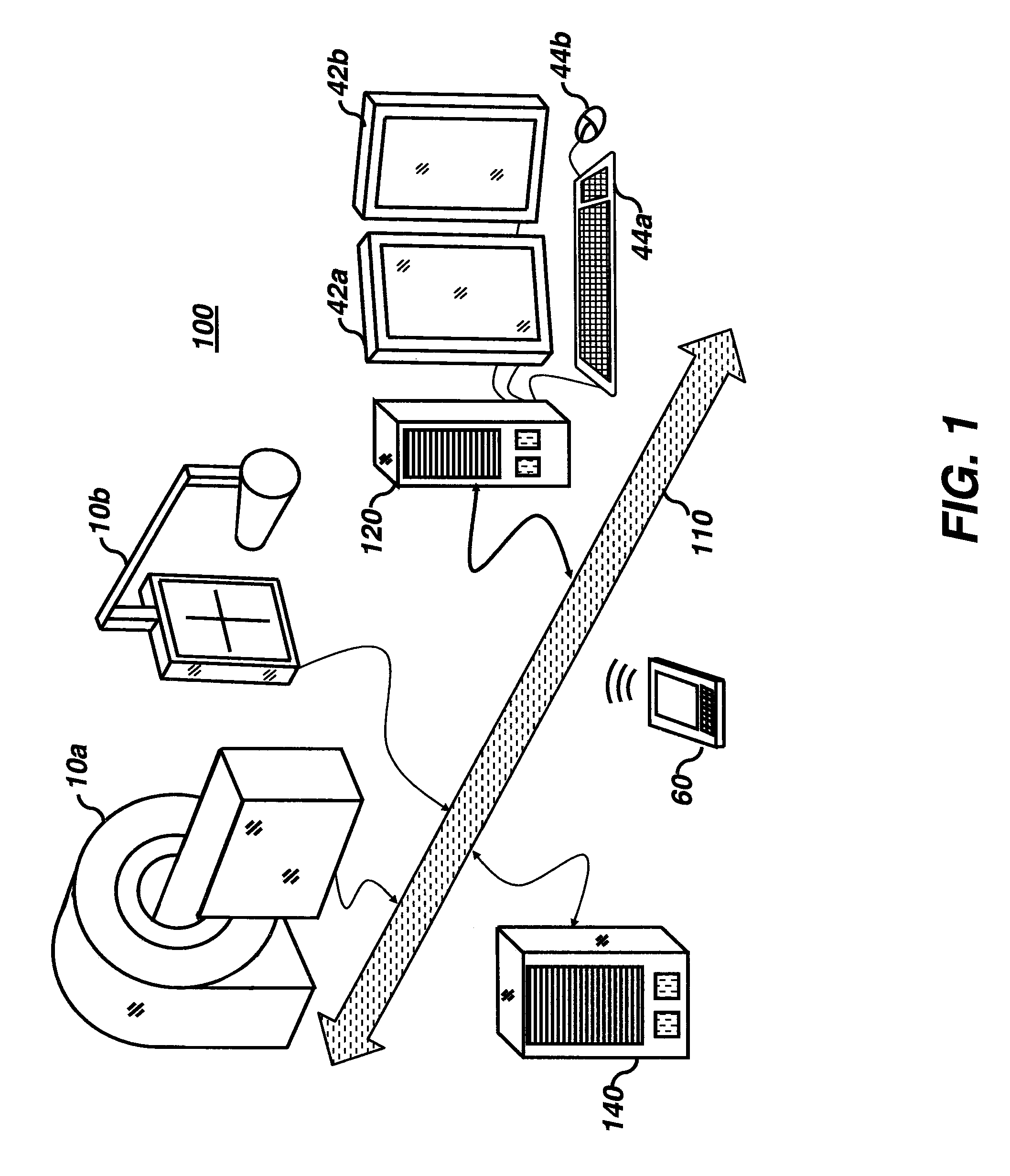
[0023]Medical imaging applications can be implemented via a picture archiving and communications systems (PACS). These systems provide a way for displaying digital images acquired using a wide variety of medical imaging modalities such as, but not limited to, projection radiography (x-ray images), computed tomography (CT images), ultrasound (US images), and magnetic resonance (MR images). Each of the above mentioned medical imaging modalities contain slightly different diagnostic information. In particular, CT and MR images when viewed and studied by a radiologist, can reveal much detail about a patient's 3-dimensional internal anatomy. To enhance the rendering of diagnostic information, computer algorithm technology can also be applied to the medical image data. This can help to detect an abnormal condition, i.e., using computer aided detection (CAD), and can help to make measurements relating to the patient's condition, i.e., computer aided measurement (CAM).

[0024]The present inve...


PUM


Abstract

A method for providing a calculated volume data value for an organ of a patient acquires image data for a first volume image series and for at least a second volume image series for the patient and displays the first volume image series. The method responds to an instruction to generate the volume data value by identifying a seed point in the first volume image series, correlating the seed point to the second volume image series, segmenting the organ in at least the second volume image series according to the correlated seed point, and calculating the volume data value for the organ using at least the segmented second volume image series. The calculated volume data value can be displayed.

Description

FIELD OF THE INVENTION[0001]The present invention relates to the field of digital imaging, and more particularly to segmenting an organ in a medical digital image and reporting the volume of the segmented organ.BACKGROUND[0002]Image segmentation is a branch of digital image processing for categorizing or classifying the picture elements of a digital image as belonging or associated with one or more class types. For medical imaging applications, image segmentation is commonly performed on the voxels (volume elements) of a 3-dimensional image data set with the classification types relating to various anatomical structures. In thoracic medical images, for example, it is convenient to segment the image voxels into classes such as bone, lung parenchyma, soft tissue, bronchial vessels, blood vessels, etc. There are many reasons to perform such a task, such as for surgical planning, treatment progress, and patient diagnosis.[0003]Among image segmentation approaches is the technique general...

Claims


Application Information

Patent Timeline
Patent Type & Authority Applications(United States)
IPC IPC(8): G06K9/00
CPCG06T7/0081G06T7/0097G06T2207/10016G06T2207/30056G06T2207/10132G06T2207/20141G06T2207/10072G06T7/11G06T7/174G06T7/187
Inventor GINDELE, EDWARD B.RAY, LAWRENCE A.
Owner CARESTREAM HEALTH INC
Why Eureka
Try Eureka