Systems and Method for Automatic Prostate Localization in MR Images Using Random Walker Segmentation Initialized Via Boosted Classifiers

a technology of random walker segmentation and automatic prostate localization, which is applied in image analysis, image enhancement, instruments, etc., can solve the problems of unique challenges of accurate prostate segmentation in mr imagery and accuracy problems

Inactive Publication Date: 2013-03-14
SIEMENS HEALTHCARE GMBH
View PDF5 Cites 26 Cited by
  • Summary
  • Abstract
  • Description
  • Claims
  • Application Information

AI Technical Summary

Problems solved by technology

Accurate prostate segmentation in MR imagery poses unique challenges.
These challenges include large variations in prostate anatomy and disease, intensity inhomogeneities, and near-field artifacts mainly induced by endorectal (ER) coils which cause accuracy problems in including the peripheral zone (PZ) of the prostate within the overall segmentation.

Method used


Image

  • Systems and Method for Automatic Prostate Localization in MR Images Using Random Walker Segmentation Initialized Via Boosted Classifiers
  • Systems and Method for Automatic Prostate Localization in MR Images Using Random Walker Segmentation Initialized Via Boosted Classifiers
  • Systems and Method for Automatic Prostate Localization in MR Images Using Random Walker Segmentation Initialized Via Boosted Classifiers

Examples


Embodiment Construction

[0034]MRI plays a key role in the diagnosis, staging and treatment monitoring for prostate cancer. Various MR modalities such as T2 MRI using endorectal (ER) coils, dynamic contrast enhanced (DCE) MRI, diffusion-weighted imaging and 3D chemical shift spectroscopy imaging contribute complementary forms of information during these processes, as described in for instance “[19] Turkbey, B., Pinto, P., Choyke, P. L.: Imaging techniques for prostate cancer: implications for focal therapy. Nature Reviews: Urology 6, 191-203 (2009).”

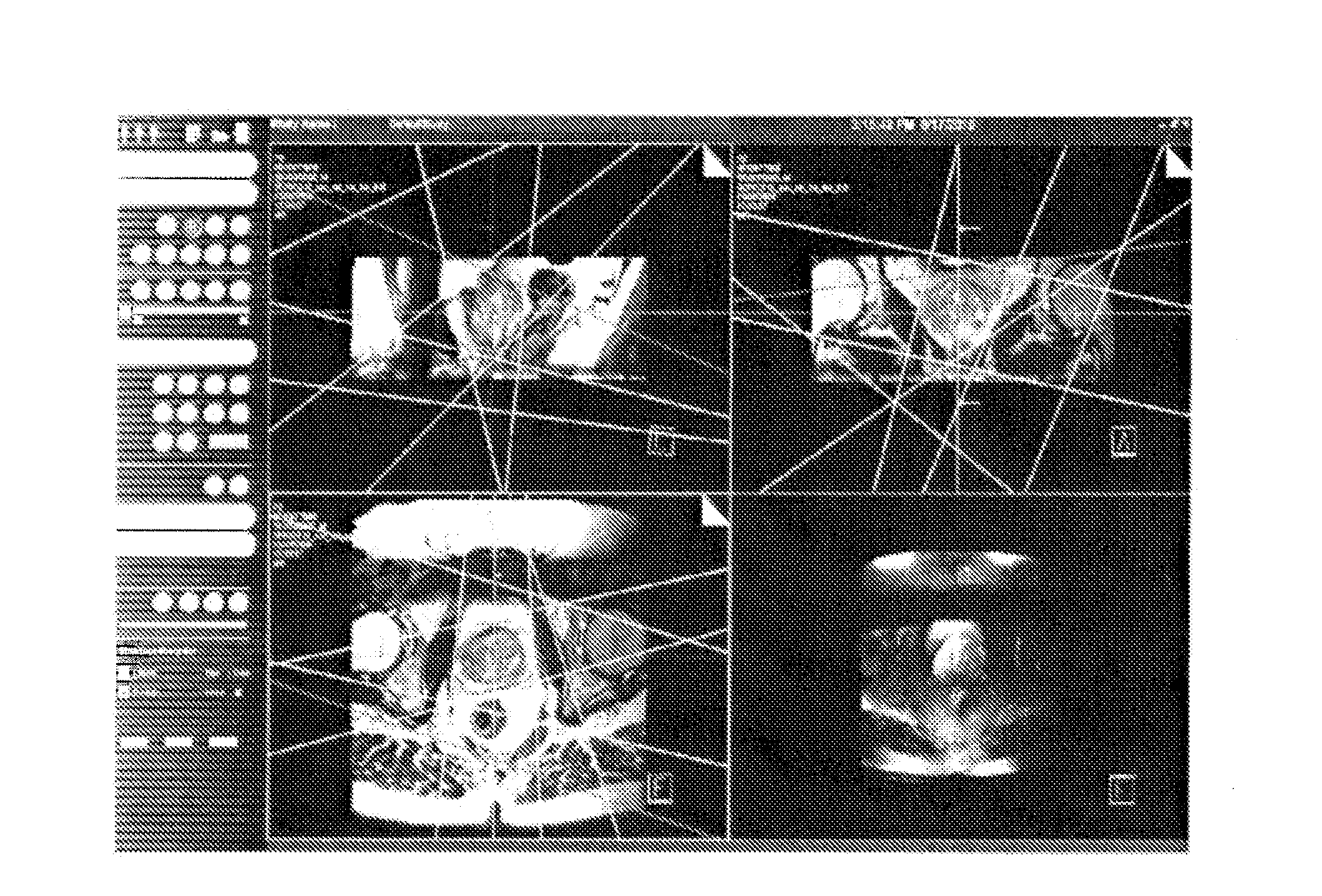
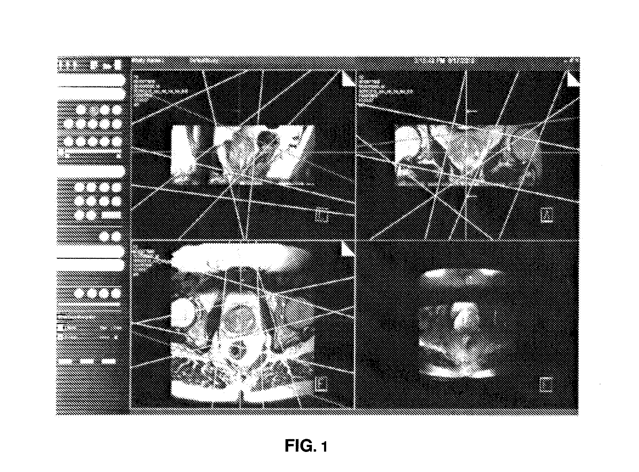
[0035]Prostate localization is a pre-requisite for optimal positioning of radio-frequency (RF) saturation bands to prevent fat contamination in 3D MR chemical shift spectroscopy as described in [14] Scheenen, T., Heijmink, S., Roell, S., de Kaa, C. H., Knipscheer, B., Witjes, J., Barentsz, J., Heerschap, A.: Three-dimensional proton MR spectroscopy of human prostate at 3 T without endorectal coil. Radiology 245(2), 507-516 (2007)” by fat surrounding the prostate...


PUM


Abstract

Automatic prostate localization in T2-weighted MR images facilitate labor-intensive cancer imaging techniques. Methods and systems to accurately segment the prostate gland in MR images are provided and address large variations in prostate anatomy and disease, intensity inhomogeneities, and artifacts induced by endorectal coils. A center of the prostate is automatically detected with a boosted classifier trained on intensity based multi-level Gaussian Mixture Model Expectation Maximization (GMM-EM) segmentations of the raw MR images. A shape model is used in conjunction with Multi-Label Random Walker (MLRW) to constrain the seeding process within MLRW.

Description

CROSS-REFERENCE TO RELATED APPLICATIONS[0001]This application claims the benefit of U.S. Provisional Application No. 61 / 445,587, filed Feb. 23, 2011, which is incorporated herein by reference.BACKGROUND OF THE INVENTION[0002]The present invention relates to methods and systems for automatic prostate localization. More specifically, it is related to automatic prostate localization using random walker segmentation initialized via boosted classifiers.[0003]Accurate prostate segmentation in MR imagery poses unique challenges. These challenges include large variations in prostate anatomy and disease, intensity inhomogeneities, and near-field artifacts mainly induced by endorectal (ER) coils which cause accuracy problems in including the peripheral zone (PZ) of the prostate within the overall segmentation.[0004]Accordingly, novel and improved systems and methods in automatic prostate segmentation are required.SUMMARY OF THE INVENTION[0005]Novel methods and systems are provided herein as a...

Claims


Application Information

Patent Timeline
Patent Type & Authority Applications(United States)
IPC IPC(8): G06K9/34G06K9/62
CPCG06K9/6226G06T7/0081G06T7/0087G06T2207/30081G06T2207/20081G06T2207/20144G06T2207/20156G06T2207/10088G06T7/11G06T7/143G06T7/194G06V10/763G06F18/2321
Inventor KHURD, PARMESHWARGRADY, LEOKAMEN, ALIDIALLO, MAMADOUGAJERA, KALPITKUMARGALL, PETERREQUARDT, MARTINKIEFER, BERTHOLDWEISS, CLIFFORD R.
Owner SIEMENS HEALTHCARE GMBH
Why Eureka
Try Eureka