OCT-Guided Femtosecond Laser to Measure a Retinal Surface for Use in Performing an Intra-Retinal Ablation

a laser and retinal surface technology, applied in laser surgery, medical science, surgery, etc., can solve the problems of retinal scar tissue formation and considerable diminution in visual acuity, and achieve the effects of minimizing “t”, minimizing any deviation of focal point, and debulking scar tissue inside the retina

Inactive Publication Date: 2013-04-25
BAUSCH & LOMB INC
View PDF2 Cites 8 Cited by
  • Summary
  • Abstract
  • Description
  • Claims
  • Application Information

AI Technical Summary

Problems solved by technology

The undesirable consequence here is the formation of retinal scar tissue.
Nevertheless, even though the retinal surface may remain functional, tachyphylaxis can eventually cause a considerable diminution in visual acuity.

Method used


Image

  • OCT-Guided Femtosecond Laser to Measure a Retinal Surface for Use in Performing an Intra-Retinal Ablation
  • OCT-Guided Femtosecond Laser to Measure a Retinal Surface for Use in Performing an Intra-Retinal Ablation

Examples


Embodiment Construction

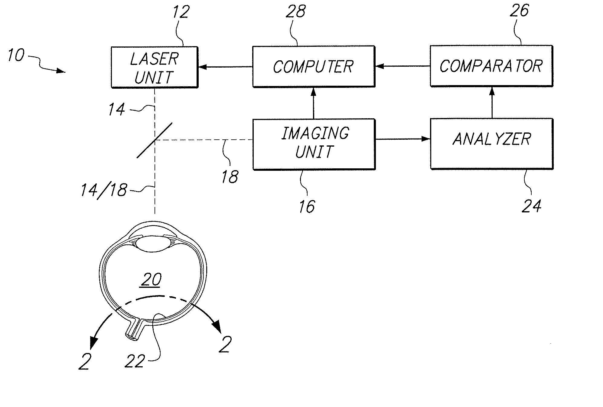
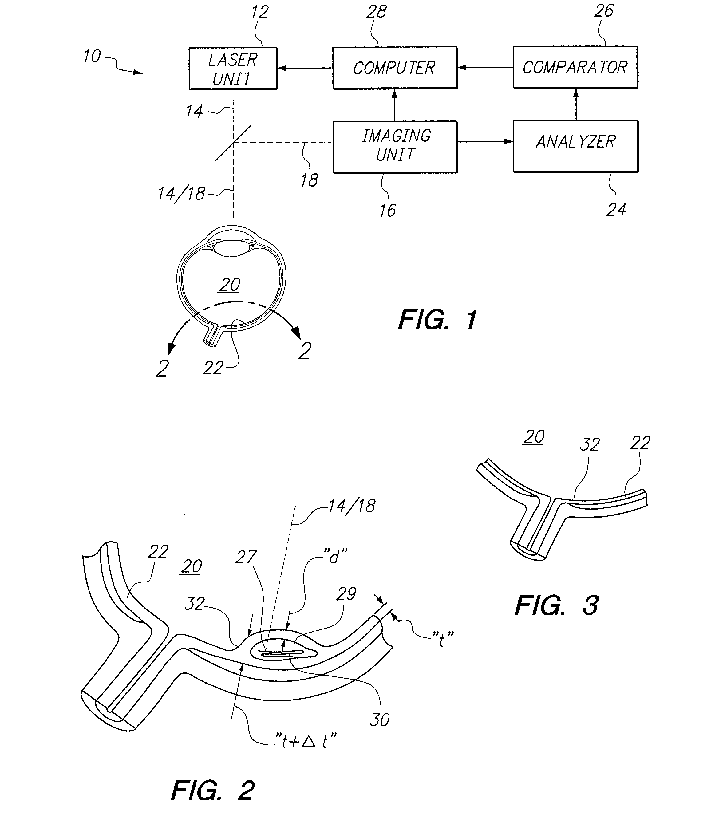
[0015]Referring initially to FIG. 1 a system for debulking retinal tissue in accordance with the present invention is shown and is generally designated 10. As shown, the system 10 includes a laser unit 12 for generating a laser beam 14. Preferably, the laser beam 14 is a pulsed femtosecond laser beam, wherein each pulse has a duration of less than about 500 femtoseconds. FIG. 1 also shows that the system 10 includes an imaging unit 16 for generating an imaging beam 18. Preferably, the imaging unit 16 is of a type well known in the pertinent art that employs Optical Coherence Tomography (OCT) techniques for the purpose of creating three dimensional images. In this case, imaging is done of the eye 20. More specifically, the imaging unit 16 is used to create an image of the retina 22 of the eye 20.

[0016]FIG. 1 also shows that the system 10 includes an analyzer 24 that is connected between the imaging unit 16 and a comparator 26. Also, the imaging unit 16 is connected directly to a comp...


PUM


Abstract

A system and method are provided for debulking scar tissue in the retina by the Laser Induced Optical Breakdown (LIOB) of scar tissue. The identification, location and extent of the scar tissue is accomplished using Optical Coherence Tomography (OCT) techniques. OCT imaging is also used to monitor the debulking procedure.

Description

[0001]This application claims the benefit of U.S. Provisional Patent Application Ser. No. 61 / 549,932, filed Oct. 21, 2011.FIELD OF THE INVENTION[0002]The present invention pertains generally to systems and methods for performing ophthalmic laser surgical procedures. More particularly, the present invention pertains to systems and methods for performing an ablation of ophthalmic tissue for the purpose of debulking scar tissue in the retina. The present invention is particularly, but not exclusively, useful as a laser surgical procedure wherein scar tissue in the retina is located and identified using Optical Coherence Tomography (OCT) techniques, and the scar tissue is then debulked by causing Laser Induced Optical Breakdown (LIOB) of the scar tissue.BACKGROUND OF THE INVENTION[0003]It sometimes happens that therapeutic injections of biologics or pharmacologics into the retina of an eye can cause a reaction from retinal tissue that is known as tachyphylaxis. The undesirable consequen...

Claims


Application Information

Patent Timeline
Patent Type & Authority Applications(United States)
IPC IPC(8): A61F9/011
CPCA61F9/00825A61F2009/00863A61F2009/00851
Inventor GRANT, ROBERT EDWARDMORDAUNT, DAVID HAYDN
Owner BAUSCH & LOMB INC
Why Eureka
Try Eureka