Automated system to create a cell smear

a cell smear and automatic technology, applied in the field of automatic system to create a cell smear, can solve the problems of low number of cells circulating and low process efficiency

Inactive Publication Date: 2013-05-30
CELLSCAPE CORP
View PDF2 Cites 7 Cited by
  • Summary
  • Abstract
  • Description
  • Claims
  • Application Information

AI Technical Summary

Benefits of technology

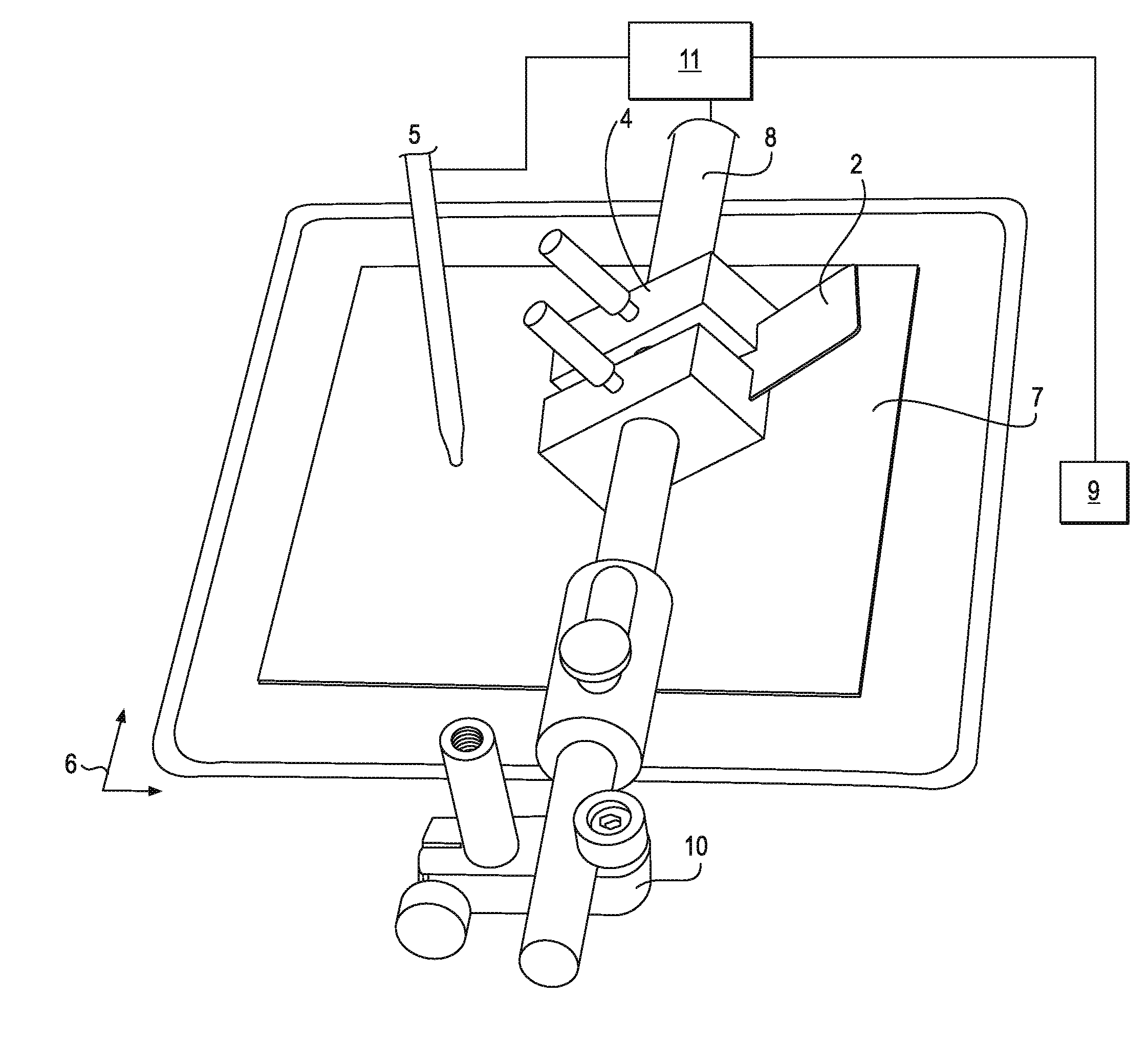
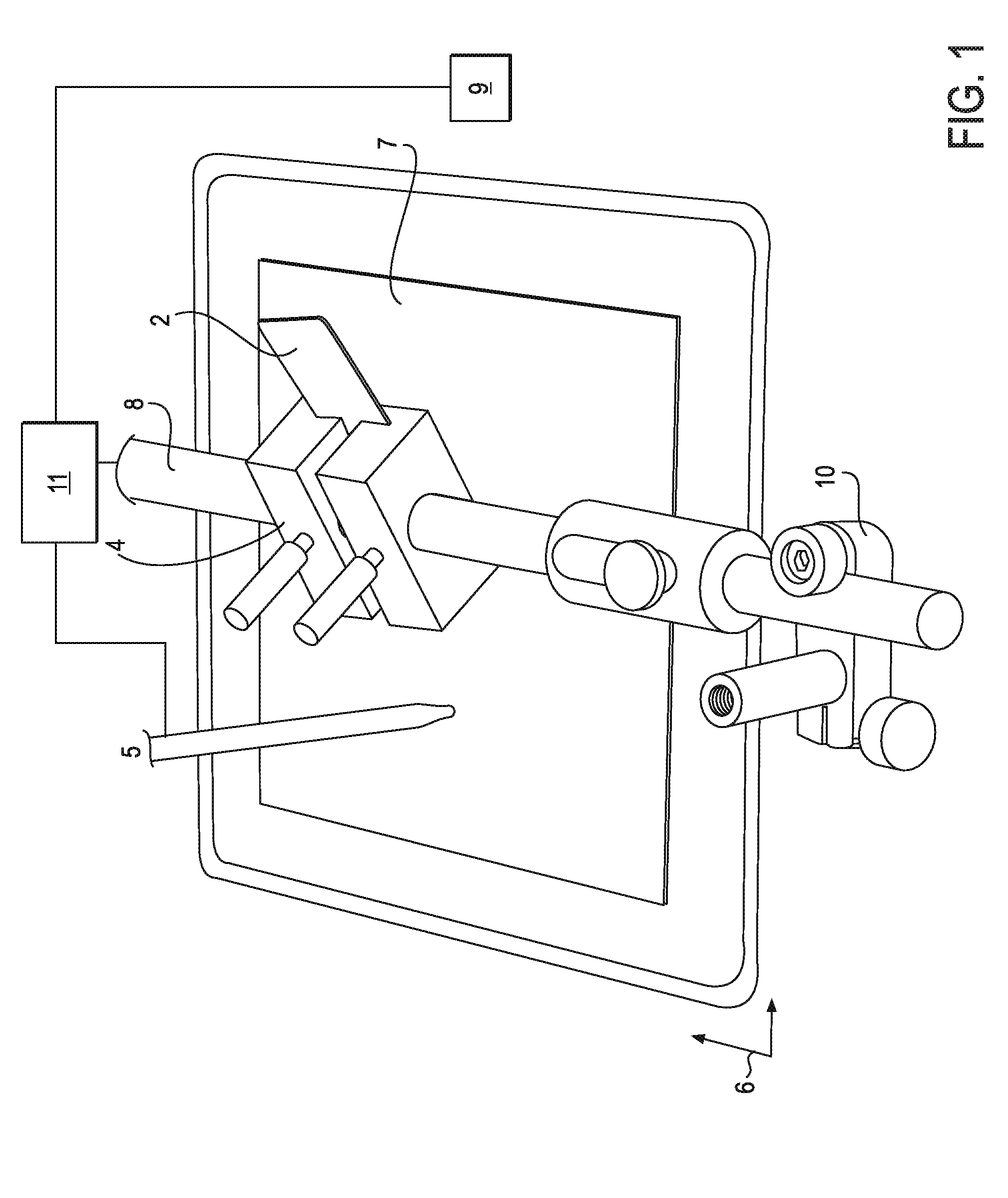
[0027]In some embodiments, the smear blade has a forward edge with a non-linear portion, such as notches, a rough surface and / or a curve. At least a portion of the smear blade may be less hydrophilic than the surface.

Problems solved by technology

However these processes do not yield the high packing, monolayer of cells of uniform high quality needed for performing Automated Digital Microscopy.
However, the numbers of cells circulating are very low compared with the number of non-fetal or non-tumorigenic cells.

Method used


Image

  • Automated system to create a cell smear
  • Automated system to create a cell smear
  • Automated system to create a cell smear

Examples


example 1

[0139]FIG. 19 shows a schematic layer of predominantly red blood cells from a whole blood smear where the vast majority of cells in the sample are red blood cells 500. This image shows a typical high density smear of red blood cells imaged with 420 nm transmitted light to highlight the cells using hemoglobin absorption. The smear parameters were 25 mm / sec smear speed, smooth edge glass smear head, styrene flexure, and 30 degree smear head angle. FIG. 22 is a photograph that shows a portion of a representative smear of the original results.

example 2

[0140]FIG. 20 shows a layer of cells enriched for cells of interest and smeared using the same parameters as described above for FIG. 19, but with a higher smear blade / substrate angle. Many white blood cells 504 are detected among the red blood cells 502. FIG. 23 is a photograph that shows a portion of a representative smear of the original results.

example 3

[0141]FIG. 21 shows a layer of cells enriched for cells of interest and smeared using the same parameters as described above for FIGS. 19 and 20, except that a medium angle smear (25 degrees) was used. White blood cells 512 are detected among red blood cells 510. Note the reduced density of the smear showing the ability to control smear density by head angle. FIG. 24 is a photograph that shows a portion of a representative smear of the original results.


PUM

PropertyMeasurementUnit
frequencyaaaaaaaaaa
areaaaaaaaaaaa
areaaaaaaaaaaa

Abstract

A method of creating a layer of cells on a surface. In some embodiments, the method includes the steps of engaging a smear tool against the surface with an engagement force; flexing a portion of the smear tool to change an orientation of the smear tool with respect to the surface; moving the smear tool along the surface through a sample comprising cells suspended in a liquid; and adhering the sample to the surface to thereby create a layer of cells. Another method according to the invention creates a layer of cells on a surface by mixing the sample prior to and/or during the smear. The invention also includes systems for implementing the methods.

Description

CROSS REFERENCE TO RELATED APPLICATIONS[0001]This application claims the benefit under 35 U.S.C. 119 of U.S. application No. 61 / 332,618, filed May 7, 2010, the disclosure of which is incorporated herein by reference.INCORPORATION BY REFERENCE[0002]All publications and patent applications mentioned in this specification are herein incorporated by reference to the same extent as if each individual publication or patent application was specifically and individually indicated to be incorporated by reference.BACKGROUND OF THE INVENTION[0003]The study of blood and its normal and diseased states dates back to blood letting in Ancient Egypt but the field really gained traction in 1624 when Anthony van Leeuwenhoek built the first microscope that was able to image blood cells. In 1770 William Hewson provided the first description of leukocytes (white blood cells) for which he has been dubbed the father of hematology. The field of hematology therefore has a very long history and techniques tha...

Claims


Application Information

Patent Timeline
Patent Type & Authority Applications(United States)
IPC IPC(8): G01N1/28
CPCC12Q1/6841C12Q1/6881G02B21/34G01N1/2813C12Q2600/158
Inventor BRODY, MICHAEL D.HALDERMAN, JONATHAN D.PARIKH, BHAIRAVISTONE, JAMES
Owner CELLSCAPE CORP
Why Eureka
Try Eureka