Image processing apparatus, method, and program

a technology of image processing and apparatus, applied in the field of image processing apparatus, method and program, can solve the problem that images cannot be said to appropriately respond to the needs

Active Publication Date: 2013-05-30
FUJIFILM CORP
View PDF8 Cites 16 Cited by
  • Summary
  • Abstract
  • Description
  • Claims
  • Application Information

AI Technical Summary

Benefits of technology

[0029]According to the image processing apparatus, method, and program of the present invention, a surface image representing an inner surface of a wall of the organ captured by an endoscope inserted in the organ is obtained, an adjacent blood vessel image is generated from a three-dimensional image representing a three-dimensional area including the organ, the adjacent blood vessel image being an image depicting a portion of the blood vessel adjacent to the wall from a viewpoint in the three-dimensional image corresponding the viewpoint of the surface image, a surrounding blood vessel image is generated from a three-dimensional image representing a three-dimensional area including a surrounding area of the organ, the surrounding blood vessel image being an image depicting the blood vessel from a viewpoint in the three-dimensional image corresponding the viewpoint of the surface image,

Problems solved by technology

In contrast, in the image for display provided by the technique described in Japanese Unexamined Patent Publication No. 2006-061274, the state of the mucosal surface of an organ may be obs

Method used


Image

  • Image processing apparatus, method, and program
  • Image processing apparatus, method, and program
  • Image processing apparatus, method, and program

Examples


Embodiment Construction

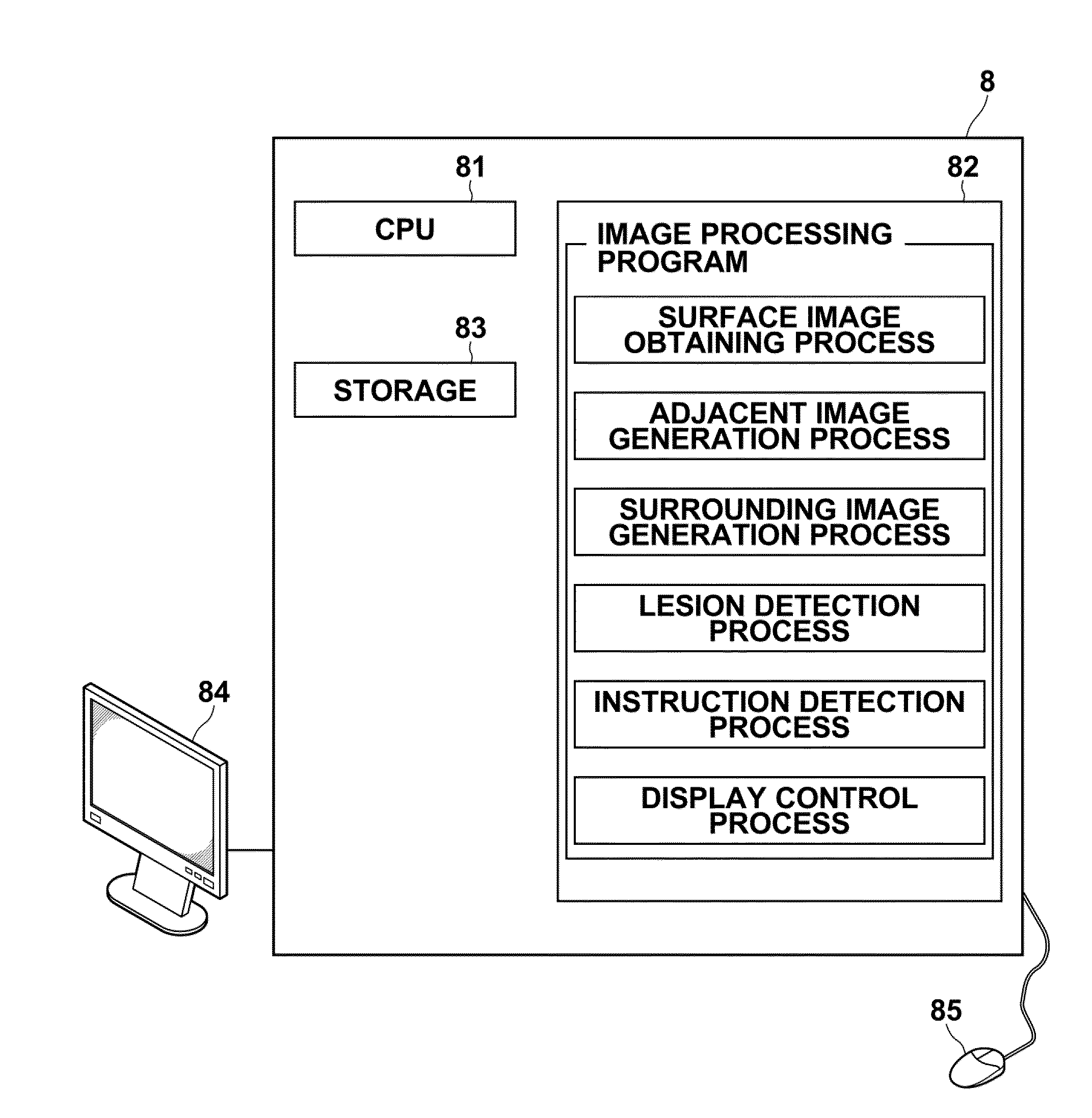
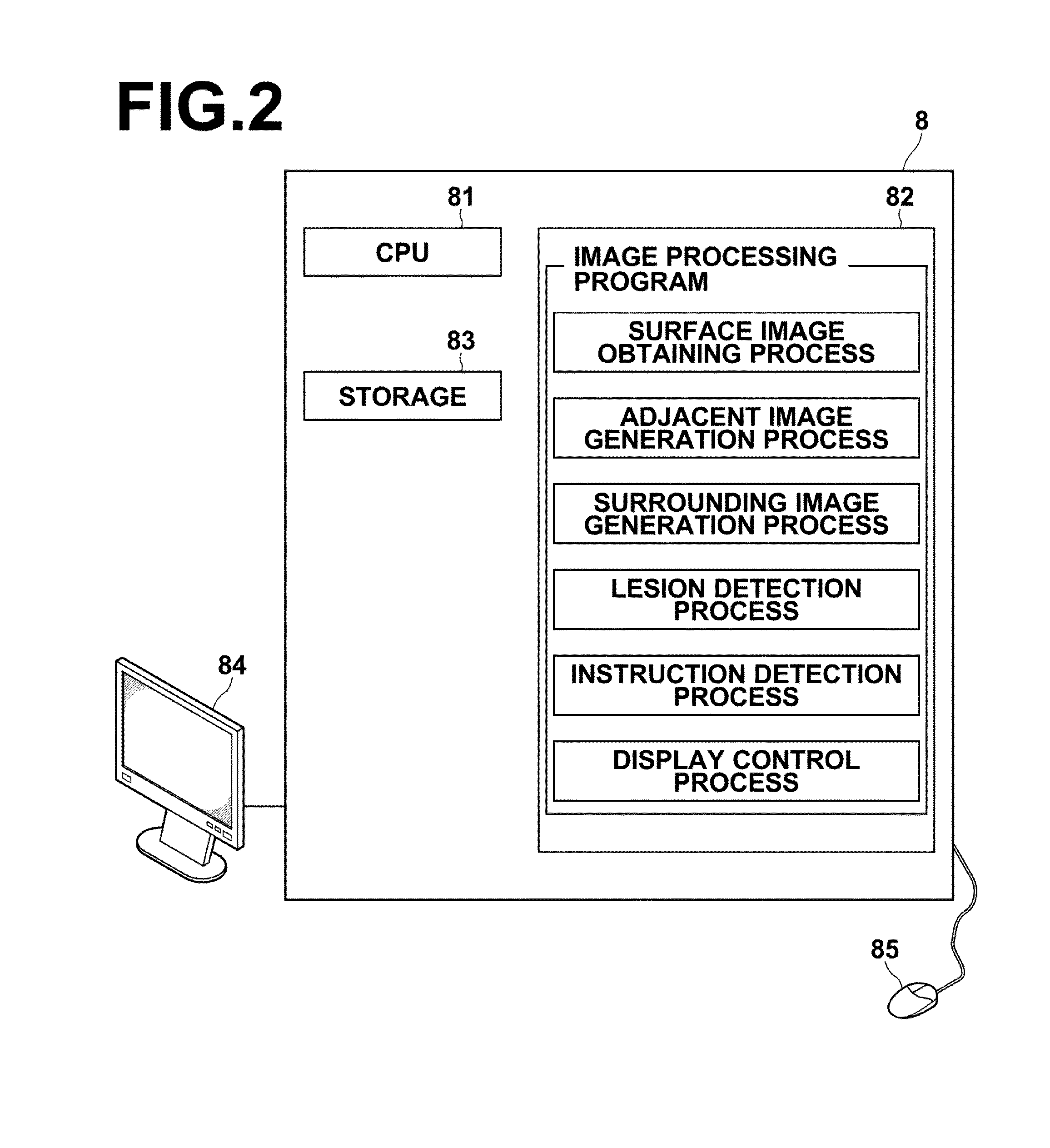
[0036]Hereinafter, embodiments of the image processing apparatus, method, and program of the present invention will be described with reference to the accompanying drawings.

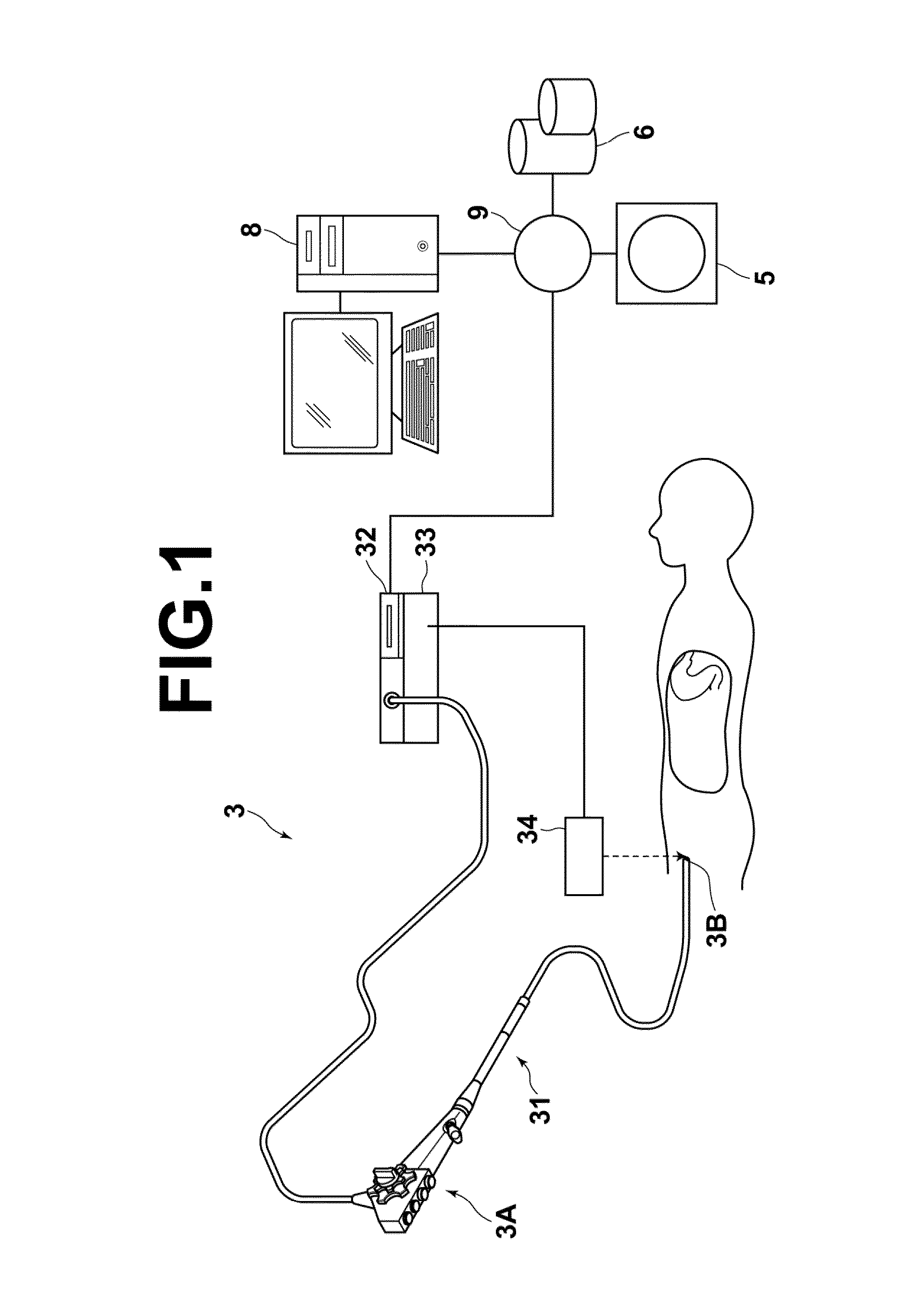
[0037]FIG. 1 is a hardware configuration diagram of a medical image processing system, illustrating an overview thereof. As illustrated in FIG. 1, an endoscope apparatus 3, a three-dimensional image capturing apparatus 5, an image storage server 6, and an image processing apparatus 8 are communicatively linked to each other via network 9 in the system.

[0038]The endoscope apparatus 3 includes an endoscope 31 that captures an image of a body cavity of a subject, a processor unit 32 that generates an image of a subject tissue in the body cavity based on a signal obtained by image capturing, a light source unit 33 that supplies light for illuminating inside of the body cavity, a position detection unit 34 that detects the position and orientation of a tip portion of the endoscope 31.

[0039]The endoscope 31 includes an...


PUM


Abstract

Obtaining a surface image captured by an endoscope inserted in a tubular organ associated with a surrounding blood vessel and representing an inner surface of a wall of the organ, generating, from a three-dimensional image representing a three-dimensional area including the organ, an adjacent blood vessel image depicting a portion of the blood vessel adjacent to the wall from a viewpoint in the three-dimensional image corresponding the viewpoint of the surface image, generating, from a three-dimensional image representing a three-dimensional area including a surrounding area of the organ, a surrounding blood vessel image depicting the blood vessel from a viewpoint in the three-dimensional image corresponding the viewpoint of the surface image, and causing the surface image, adjacent blood vessel image, and surrounding blood vessel image to be displayed in this order on a display unit.

Description

TECHNICAL FIELD[0001]The present invention relates to an image processing apparatus, method, and program for displaying an image for observing an organ, such as a large intestine or a stomach.BACKGROUND ART[0002]Heretofore, a technology for diagnosing and treating an organ of a patient, such as a large intestine or a stomach, using a real endoscopic image captured by an endoscope inserted in the organ has been known. Further, a technology for generating a virtual endoscopic image from a three-dimensional volume image obtained by CT imaging or the like and displaying the generated image has also been known.[0003]Japanese Unexamined Patent Publication No. 2006-061274 proposes a method in which the position and orientation of an endoscope inserted into an organ are detected, then a virtual endoscopic image having the same field of view as the real endoscopic image is generated based on the detected position and orientation, and the real endoscopic image obtained by the endoscope is tex...

Claims


Application Information

Patent Timeline
IPC IPC(8): A61B1/00
CPCA61B1/0005G06T2207/30092G06T15/08G06T19/00G06T2210/41G06T2219/028A61B1/00009A61B6/504A61B6/5247A61B1/31G06T2207/30101G06T7/0012G06T2207/10068G06T2207/30028A61B1/04A61B1/000094
Inventor ITAI, YOSHINORI
Owner FUJIFILM CORP
Why Eureka
Try Eureka