2d3d registration for mr-x ray fusion utilizing one acquisition of mr data

a technology of mr-x ray fusion and mr volume, applied in the field of medical imaging, can solve the problems of difficult drr-based method for registering mr volume, and the typical non-delineation of anatomic structures

Inactive Publication Date: 2013-07-25
SIEMENS CORP
View PDF3 Cites 39 Cited by
  • Summary
  • Abstract
  • Description
  • Claims
  • Application Information

AI Technical Summary

Problems solved by technology

However, anatomic structures are typically not delineated by fluoroscopy because soft tissues are not distinguishable by X-rays.
A DRR-based method for registering MR volume is much more difficult, because the physics for MR and X-ray imaging is completely different.

Method used


Image

  • 2d3d registration for mr-x ray fusion utilizing one acquisition of mr data
  • 2d3d registration for mr-x ray fusion utilizing one acquisition of mr data
  • 2d3d registration for mr-x ray fusion utilizing one acquisition of mr data

Examples


Embodiment Construction

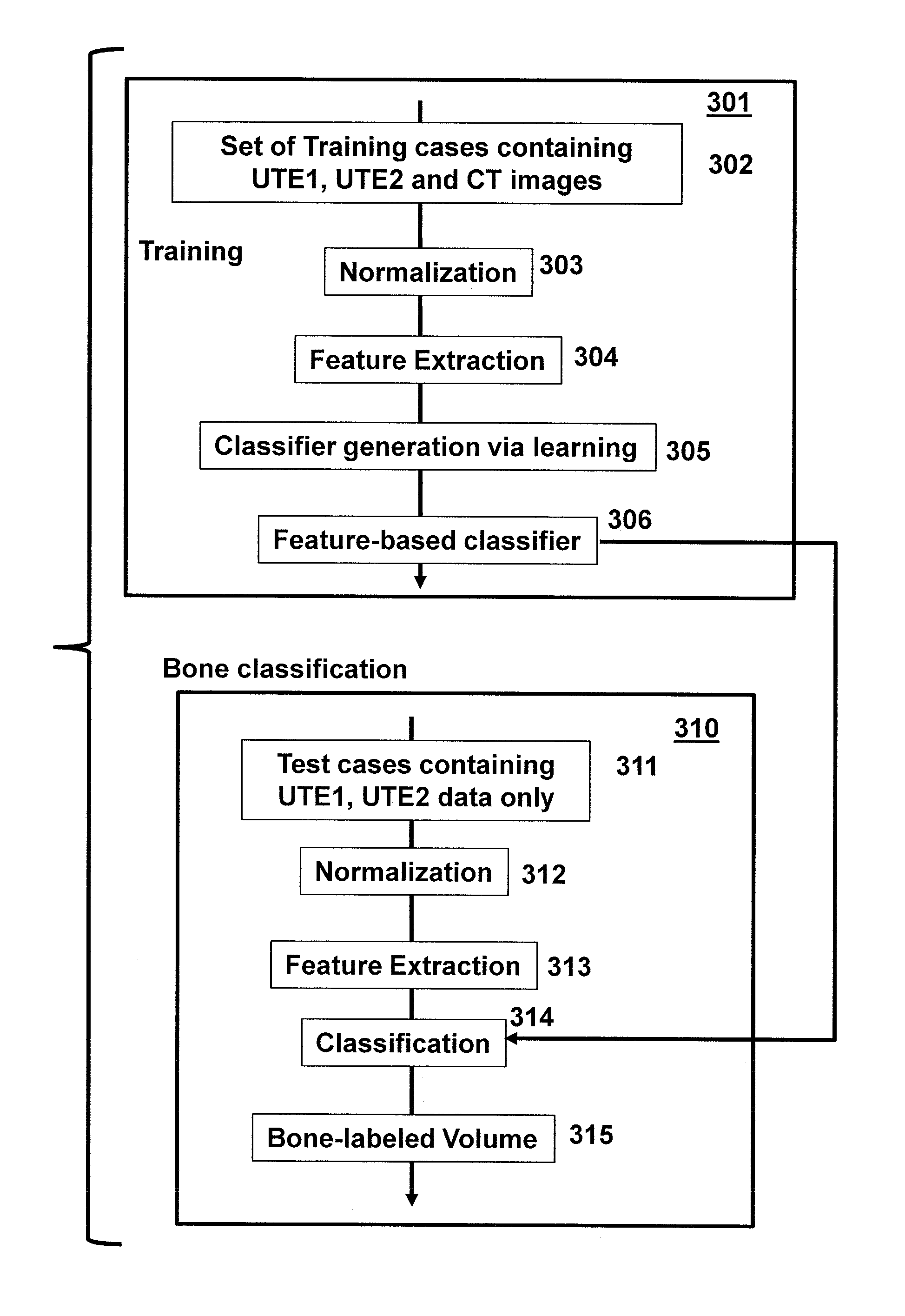
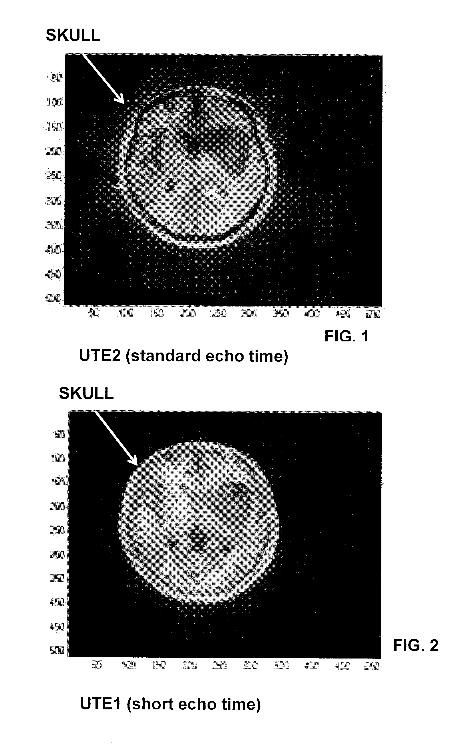
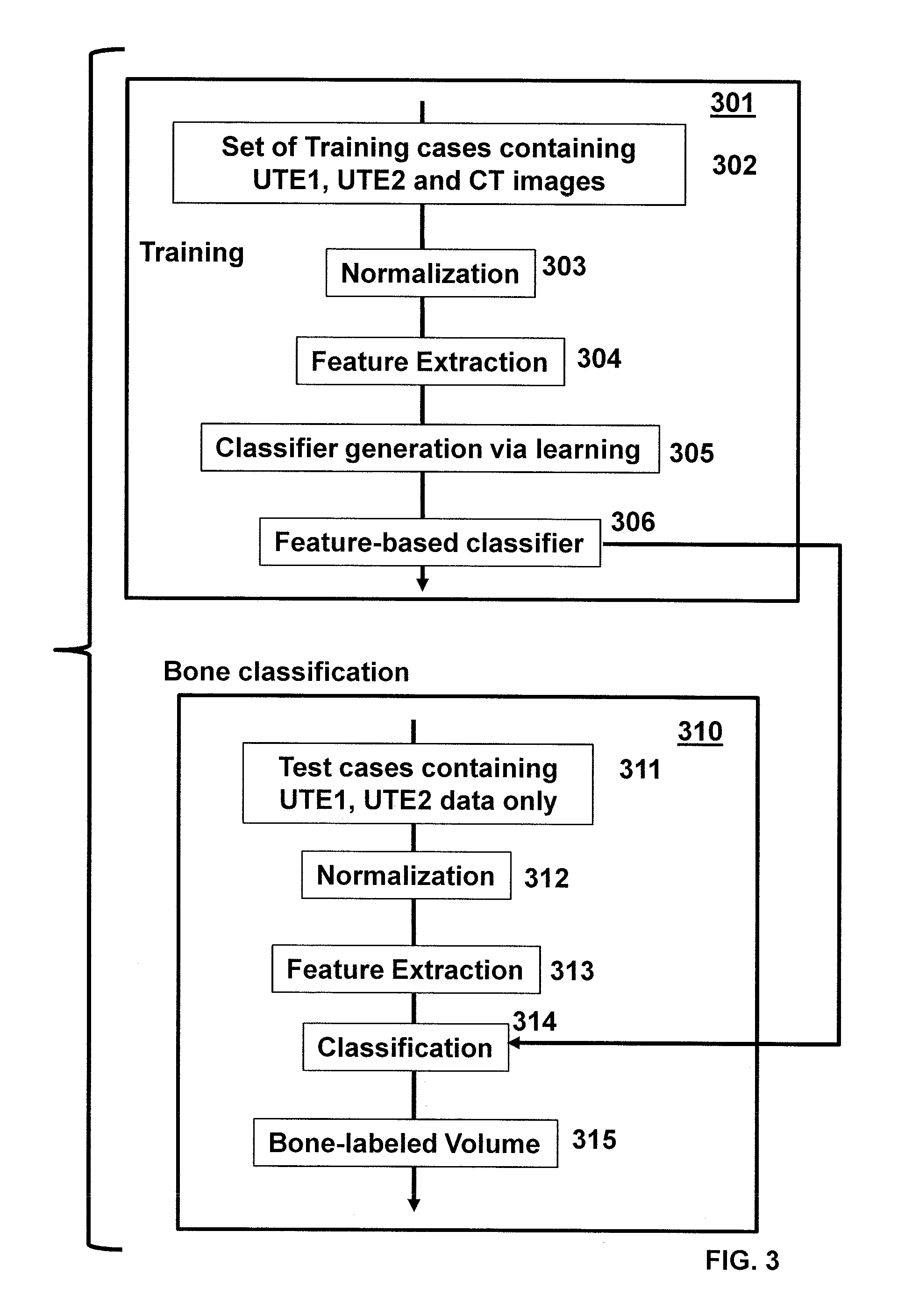
[0035]It is known that a DRR-based method for registering an MR volume is much more difficult than registering a CT volume, because the physics for MR and X-ray imaging is completely different. For example, the bony structure is usually not picked up well by MR using the standard protocol and can be confused with air or soft tissues. In particular, what is typically seen on MRI is the bone marrow or phrased in another way: the fat mixed into a spongy matrix. The outer / hard bone shell (cortical bone) surrounding the matrix is not seen with standard MR because there simply is no signal. For registration purpose, the diminished bony structures in MR volume do not correspond well to the highly opaque bony structures showed in the X-ray image, which can be misleading and lead to wrong registration.

[0036]As an aspect of the present invention a 2D3D registration technique for aligning MR volumes with X-ray images is provided by generating DRRs using one specialized MR acquisition, named ul...


PUM


Abstract

Systems and methods for 2D3D registration of apply MR volumes and X-ray images using DRR techniques. A bone classifier is trained from co-registered UTE1, UTE2 and CT prior images. Dual-echo MR UTE1 and UTE2 images are acquired from a patient. The bone structure of the patient is classified and a labeled segmentation is generated. A DRR image is generated from the labeled segmentation and is registered with an X-ray image of the patient. The registration methods are implemented on a processor based system.

Description

BACKGROUND OF THE INVENTION[0001]The present invention relates generally to medical imaging and more particularly to 2D3D registration of MR volumes with X-ray images.[0002]2D X-ray fluoroscopy has been a preferred modality routinely used for interventional and hybrid medical procedures. It can provide real-time monitoring of the procedure and visualization of the device location. However, anatomic structures are typically not delineated by fluoroscopy because soft tissues are not distinguishable by X-rays. In order to augment the view of the anatomies and help the doctor navigate the device to the target area, pre-operative high quality computed tomography (CT) and / or magnetic resonance (MR) volumes can be fused with the intra-operative fluoroscopic images, for which 2D3D registration of the coordinate systems of the two modalities is needed.[0003]One technique for 2D3D registration between CT volumes and X-ray images is based on digitally reconstructed radiographs (DRRs), which si...

Claims


Application Information

Patent Timeline
Patent Type & Authority Applications(United States)
IPC IPC(8): A61B5/055A61B6/00
CPCA61B5/055A61B6/032A61B6/5247G06T7/0028A61B5/7425G06T2207/30016G06T7/0081G06T2207/10088G06T2207/10116G06T2207/10124G06T2207/30008A61B5/0035G06T7/11G06T7/33
Inventor LIAO, RUIREISMAN, JAMES G.CHEFD'HOTEL, CHRISTOPHESHEA, STEVEN MICHAEL
Owner SIEMENS CORP
Why Eureka
Try Eureka