Automated quantification of intravascular embolization success

a technology of intravascular embolization and automatic quantification, which is applied in the field of imaging systems, can solve the problems of difficult to identify the exact outline of a tumor, difficult to relate and compare corresponding tumor regions within images before & after treatment, and complicated approach to outcome control. achieve the effect of increasing the visibility of the tissue of interes

Inactive Publication Date: 2013-08-15
KONINKLIJKE PHILIPS ELECTRONICS NV
View PDF2 Cites 3 Cited by
  • Summary
  • Abstract
  • Description
  • Claims
  • Application Information

AI Technical Summary

Benefits of technology

[0021]For improving the results of the method according to the invention the first image and the second image are obtained in a late arterial phase each, hence increasing the visibility of the tissue of interest.

Problems solved by technology

Furthermore, this approach of outcome control is complicated by the general difficulty in defining e.g. an exact outline of a tumor, by the occurring patient movement between pre- and post-treatment scans, and by the different appearance of the untreated and embolized tumors.
In particular, identifying the exact outline of a tumor is usually more difficult in the post-interventional scan due to its embolized state and the consequent under-perfusion.
These issues make it difficult to relate and compare corresponding tumor regions within images before & after the treatment.

Method used


Image

  • Automated quantification of intravascular embolization success
  • Automated quantification of intravascular embolization success
  • Automated quantification of intravascular embolization success

Examples


Embodiment Construction

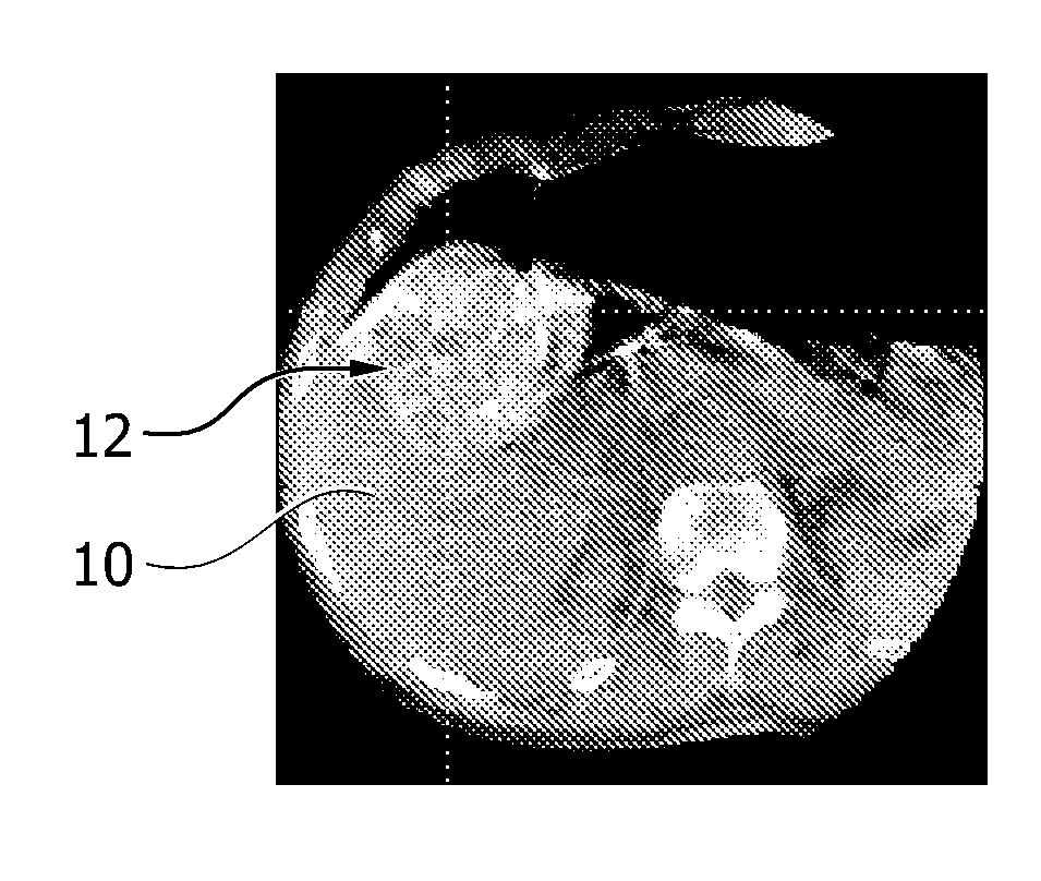
[0044]A first exemplary embodiment will be described in the following. FIG. 1 schematically shows a device 2 for automatically quantifying intravascular embolization success according to the first embodiment of the invention. The device 2 comprises a registration unit 4 which is provided with image data ID. This image data comprises a first image obtained before an intravascular embolization treatment is conducted and a second image obtained after an intravascular embolization treatment is conducted. The registration unit 4 registers the first image and the second image in order to compensate for patient / organ motion occurring between the acquisition time of the first image and the second image and provides registered image data RID. Registering itself may include selecting one or more reference points of the first image and of the second image and aligning these reference points relative to each other, thereby turning and stretching at least one of the first image and the second im...


PUM


Abstract

The present invention relates to a device (2) for automatically quantifying intravascular embolization success, comprising a registration unit (4) adapted for registering a first image and a second image, a segmentation unit (6) adapted for segmenting a tissue of interest in the first image and in the second image and an evaluation unit (8) for evaluating a deviation of perfusion of the tissue of interest by comparing the first image and the second image. The first image is obtained before an interventional treatment, whereas the second image is obtained after such a treatment. Evaluating may comprise comparing the segments of the first and the second images and thus providing a quantitative measure for a perfusion deviation of the tissue, e.g. the perfusion deviation of a tumorous tissue before and after an embolization treatment.

Description

FIELD OF THE INVENTION[0001]The invention relates to imaging systems which can be used for obtaining image data of tissues. In particular, the invention relates to a device and a method for automatically quantifying intravascular embolization success and to an imaging system capable of conducting such a method.BACKGROUND OF THE INVENTION[0002]Transcatheter targeted chemoembolic drug delivery today represents the mainstay treatment of liver cancer, requiring intensive support by X-ray imaging with catheterization laboratory systems. Performing and analyzing a dual phase scan prior to an actual treatment, which dual phase scan comprises imaging during both the early-arterial and late-arterial phases of enhancement under utilization of an intra-arterially injected contrast agent for enhancement of vessels or tissue, e.g. tumorous tissue, the information from the dual phases allows visualization of e.g. a tumor-feeding vessel tree and a tumor itself. This may thereby facilitate identify...

Claims


Application Information

Patent Timeline
Patent Type & Authority Applications(United States)
IPC IPC(8): A61B5/00
CPCG06T7/0014G06T2207/10081G06T2207/10096A61B6/507G06T2207/30096A61B5/0033G06T2207/30056A61B5/0036
Inventor WIESNER, STEFFEN GUNTHERBERTRAM, MATTHIASLIN, MING DE
Owner KONINKLIJKE PHILIPS ELECTRONICS NV
Why Eureka
Try Eureka