
Method and System for Joint Multi-Organ Segmentation in Medical Image Data Using Local and Global Context

a multi-organ and image data technology, applied in image analysis, image enhancement, instruments, etc., can solve the problems of computational demands and efficiency decline of level set methods, and achieve the effect of efficient multiple organ segmentation

Active Publication Date: 2013-08-29
SIEMENS HEALTHCARE GMBH
View PDF5 Cites 24 Cited by
  • Summary
  • Abstract
  • Description
  • Claims
  • Application Information

AI Technical Summary

Benefits of technology

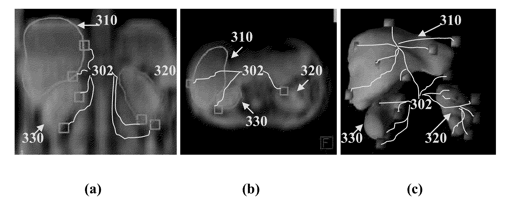
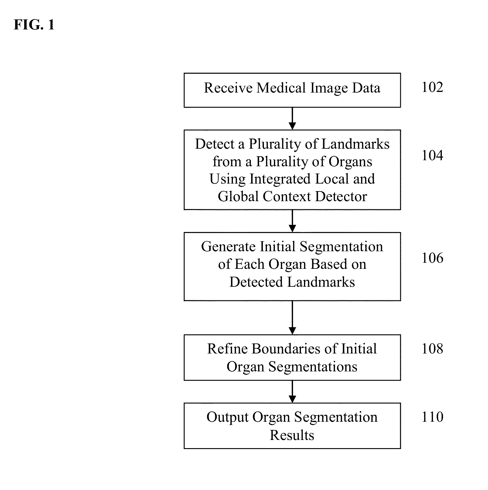
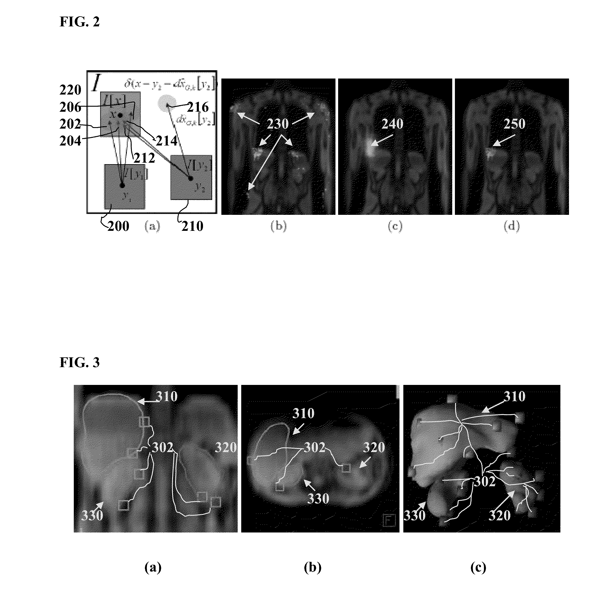
The present invention provides a method and system for segmenting multiple organs in medical images using both local and global context. This approach integrates information from both sources to improve the accuracy of organ segmentation. The global context helps to hypothesize the likely location of organ landmarks, which are then evaluated by the local discriminative classifier. The non-parametric representation of global image context models correlations in the target shape for multiple organs, allowing landmarks of multiple organs to be jointly localized. Additionally, the method imposes a statistical shape constraint on allowable reconstructed shapes, which helps to filter out poorly detected landmarks. Overall, this approach enhances the accuracy of segmenting multiple organs in medical images.

Problems solved by technology

However, level set methods are computationally demanding and typically require an accurate initialization so as not to fall into a local minimum.
Discriminative learning-based methods are an alternative approach to such level set segmentations, but learning-based methods typically treat the initialization of each organ as an independent problem.
While solving a single organ segmentation problem with learning-based methods can be fast, in order to achieve multi-object segmentation, a tree-like search structure has to be imposed on the detection order of the structures, resulting in a decrease in efficiency.

Method used


Image

  • Method and System for Joint Multi-Organ Segmentation in Medical Image Data Using Local and Global Context
  • Method and System for Joint Multi-Organ Segmentation in Medical Image Data Using Local and Global Context
  • Method and System for Joint Multi-Organ Segmentation in Medical Image Data Using Local and Global Context

Examples


Embodiment Construction

[0013]The present invention relates to a method and system for segmenting multiple organs in medical images using a combination of local and global context. Embodiments of the present invention are described herein to give a visual understanding of the multi-organ segmentation method. A digital image is often composed of digital representations of one or more objects (or shapes). The digital representation of an object is often described herein in terms of identifying and manipulating the objects. Such manipulations are virtual manipulations accomplished in the memory or other circuitry / hardware of a computer system. Accordingly, is to be understood that embodiments of the present invention may be performed within a computer system using data stored within the computer system.

[0014]Embodiments of the present invention jointly segment multiple organs in medical image data, such as magnetic resonance (MR), computed tomography (CT), ultrasound, X-ray, etc. Embodiments of the present in...


PUM


Abstract

A method and system for segmenting multiple organs in medical image data is disclosed. A plurality of landmarks of a plurality of organs are detected in a medical image using an integrated local and global context detector. A global posterior integrates evidence of a plurality of image patches to generate location predictions for the landmarks. For each landmark, a trained discriminative classifier for that landmark evaluates the location predictions for that landmark based on local context. A segmentation of each of the plurality of organs is then generated based on the detected landmarks.

Description

[0001]This application claims the benefit of U.S. Provisional Application No. 61 / 604,200, filed Feb. 28, 2012, the disclosure of which is herein incorporated by reference.BACKGROUND OF THE INVENTION[0002]The present invention relates to segmenting multiple organs in medical images more particularly, to joint segmentation of multiple organs by fusing local and global context.[0003]Algorithms for segmenting anatomical structures in medical images are typically targeted to segmenting individual structures. When the problem is posed as the joint segmentation of multiple organs, constraints, such as a non-overlapping constraint, may be formulated between the organs, and the combined formulation allows for a richer prior model on the joint shape of the multiple structures of interest. Such multi-organ segmentation is typically posed with atlas-based or level set-based formulations due to the ease in which geometric constraints can be modeled using such formulations.[0004]However, level se...

Claims


Application Information

Patent Timeline
Patent Type & Authority Applications(United States)
IPC IPC(8): G06K9/34
CPCG06K9/34G06T7/0081G06T2207/10088G06T2207/20076G06T2207/30084G06T2207/20124G06T2207/20164G06T2207/30056G06T2207/20081G06T7/11
Inventor LAY, NATHANBIRKBECK, NEILZHANG, JINGDANGUEHRING, JENSZHOU, SHAOHUA KEVIN
Owner SIEMENS HEALTHCARE GMBH
Learn More