Device and method for determining the presence of middle ear fluid

a technology of fluid in the middle ear and a device, applied in the field of otolaryngology, can solve the problems of otitis media (ear infection, ome, hearing loss, etc.) and recurrent ear infections that often follow

Active Publication Date: 2013-10-31
THE CLEVELAND CLINIC FOUND +1
View PDF4 Cites 29 Cited by
  • Summary
  • Abstract
  • Description
  • Claims
  • Application Information

AI Technical Summary

Problems solved by technology

Otitis media (ear infection) is a common problem that bears significant health implications.
Persistence of a MEE can result in hearing loss and recurrent otitis media with effusion (OME).
However, recurrent ear infections often ensue in OME as the presence of effusion creates a fertile environment for bacterial growth.
However, inflammatory stimuli, such as bacteria, virus, and allergy, may cause excessive production, increased viscosity, or impaired drainage of the mucus.
MEEs may persist for prolonged durations, especially in children, resulting in hearing loss and recurrent ear infections.
However, these both have limitations in diagnostic accuracy and are dependent on the practitioner's experience, warranting consideration of alternate technologies.
Therefore, the tympanometry probe placed in the external ear canal must achieve a tight seal, which is difficult to obtain in young children.

Method used


Image

  • Device and method for determining the presence of middle ear fluid
  • Device and method for determining the presence of middle ear fluid
  • Device and method for determining the presence of middle ear fluid

Examples


example

Device Design

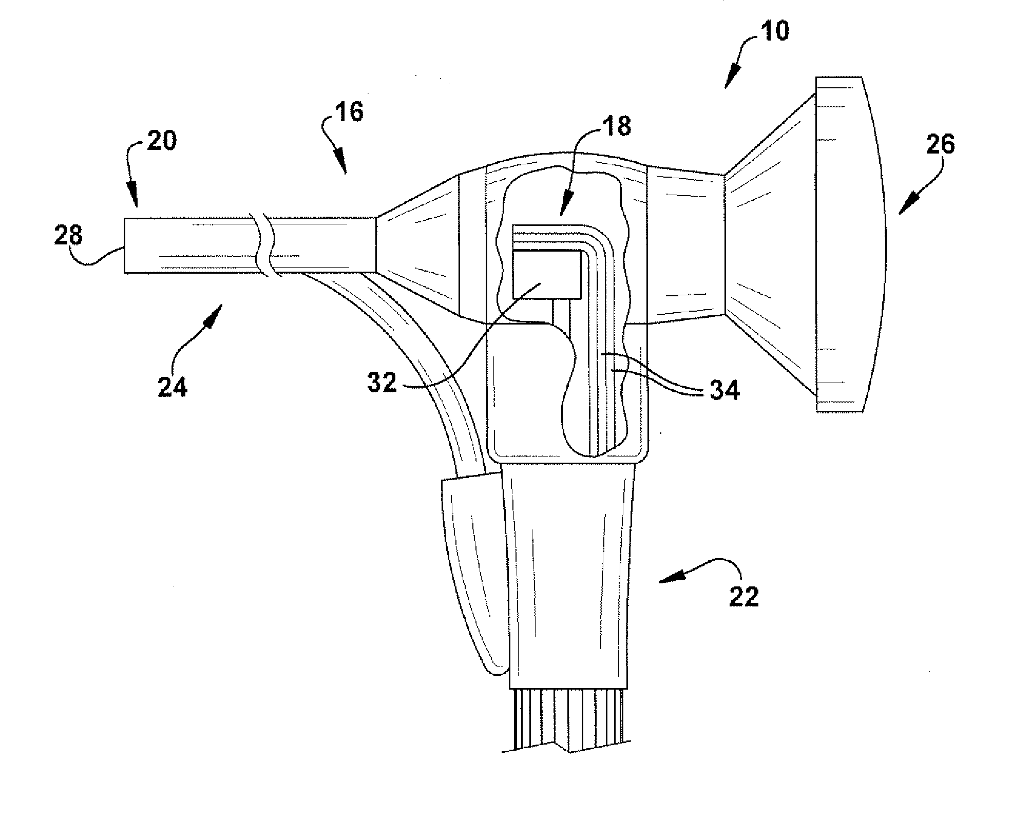
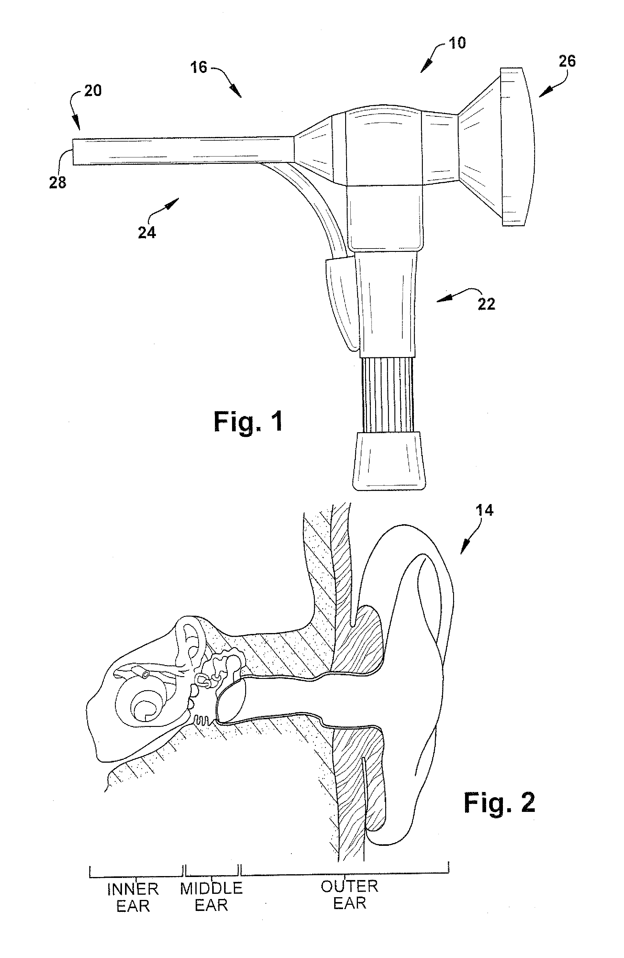
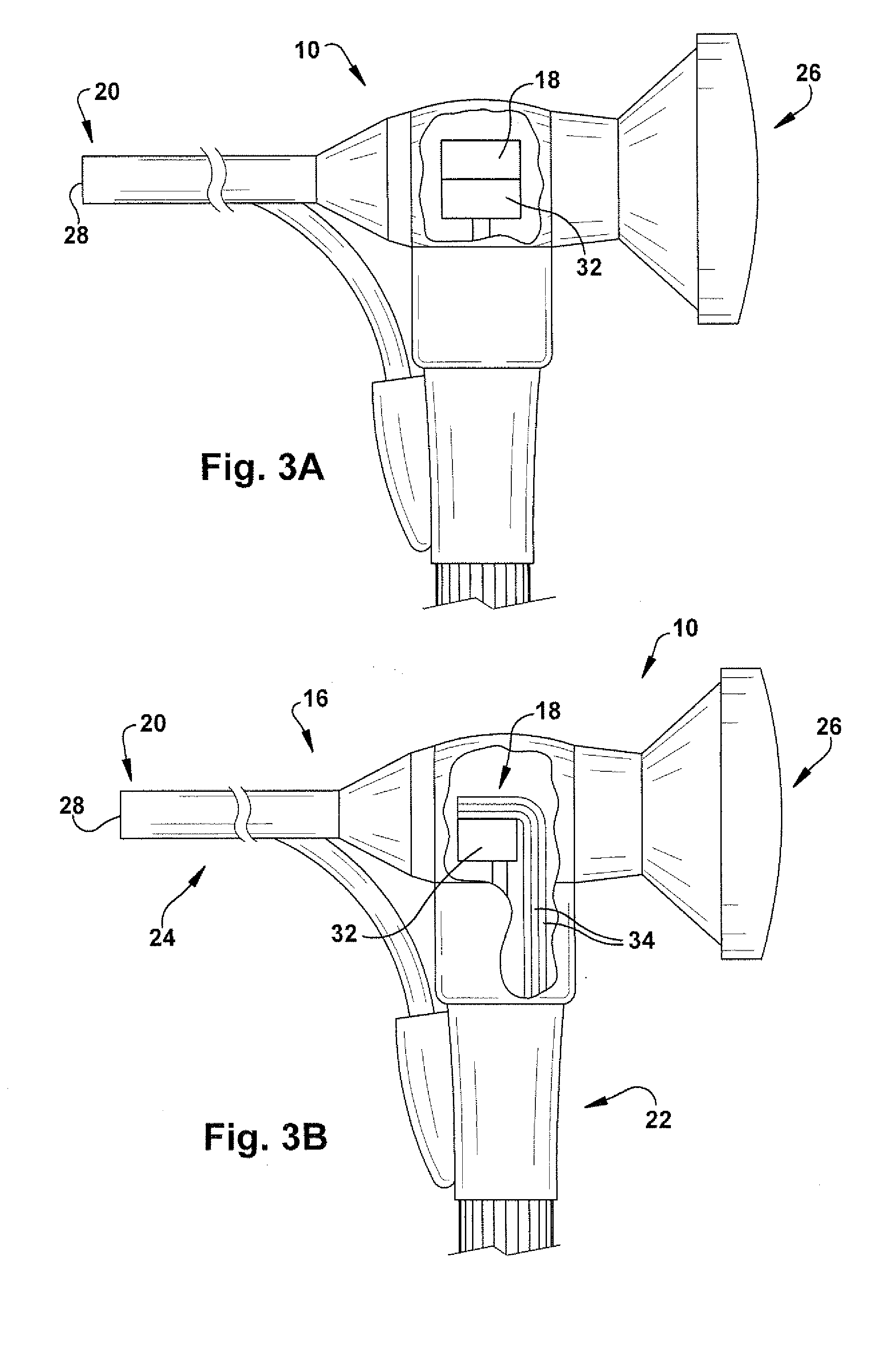
[0051]Initial device design centered on the addition of a laser diode module and mount to an existing open otoscope (Welch Allyn, Skaneateles Falls, N.Y.). For the purpose of the present invention, an open otoscope easily allows positioning and adjustment of a tightly focused beam. A simple mount was built and attached to the otoscope near the light source to secure the laser diode module.

[0052]This design allowed convenient replacement of the laser diode module so that different wavelengths (colors) may be tested on the same prototype. Also, the mount allowed accurate and stable positioning of the laser diode module so that the beam could be aimed through the opening of the speculum and illuminate onto the tympanic membrane surface. For purposes of this prototype, the laser diode module was attached to the mount by double-sided tape, and power was achieved via an external battery source. Power for the otoscope incandescent bulb was from a separate power supply built in...


PUM


Abstract

A device for determining the presence of abnormal fluid in a middle ear of a subject includes an elongated probe, a first light source housed within the elongated probe, and a second light source housed within the elongated probe. The elongated probe includes a distal end for inspection of an ear. The First light source is configured to convey an optical beam through a tympanic membrane associated with the middle ear of the subject, without puncturing the tympanic membrane. The second light source is configured to convey light through the distal end of the elongated probe and illuminate the tympanic membrane.

Description

RELATED APPLICATION[0001]This application claims the benefit of U.S. Provisional Patent Application Ser. No. 61 / 410,114, filed Nov. 4, 2010, the entirety of which is hereby incorporated by reference for all purposes.TECHNICAL FIELD[0002]The present invention relates generally to the field of otolaryngology, and more specifically to a device and method for determining the presence of fluid in the middle ear of a subject.BACKGROUND OF THE INVENTION[0003]Otitis media (ear infection) is a common problem that bears significant health implications. It is the most common cause of deafness in children in the developed world, affecting up to 80% of preschool children at some time. Second only to upper respiratory infections, it is the most common indication for outpatient antibiotic use in children. Further, it is the most common reason for a child to visit the pediatrician. In the United States, the annual cost of medical and surgical treatment of otitis media is estimated at $5 billion.[00...

Claims


Application Information

Patent Timeline
Patent Type & Authority Applications(United States)
IPC IPC(8): A61B1/227
CPCA61B1/227A61B1/063A61B1/0638A61B5/0075A61B5/0084A61B5/12A61B5/6817A61B1/0684
Inventor SETH, RAHULFORD, MATTHEW R.ROLLINS, ANDREW M.KRAKOVITZ, PAUL K.
Owner THE CLEVELAND CLINIC FOUND
Why Eureka
Try Eureka