
Noninvasive physiological sensor cover

Inactive Publication Date: 2013-11-07
JPMORGAN CHASE BANK NA
View PDF2 Cites 491 Cited by
  • Summary
  • Abstract
  • Description
  • Claims
  • Application Information

AI Technical Summary

Benefits of technology

The patent text is discussing a problem with optical sensors in clinical settings. These sensors can generate false readings and cause noise if they are not in use and can also be exposed to infectious agents and other contaminants, which can damage them. The solution proposed is a sensor cover that prevents or reduces false readings and also protects the sensor components during shipment and prior to use. The cover has a pattern that can be easily seen on a surface such as a floor of a clinical setting. Overall, this invention improves the accuracy and reliability of optical sensors in clinical settings.

Problems solved by technology

While attached, the sensor can generate false readings by detecting ambient light even though the sensor is not in use.
The sensor can also cause the monitor to emit alarms or otherwise make noise due to false readings, which can be distracting to medical personnel.
Further, in certain embodiments, a sensor cover decreases the likelihood of contamination by keeping covered portions of the sensor clean.
Sensors in hospitals and other clinical environments are subject to exposure to infectious agents, dust, or other foreign matter from depositing on sensor components.

Method used


Image

  • Noninvasive physiological sensor cover
  • Noninvasive physiological sensor cover
  • Noninvasive physiological sensor cover

Examples


Embodiment Construction

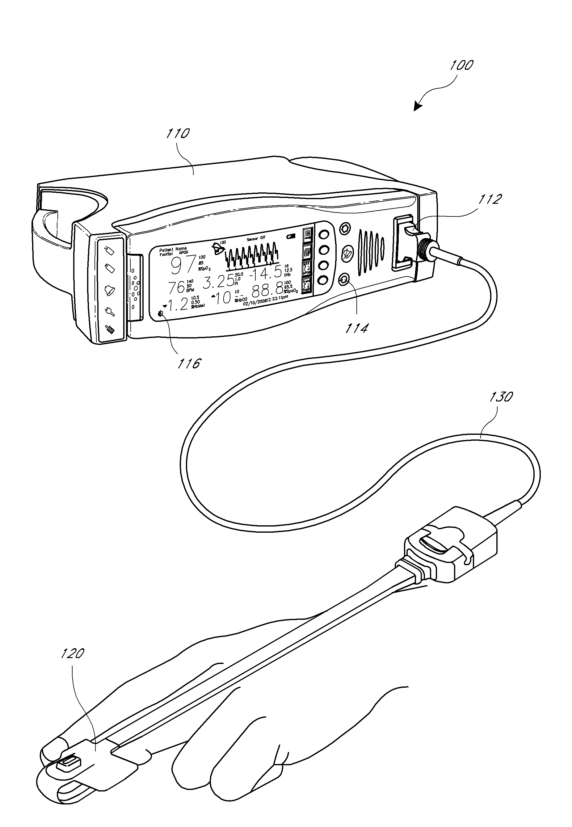
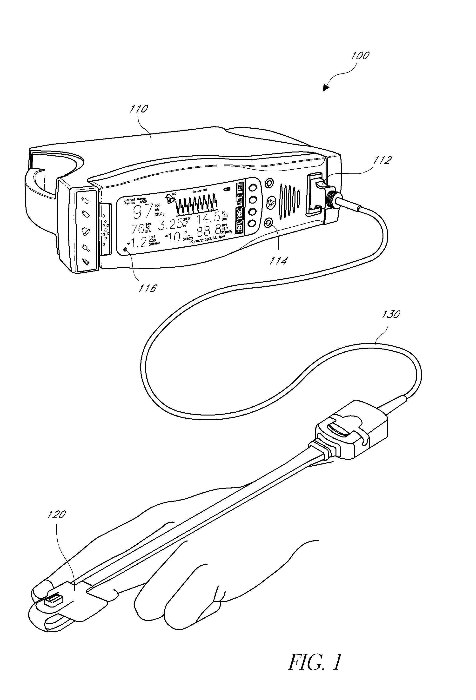
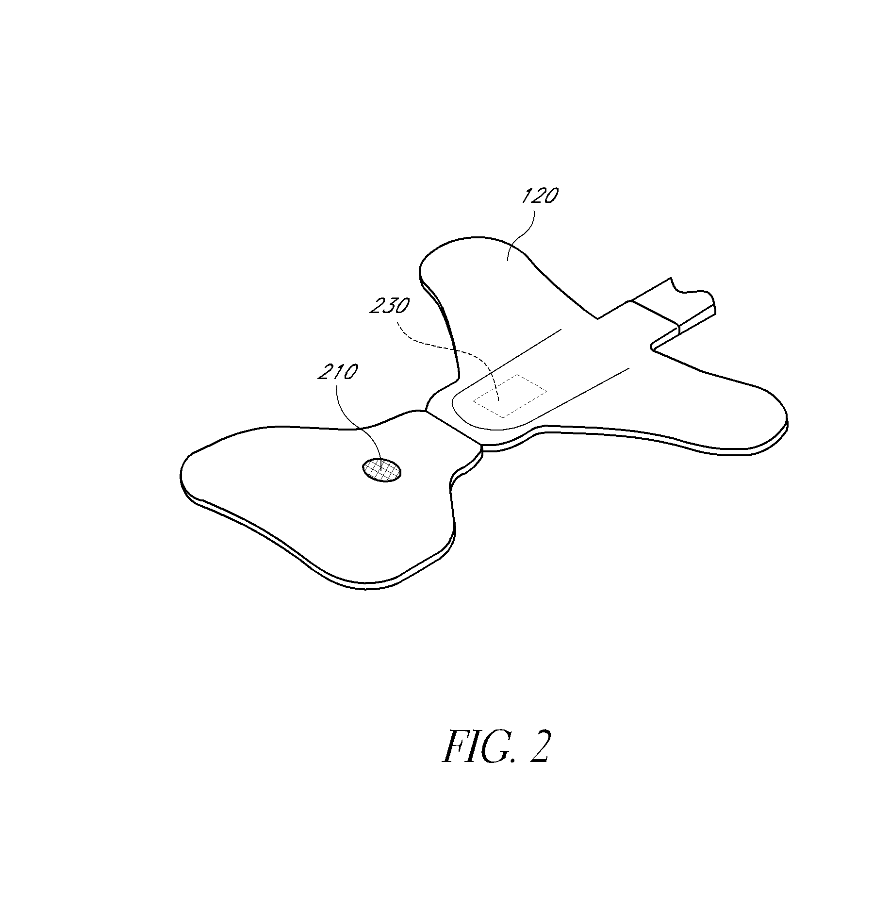
[0015]A sensor cover according to embodiments of the disclosure is capable of being used with a noninvasive physiological sensor. Certain embodiments of the sensor cover reduce or eliminate false readings from the sensor when the sensor is not in use. Some embodiments of the sensor cover can have patterns that aid seeing the sensor cover on a floor of a clinical settings, such as operating rooms, emergency rooms, post anesthesia care units, critical care units, outpatient surgery and physiological labs, to name a few. Further, embodiments of the sensor cover prevent contamination of the sensor. Additionally, embodiments of the sensor cover can prevent damage to the sensor.

[0016]The tissue site of the illustrated embodiments is a finger and the following description therefore refers specifically to the tissue site as a finger for the purposes of clarity. This is not intended to be limiting and, as described herein, the sensor cover of certain embodiments can be used with sensors atta...


PUM


Abstract

A sensor cover according to embodiments of the disclosure is capable of being used with a noninvasive physiological sensor, such as a pulse oximetry sensor. Certain embodiments of the sensor cover reduce or eliminate false readings from the sensor when the sensor is not in use, for example, by blocking a light detecting component of a pulse oximeter sensor when the pulse oximeter sensor is active but not in use. In certain embodiment, the sensor cover has a pattern that allows it to be more easily seen on a surface such as a floor. Further, embodiments of the sensor cover prevent contamination of the sensor. Additionally, embodiments of the sensor cover can prevent damage to the sensor.

Description

CROSS-REFERENCE TO RELATED APPLICATIONS[0001]This application claims the benefit under 35 U.S.C. §119(e) as a nonprovisional of U.S. Provisional Application No. 61 / 641,611, filed May 2, 2012, titled NON-INVASIVE PHYSIOLOGICAL SENSOR COVER, the entirety of which is incorporated herein by reference.FIELD OF THE DISCLOSURE[0002]The present disclosure relates to a sensor for measuring oxygen content in the blood, and, in particular, relates to an apparatus and method for preventing sensor activity when the sensor is not in use.BACKGROUND OF THE DISCLOSURE[0003]Noninvasive physiological sensors are applied to the body for monitoring or making measurements indicative of a patient's health. One application for a noninvasive physiological sensor is pulse oximetry, which provides a noninvasive procedure for measuring the oxygen status of circulating blood. Oximetry has gained rapid acceptance in a wide variety of medical applications, including surgical wards, intensive care and neonatal uni...

Claims


Application Information

Patent Timeline
IPC IPC(8): A61B5/1455A61B5/0205
CPCA61B5/14552A61B5/0205A61B5/6898A61B5/6816A61B5/6819A61B5/6826A61B2562/18
Inventor O'NEIL, CHARLES D.SCHMIDT, JOHNBARKER, NICHOLAS EVAN
Owner JPMORGAN CHASE BANK NA
Learn More