Magnetic resonance using quazi-continuous RF irradiation

a quazi-continuous rf and magnetic resonance technology, applied in the field of magnetic resonance imaging, can solve the problems of insufficient selective saturation prior to the actual acquisition of image data, insufficient contrast, disadvantageous suboptimal apt/cest detection sensitivity, etc., to achieve the effect of maintaining or minimizing the sar contribution and improving the homogeneity of the rf b1 field

Active Publication Date: 2013-12-26
KONINKLIJKE PHILIPS ELECTRONICS NV
View PDF4 Cites 18 Cited by
  • Summary
  • Abstract
  • Description
  • Claims
  • Application Information

AI Technical Summary

Benefits of technology

[0025]The RF power amplifiers associated with different groups of coil elements can be activated during non-overlapping or partly overlapping time slots according to the invention. The amplitudes and / or the phases of the RF signals supplied to the RF coil arrangement via the individual RF power amplifiers can be controlled in such a manner that the instantaneous and / or the time-integrated homogeneity of the RF magnetic field distribution of the at least one long RF pulse is optimized. In the case of a non-overlapping activation of the different RF power amplifiers, the incoherent sum (i.e. the modulus) of the RF signals generated during the different activation periods provides sufficient homogeneity of the RF magnetic field distribution of the long or quasi continuous-wave RF pulse. In the case of a partly overlapping activation of the individual RF power amplifiers, the coherent superposition of the RF signals generated via the different channels has to be considered. This enables the application of RF shimming as in conventional multi-channel parallel transmission systems
[0031]According to a preferred embodiment, all RF power amplifiers of the MR device are high-power / low-duty-cycle amplifiers. This means that according to the specifications of each individual RF power amplifier, the RF duty cycle is limited to a maximum of e.g. 50%. The maximum pulse duration is limited to e.g. 250 ms. On the other hand, the high-power / low-duty-cycle RF power amplifiers are able to deliver RF pulses having a RF peak power of several kW or even more than 10 kW as required for most MR imaging sequences. The time-multiplexed activation of the high-power / low-duty-cycle RF power amplifiers enables the generation of long or quasi continuous-wave RF pulses which are beyond the specification of a single high-power / low-duty-cycle RF power amplifier. In this embodiment, no low-power / high-duty-cycle mode of the RF power amplifiers needs to be provided, leading to cost reduction in the RF transmit chain.

Problems solved by technology

In some medical applications, the difference in MR signal intensity from standard MR protocols, i.e. the contrast, between different tissues might not be sufficient to obtain satisfactory clinical information.
A problem of all known APT / CEST MR imaging techniques is that the selective saturation prior to the actual acquisition of image data takes a comparably long time.
When implemented on MR imaging systems which are in clinical use at present, the APT / CEST detection sensitivity is disadvantageously suboptimal due to the hardware constraints of the standard RF power amplifiers commonly used in those systems.
Such standard RF power amplifiers are designed to deliver high-power short RF pulses for imaging applications, wherein the maximum length of the RF pulses as well as the RF duty cycle are limited because of the heat dissipation within the electronics of the RF power amplifier.
This is not suitable for effective APT / CEST MR imaging which actually requires continuous-wave RF irradiation or pulse trains with a RF duty cycle of 70 to 100% over a time interval of 2-5 seconds for obtaining sufficient saturation of the proton magnetization, albeit at a lower RF power than required for the short RF pulses used for MR image-acquisition.
This disadvantageously leads to a substantially reduced APT / CEST signal as compared to the CEST signal that could be obtained with optimal saturation.
A further disadvantage of the saturation sequences conventionally applied in APT / CEST MR imaging is that the ability to quantify the CEST effect may be compromised due to a spatially inhomogeneous saturation effect of the saturation RF pulse.
This is particularly a problem at high static magnetic field strength of 3 Tesla or more.
The inhomogeneity of the B1-field results in non-linear distortions of the CEST signal intensity.
A quantitative analysis of the CEST effect, which would be desirable for various applications, is difficult to achieve under such conditions.

Method used


Image

  • Magnetic resonance using quazi-continuous RF irradiation
  • Magnetic resonance using quazi-continuous RF irradiation
  • Magnetic resonance using quazi-continuous RF irradiation

Examples


Embodiment Construction

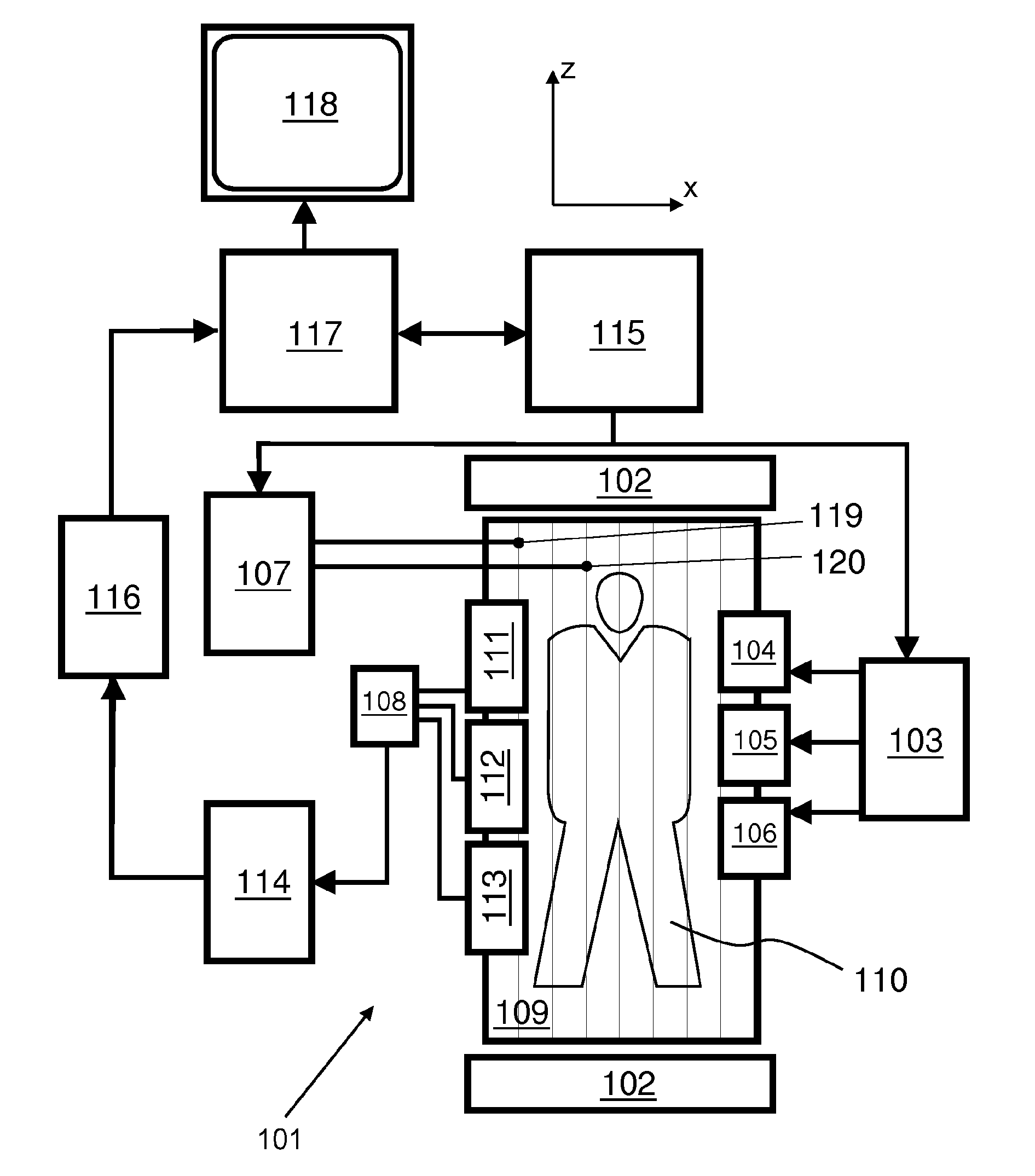
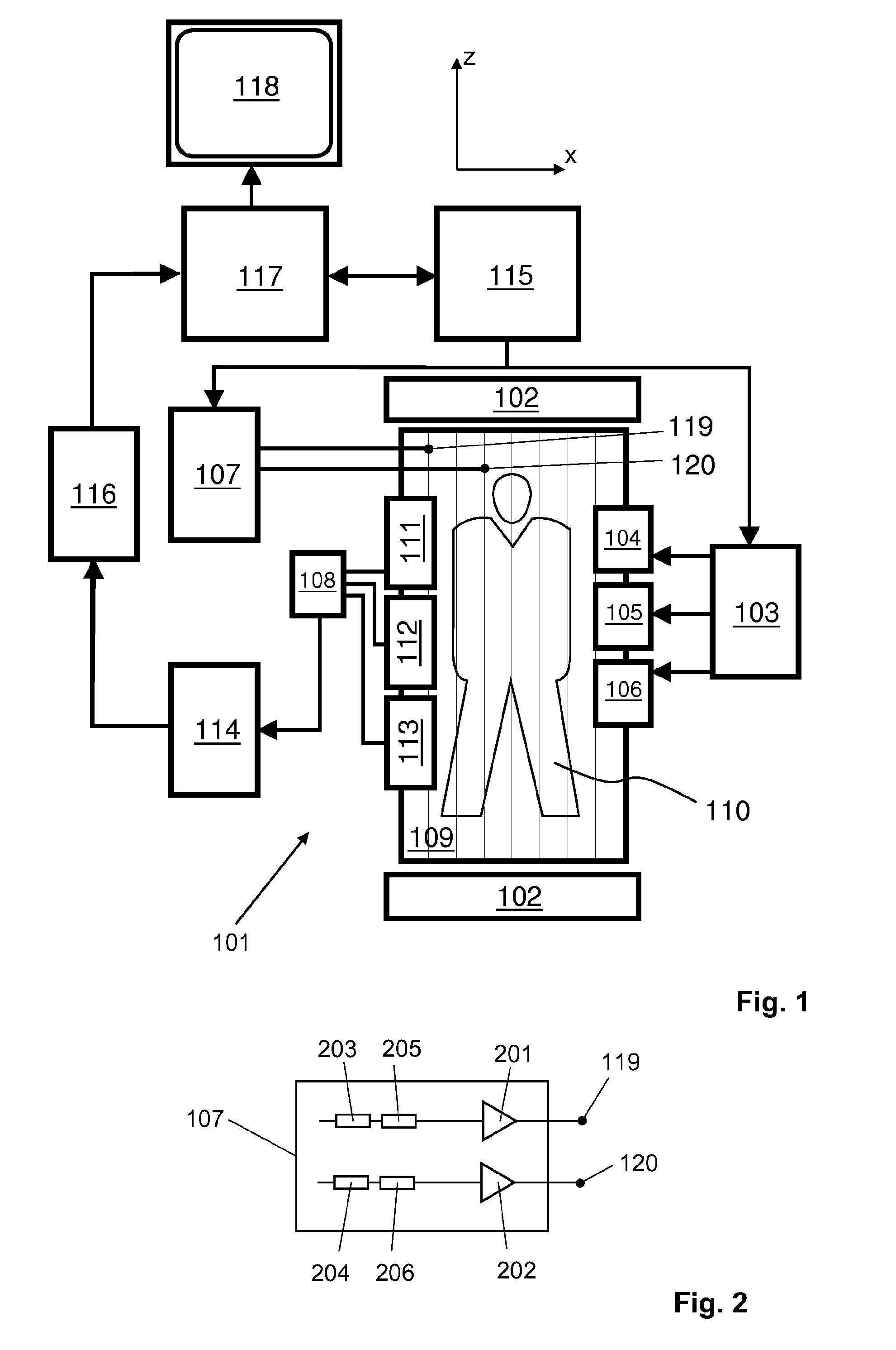
[0043]With reference to FIG. 1, a MR device 101 is shown. The device comprises superconducting or resistive main magnet coils 102 such that a substantially uniform, temporally constant main magnetic field is created along a z-axis through an examination volume.

[0044]A magnetic resonance generation and manipulation system applies a series of RF pulses and switched magnetic field gradients to invert or excite nuclear magnetic spins, induce magnetic resonance, refocus magnetic resonance, manipulate magnetic resonance, spatially and otherwise encode the magnetic resonance, saturate spins, and the like to perform MR imaging.

[0045]More specifically, a gradient pulse amplifier 103 applies current pulses to selected ones of whole-body gradient coils 104, 105 and 106 along x, y and z-axes of the examination volume. A multi-channel transmission unit 107 transmits RF pulses or pulse packets via two RF drive ports 119, 120 to a whole-body volume RF coil 109 to transmit RF pulses into the examin...


PUM


Abstract

The invention relates to a method of MR imaging of at least a portion of a body (110) of a patient placed in an examination volume of a MR device, the method comprising the steps of:—subjecting the portion of the body (110) to an imaging sequence comprising at least one RF pulse, the RF pulse being transmitted toward the portion of the body (110) via a RF coil arrangement (109) to which RF signals are supplied by two or more RF power amplifiers the RF power amplifiers being activated alternately during the imaging sequence in a time-multiplexed fashion, wherein the imaging sequence requires a RF duty cycle and / or a RF pulse duration exceeding the specification of at least one of the RF power amplifiers;—acquiring MR signals from the portion of the body (110); and—reconstructing a MR image from the acquired MR signals. Moreover, the invention relates to a method of MR spectroscopy involving the alternating use of RF power amplifiers in a time-multiplexed fashion.

Description

TECHNICAL FIELD[0001]The invention relates to the field of magnetic resonance (MR) imaging. It concerns a method of MR imaging and a method of MR spectroscopy. The invention also relates to a MR device and to a computer program for a MR device.BACKGROUND OF THE INVENTION[0002]Image-forming MR methods which utilize the interaction between magnetic fields and nuclear spins in order to form two-dimensional or three-dimensional images are widely used nowadays, notably in the field of medical diagnostics, because for the imaging of soft tissue they are superior to other imaging methods in many respects, do not require ionizing radiation and are usually not invasive.[0003]According to the MR method in general, the body of the patient to be examined is arranged in a strong, uniform magnetic field whose direction at the same time defines an axis (normally the z-axis) of the co-ordinate system on which the measurement is based. The magnetic field produces different energy levels for the indi...

Claims


Application Information

Patent Timeline
Patent Type & Authority Applications(United States)
IPC IPC(8): G01R33/54G01R33/32
CPCG01R33/54G01R33/32G01R33/34076G01R33/3614G01R33/5605G01R33/5612G01R33/5659
Inventor KEUPP, JOCHENVAN DEN BRINK, JOHAN SAMUELHARVEY, PAUL ROYSTON
Owner KONINKLIJKE PHILIPS ELECTRONICS NV
Why Eureka
Try Eureka