Method and system of automated detection of lesions in medical images

a technology of medical images and automated detection, applied in the field of computerized processing of medical images, can solve the problems of inconsistent diagnosis of ultrasound images, inability to achieve negative predictive values attainable by highly experienced experts, and inability to achieve negative predictive values attainable by less experienced radiologists

a technology of medical images and automated detection, applied in the field of computerized processing of medical images, can solve the problems of inconsistent diagnosis of ultrasound images, inability to achieve negative predictive values attainable by highly experienced experts, and inability to achieve negative predictive values attainable by less experienced radiologists

US20130343626A1Inactive Publication Date: 2013-12-26SALIENT IMAGING

Image

  • Method and system of automated detection of lesions in medical images
  • Method and system of automated detection of lesions in medical images
  • Method and system of automated detection of lesions in medical images

Examples


Embodiment Construction

[0032]The description which follows and the embodiments described therein are provided by way of illustration of an example, or examples, of particular embodiments of the principles of the present invention. These examples are provided for the purposes of explanation, and not limitation, of those principles and of the invention. In the description which follows, like parts are marked throughout the specification and the drawings with the same respective reference numerals.

[0033]The present invention generally relates to a system and method of processing medical images. In particular, the invention relates to detection of lesion candidates in ultrasound medical images.

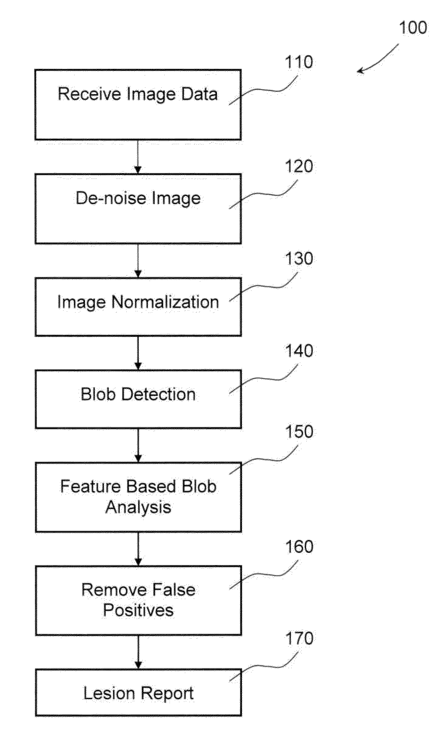
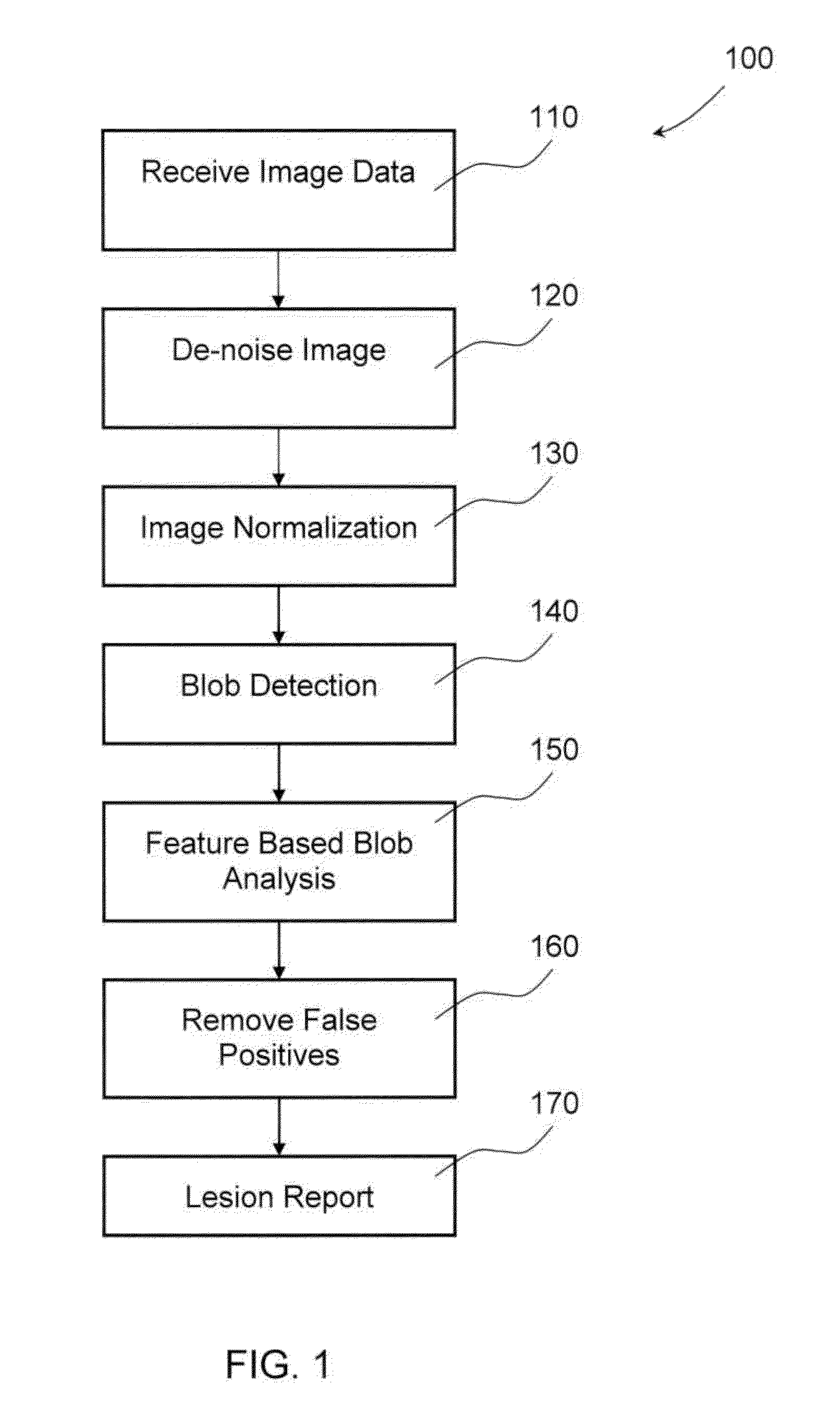
[0034]In one embodiment a sequence of image processing routines are applied to an input image, such as a single breast ultrasound image (or volume data set), to detect and classify each lesion candidate that might require further diagnostic review. FIG. 1 is a flow chart that provides an overview of the process 100.

[003...


PUM


Abstract

The invention provides a system and method for processing medical images. Input medical images are normalized first, utilizing pixel intensities of control point tissues, including subcutaneous fat. Clustered density map and malignance probability map are generated from a normalized image and further analyzed to identity regions of common internal characteristics, or blobs, that may represent lesions. These blobs are analyzed and classified to differentiate possible true lesions from other types of non-malignant masses often seen in medical images.

Description

CROSS-REFERENCE TO RELATED APPLICATIONS[0001]This application is a continuation of U.S. patent application Ser. No. 12 / 643,337, filed Dec. 21, 2009, which claims priority from U.S. Provisional Application No. 61 / 139,723 filed on Dec. 22, 2008, wherein the contents of each hereby incorporated by reference.FIELD OF INVENTION[0002]The invention relates generally to the field of computerized processing of medical images. In particular, the invention relates to identification of tissue layers in medical images, automated detection of lesions in medical images and normalization of pixel intensities of medical images.BACKGROUND OF INVENTION[0003]Cancer is recognized as a leading cause of death in many countries. It is generally believed that early detection and diagnosis of cancer and therefore early treatment of cancer help reducing mortality rate. Various imaging techniques for detection and diagnosis of cancer, such as breast cancer, ovarian cancer, and prostate cancer, have been develo...

Claims


Application Information

Patent Timeline
26 Dec 2013
Publication
US20130343626A1
IPC
A61B5/00
CPC
A61B5/0033; A61B5/4312; A61B5/7203; A61B5/7264; G06T7/0012; G06T2207/10132; G06T2207/20192; G06T2207/30096
Inventors
RICO, DAN; CHUNG, DESMOND RYAN