Magnetic Imaging Device To Inventory Human Brain Cortical Function

a magnetic imaging and cortical function technology, applied in the field of medical imaging, can solve the problems of large imaging problem, large complexity, unpredictable signal attenuation by tissues,

Inactive Publication Date: 2014-01-02
FORD JOHN P
View PDF12 Cites 23 Cited by
  • Summary
  • Abstract
  • Description
  • Claims
  • Application Information

AI Technical Summary

Problems solved by technology

Even though neurons constitute only about 10% of the brain cell population, this complexity presents a huge imaging problem.
A functional three-dimensional image of a volume with this complexity would be almost impossible both to display and to understand.
A limitation for an electroencephalogram (EEG), which is sensitive to a millisecond time frame, is the problem of unpredictable signal attenuation by the tissues that surround the brain, a problem which does not exist for the magnetic field generated by the electric impulses in the brain.
EEG signals do not attenuate predictably, however, such that both near and far signals are comingled (see, for example, Gallen et al., “Magnetoencephalography and Magnetic Source Imaging: Capabilities and Limitations”, Functional Neuroimaging, Vol. 5, pp.
This problem is particularly true of multiple current sources (e.g., both primary and secondary cortical sources).
A significant challenge in MEG, however, is the filtering out of environmental magnetic noise, which may be considerably higher (six or greater orders of magnitude) than the brain-generated magnetic fields of interest.
First, magnetically-shielded rooms are very expensive.
Second, the cost of a 300-sensor SQUID is exorbitant, at a cost of about $10,000 per SQUID sensor.
Third, solving the virtually infinitely complex inverse problem (i.e., identifying the location of the relevant current sources) has not been accomplished and may not be possible.

Method used


Image

  • Magnetic Imaging Device To Inventory Human Brain Cortical Function
  • Magnetic Imaging Device To Inventory Human Brain Cortical Function
  • Magnetic Imaging Device To Inventory Human Brain Cortical Function

Examples


Embodiment Construction

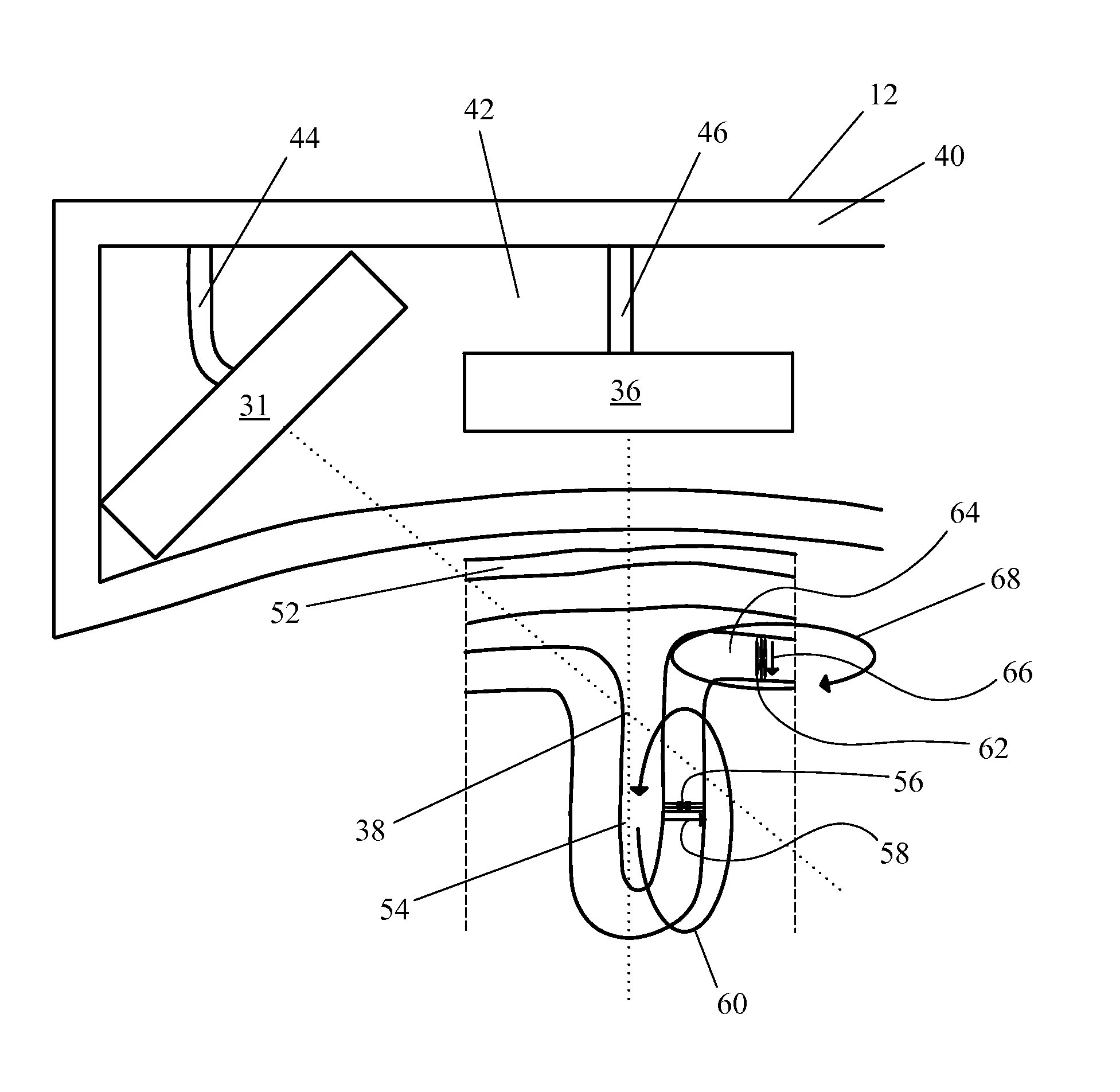
[0030]To achieve adequate data homogenization in order to render the content of the collected data understandable without degrading it, in the current apparatus and methods the data collection is limited to neural transmission originating in the most superficial neurons lining the sulci of the relevant gyms of the human cortex. Also, the output is presented as a contour map and no attempt is made to determine the underlying dipole or current structure.

[0031]Systems and methods include a number of major simplifications over prior art systems and methods. First, the system preferably uses a single wire Faraday cage. The Faraday cage is a wire enclosure formed by a mesh of conducting material and blocks external static and non-static electric fields by canceling out their effects on the interior of the cage. The Faraday cage surrounds the human subject and SQUID equipment.

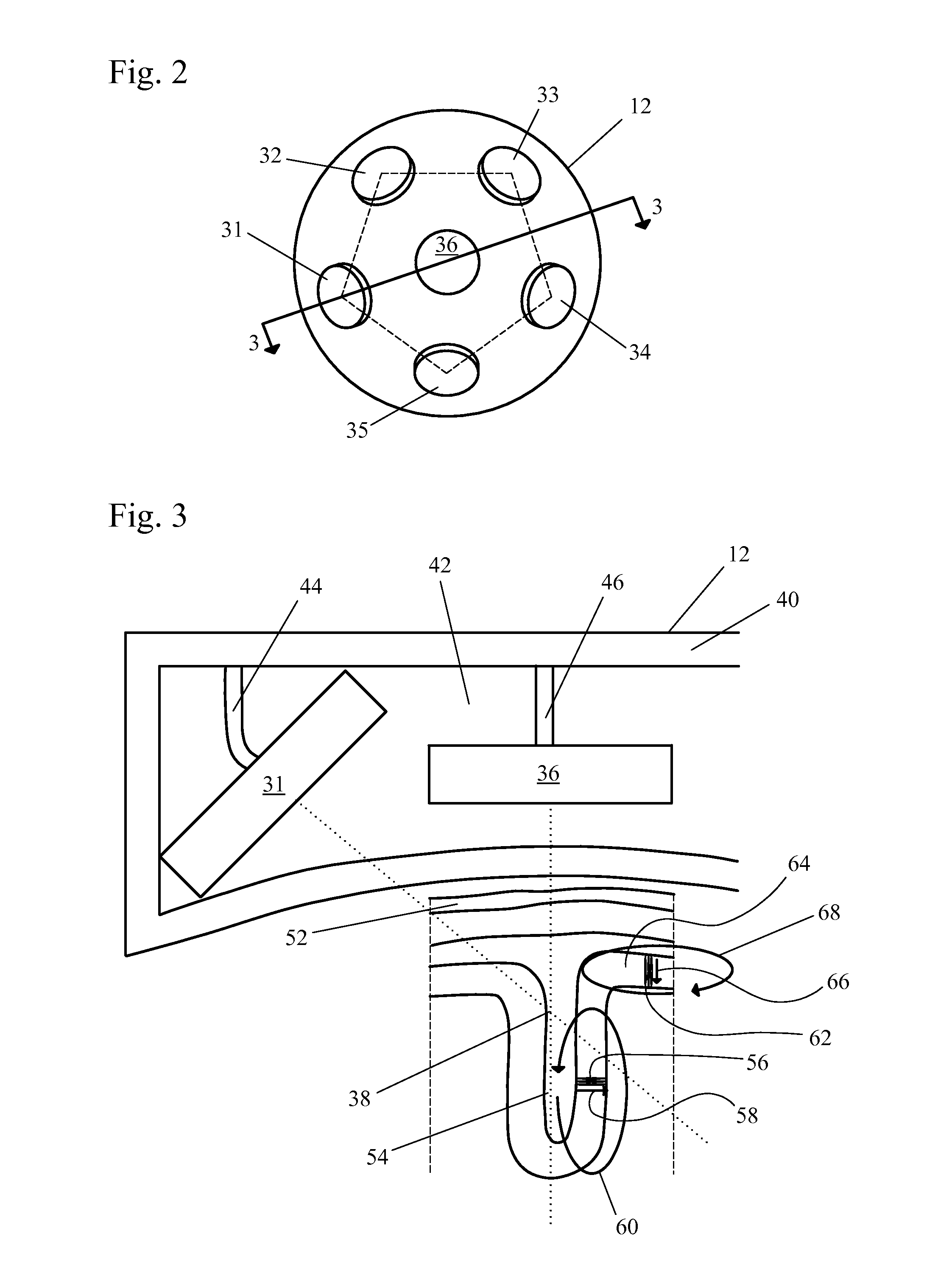
[0032]Second, far fewer SQUID detectors are required, which reduces the equipment cost. In some embodiments, about ...


PUM


Abstract

Systems and methods detect electrical activity in the human brain in the form of the generated magnetic fields and map the magnetic field strength to the surface of the cerebral cortex. By mapping the measured magnetic fields to the sulcal-defined gyrus of the cerebral cortex rather than to its three-dimensional volume, the data complexity and the resulting image are dramatically reduced. The device allows an inventory of brain function, including, but not limited to, vision, sound, sensory, and cognition, with the sensors placed over the corresponding region or regions of interest of the brain.

Description

REFERENCE TO RELATED APPLICATIONS[0001]This application claims one or more inventions which were disclosed in Provisional Application Number 61 / 666,171, filed Jun. 29, 2012, entitled “MAGNETIC IMAGING DEVICE TO INVENTORY HUMAN BRAIN CORTICAL FUNCTION”. The benefit under 35 USC §119(e) of the United States provisional application is hereby claimed, and the aforementioned application is hereby incorporated herein by reference.BACKGROUND OF THE INVENTION[0002]1. Field of the Invention[0003]The invention pertains to the field of medical imaging. More particularly, the invention pertains to systems and methods of detecting electrical activity in the brain.[0004]2. Description of Related Art[0005]The adult human cerebral cortex has an average volume in the range of about 870 to 970 cm3 and includes about 1010 neurons forming about 1014 neural connections. Even though neurons constitute only about 10% of the brain cell population, this complexity presents a huge imaging problem. A function...

Claims


Application Information

Patent Timeline
Patent Type & Authority Applications(United States)
IPC IPC(8): A61B5/04A61G13/12
CPCA61B5/04008A61G13/12A61B5/162A61B5/4088A61B2576/026A61B6/04A61B5/0042A61B5/7282A61B5/05A61B5/4064A61B5/6803A61B2562/0223G16H30/40A61B5/246A61B5/245
Inventor FORD, JOHN P.
Owner FORD JOHN P
Why Eureka
Try Eureka