System and method for generating and displaying a 2d projection from a 3D or 4d dataset

a technology of a system is applied in the field of generating and displaying a 2d projection from a 3d or 4d dataset, which can solve the problems of additional effort and cost, and achieve the effects of convenient and comfortable viewing, simplified procedure, and convenient viewing

Inactive Publication Date: 2014-01-16
KONINKLJIJKE PHILIPS NV +1
View PDF2 Cites 12 Cited by
  • Summary
  • Abstract
  • Description
  • Claims
  • Application Information

AI Technical Summary

Benefits of technology

[0010]Such, a system and method by which so-called standard angiographic views can be generated using a 3D or 4D reconstructed image of the object of interest. These standard angiographic views can be individually customized by a physician; however, the standard angiographic views can also be firmly defined and carried out in the method and / or by the system. One example is the reconstruction of coronary angiograms from rotational angiography sequences. Once the 3D image is created, it can be forward projected into the individually user-defined or commonly given standard views for live presentation during the procedure. These standard views, which more closely mimic what the physician is accustomed to see, will be more readily accepted by the interventional community. The physician is thus given an easy way to utilize the 3D reconstruction in room using views which he would typically acquire if the 3D reconstruction were not available. The operator now has the opportunity to quickly assess the potential diagnostic quality of these standard views and deviate from them if deemed necessary, for example to reduce overlap and / or foreshortening.
[0011]With the system and method described beforehand, the procedure of generating a complete set of pictures of the coronary artery tree to be observed is improved and simplified, and the data collected from the coronary artery tree are made more suitable and comfortable for viewing by the physician.

Problems solved by technology

However, physicians are typically accustomed to viewing static, that means not rotating, images of the coronary artery tree from certain standard, that means usual viewing angles and therefore, even if a 3D dataset is available, it may be underutilized such that additional static shots viewing the coronary artery tree in a two-dimensional manner may be acquired by the physician.
This makes the procedure of generating a complete set of pictures of the coronary artery tree to be observed more complicated and time consuming, means additional discomfort to the patient and such leads to additional effort and costs.

Method used


Image

  • System and method for generating and displaying a 2d projection from a 3D or 4d dataset
  • System and method for generating and displaying a 2d projection from a 3D or 4d dataset
  • System and method for generating and displaying a 2d projection from a 3D or 4d dataset

Examples


Embodiment Construction

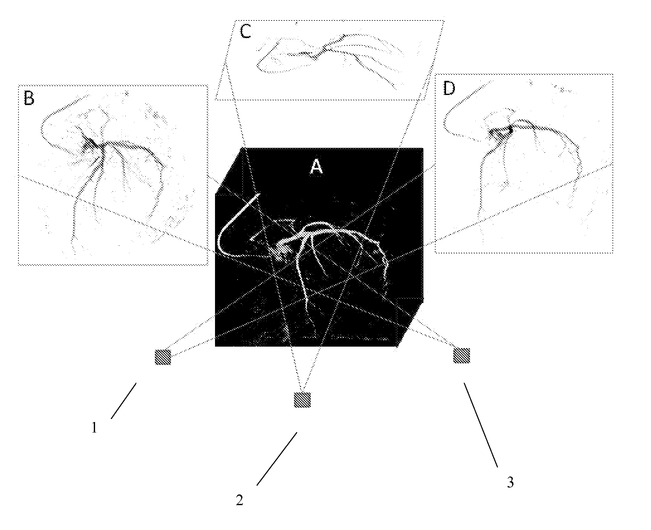
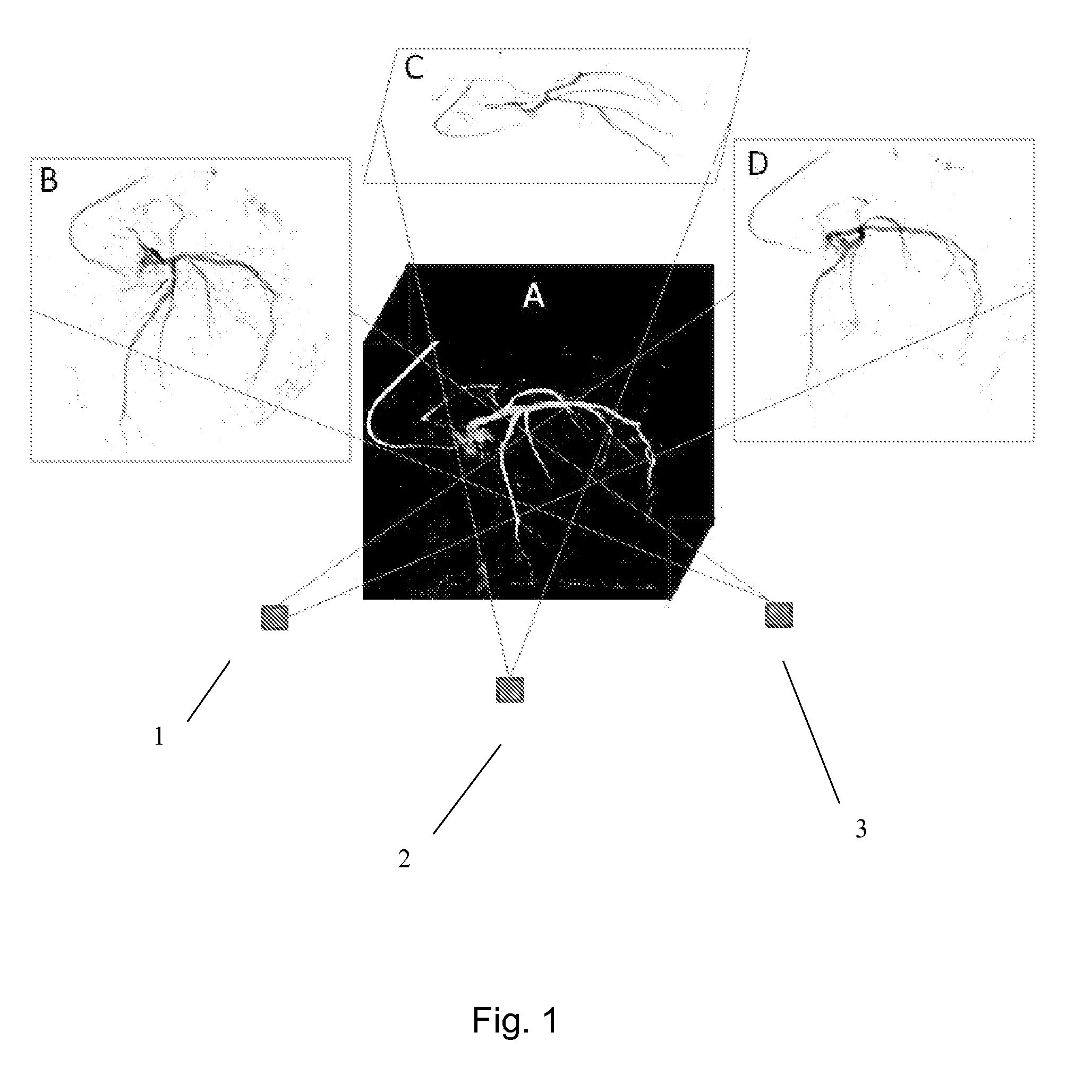
[0018]In FIG. 1, a 3D reconstructed image of a left coronary artery of a human heart serving as an object of interest in an embodiment of a system and method according to the invention is shown and denoted by reference numeral A. This 3D image can be derived from a single 3D dataset of the object of interest; however, it can also be understood as one out of a number of 3D datasets being derived from the object to be represented one by one at defined time intervals, the number of 3D datasets forming a 4D dataset. To generate different 2D projections from this given dataset representing the 3D view of the object of interest, that is the left coronary artery, different views denoted as view 1, view 2 and view 3 and marked with reference numerals 1, 2 and 3, respectively, are defined according to the desired perspectives from which the 3D reconstructed image is intended to be viewed. Then, based on these views 1, 2 and 3, respectively, three 2D projections are generated and displayed; t...


PUM


Abstract

A system and method is described by which so-called standard angiographic views can be generated using a 3-or 4-D reconstructed image of the object of interest. One preferred example is the reconstruction of coronary angiograms from rotational angiography sequences. Once the 3D image is created, it can be forward projected into the user-defined “standard” views for live presentation during the procedure. It is anticipated that these standard views, which more closely mimic what a physician is accustomed to see, will be more readily accepted by the interventional community.

Description

FIELD OF THE INVENTION[0001]The invention is related to a system for generating and displaying a 2D projection from a 3D or 4D dataset. The invention is further related to a method for generating and displaying such a 2D projection from a 3D or 4D dataset of the kind mentioned.BACKGROUND OF THE INVENTION[0002]Methods have become available which permit the incorporation of volumetric or three-dimensional images, further abbreviated as 3D images, of coronary artery trees of the heart, also denoted as vessel trees, into cardiac catheterization laboratory during surgical interventional procedures, which, in the context of the present application, hereinafter means and reads as so-called minimally invasive interventional procedures. For instance, previously acquired data from cardiac computed tomography, further abbreviated as CT, or magnetic resonance imaging, further abbreviated as MRI, can be imported for use during the surgical interventional procedure, or a new technique which can p...

Claims


Application Information

Patent Timeline
Patent Type & Authority Applications(United States)
IPC IPC(8): G06T11/00
CPCG06T11/003G06T11/008G06T15/08G06T19/003G06T2211/404
Inventor NEUBAUER, ANNE MORAWSKIDEN HARTOG, WILLEM FREDERIKCARROLL, JOHN DOUGHERWINK, ONNOSCHOONENBERG, GEERT ANTONIUS FRANCISCUSCHEN, SHIUH-YUNG JAMESGRASS, MICHAEL
Owner KONINKLJIJKE PHILIPS NV
Why Eureka
Try Eureka