Anastomosis clipping tool with half-loop clip

a clipping tool and clipping tool technology, applied in the field of apparatus, system and method, can solve the problems of unusable standard tools, few surgeons who are able to perform, and pediatric surgery faces access and space challenges not found in adult patients, so as to minimize the dependence on surgeon dexterity and experience, and reduce the time of procedure and operating cost

Active Publication Date: 2014-03-27
CHILDRENS NAT MEDICAL CENT
View PDF7 Cites 29 Cited by
  • Summary
  • Abstract
  • Description
  • Claims
  • Application Information

AI Technical Summary

Benefits of technology

[0013]While the above patents propose methods of achieving more efficient MIS suturing, none address the difficulty with subsequent knot tying. Thus, there is a need for a fully automated / assisted laparoscopic anastomosis device that can reduce procedure time and operating costs. The device will also be of interest to surgeons as it would minimize dependence on a surgeon's dexterity and experience for producing an efficacious anastomosis.
[0014]To our knowledge, there are currently no devices or patents that deliver a flexible fastener, such as a clip, or a staple, or a T-suture that behaves like suture but can be closed with a simple holder, such as a clasp, a stopper, or a clamp. While there is a commercial product that can be used on suture to replace knots (LAPRA-TY®), the present invention combines the functions of tissue piercing and fastening, joining lumen together, and securing a holder, which obviates the need to employ multiple tools to perform this complex portion of an anastomosis procedure. Use of a holder that is a half-loop clip is also novel.
[0015]Laparoscopic anastomosis surgery involves complicated, technically-demanding movements, and often requires the surgeon to tie knots in single-suture stitches and make frequent instrument changes during the procedure. A fastening device is intended to provide surgeons a way to more easily and efficiently join two lumen by placing intermittent fasteners, rather than sutures that require complicated knot-tying and instrument changes, to achieve the same closures. The present surgical device has the potential to provide the means for increasing procedure efficiency and decreasing operating times, and is consistent with our goal to make pediatric surgery more precise, less invasive, and safer.
[0016]The anastomosis segment of a minimally invasive procedure for pediatric patients requires intense concentration and dexterity, and often takes more than an hour to perform. Because it most often occurs near the end of a procedure, surgeon fatigue plays a role in the efficacy of the anastomosis. The present surgical device provides the surgeon assistance with this complex manual task by reducing the frequency of instrument changes, and eliminating the need to place multiple serial sutures that require difficult knot tying and cutting suture.
[0017]A goal of the fastening device disclosed herein is to minimize a number of necessary trocars in order to reduce patient pain. Another goal of the present invention is to reduce suturing time and frequency of instrument changes in order to increase procedure efficiency and reduce surgeon fatigue.
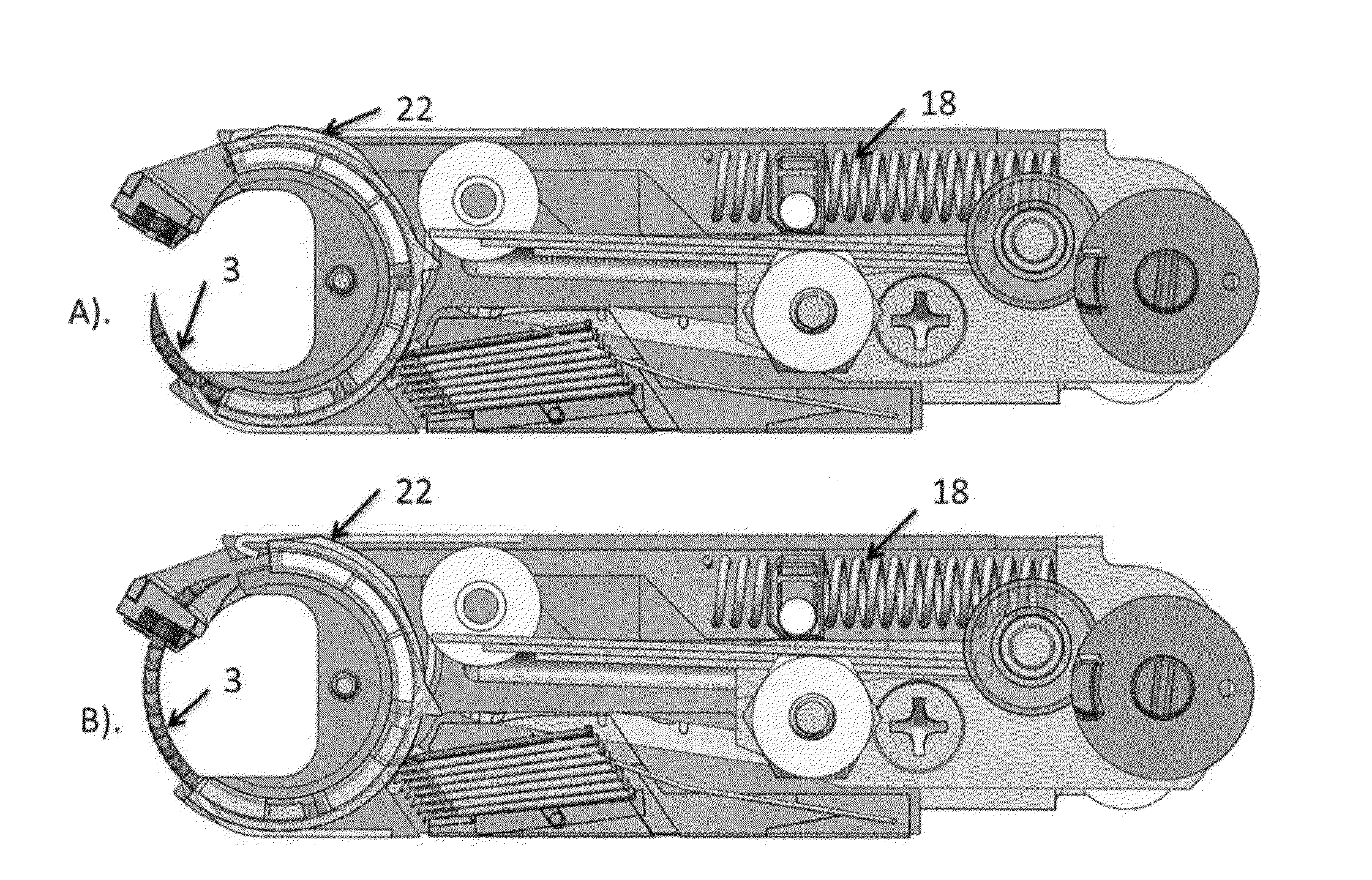
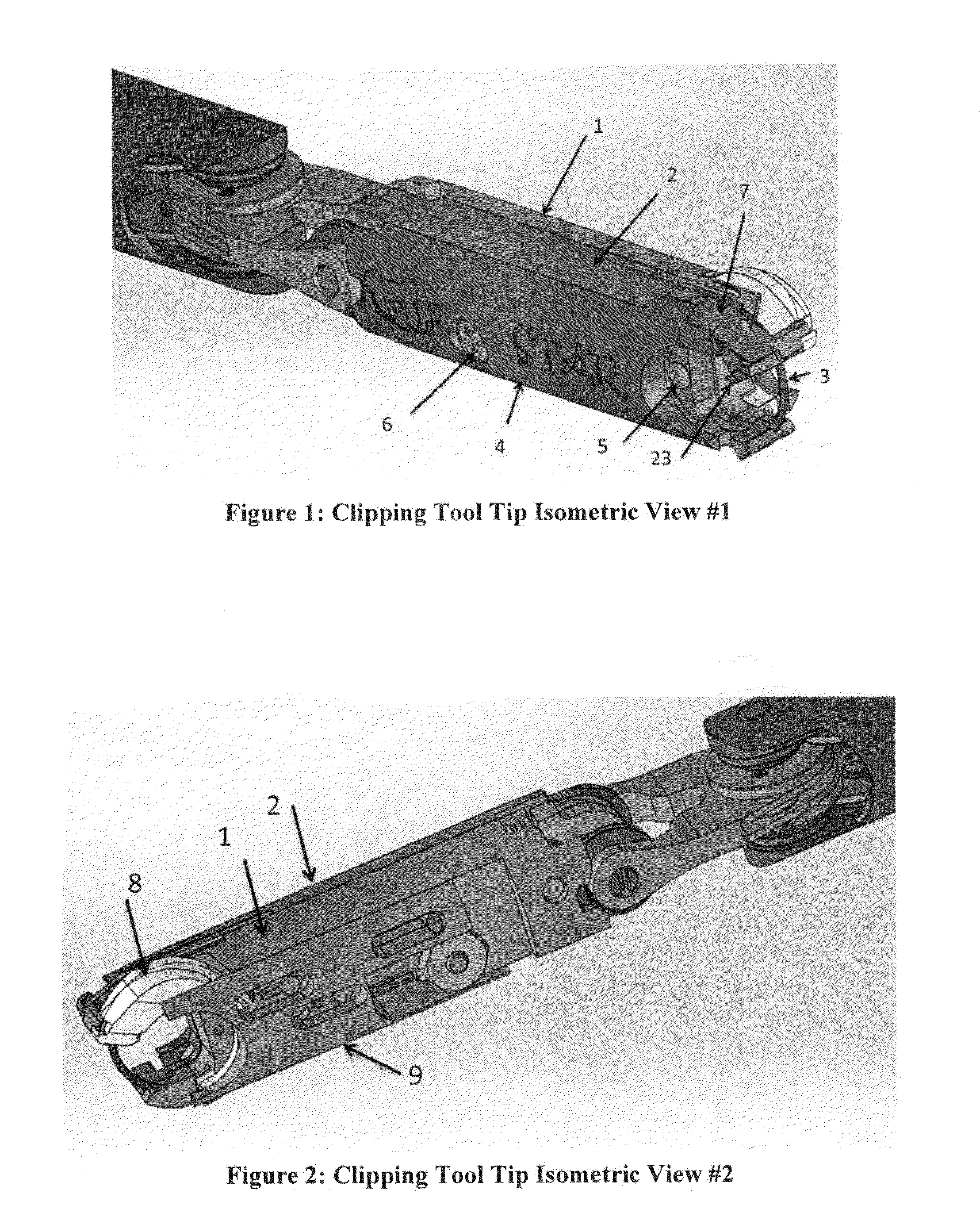
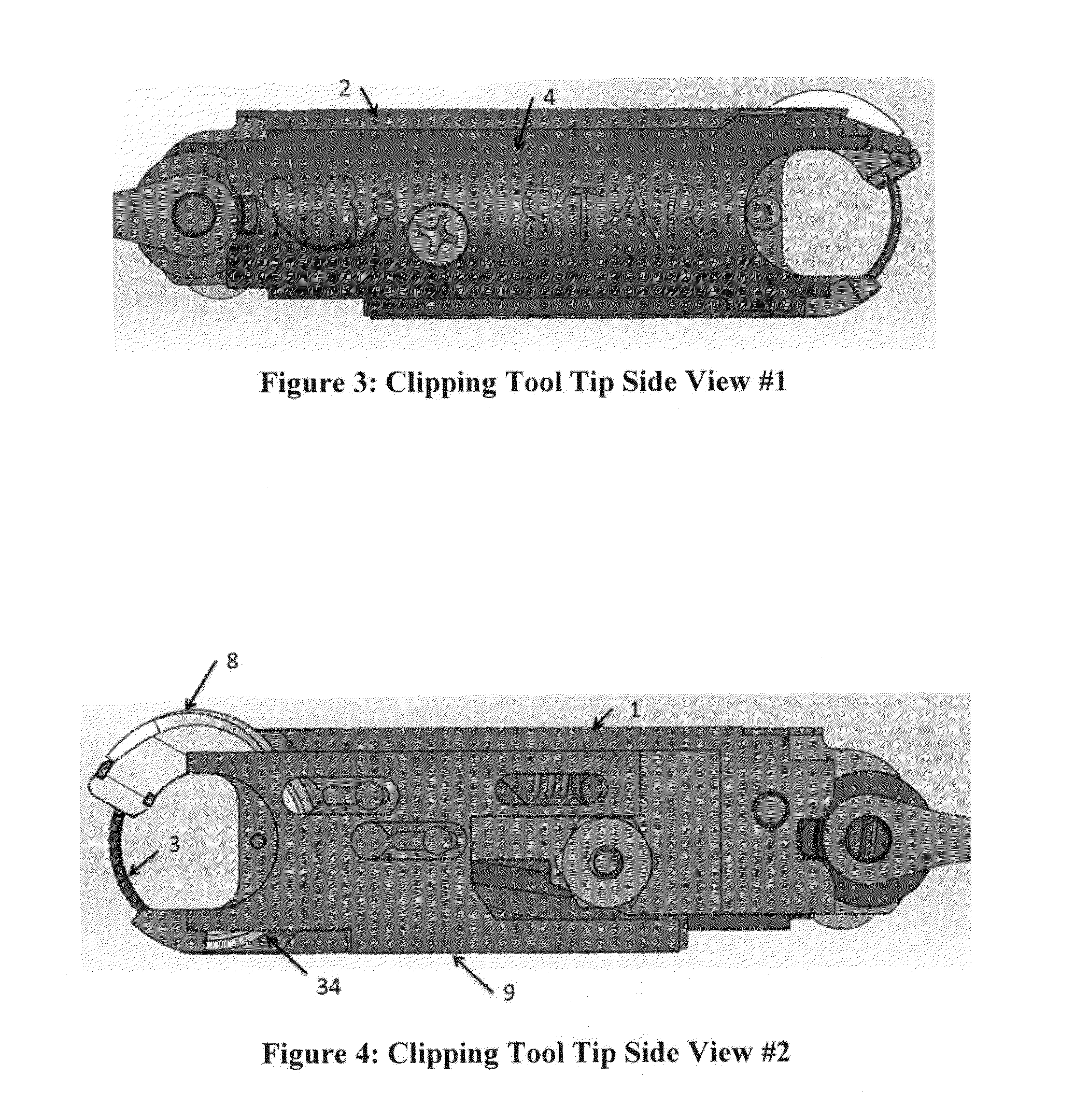
[0030]In an aspect of the disclosure, the tool further includes a tool tip body, a shaft, and a mechanism. The tool tip body includes the gripper, the needle, the actuator, the fastener cartridge, the holder cartridge, and the holder applier. The mechanism joins the tool tip body to the shaft, and the mechanism allows the tool tip body to be movable with at least two degrees of freedom.

Problems solved by technology

There are many procedures for pediatric patients that include minimally invasive techniques, but there are few surgeons who are able to perform them due to the challenges they present.
Because the patient's anatomy is so small, pediatric surgery faces access and space challenges not found in adult patients.
The tiny volume available for tool movement and accommodation when using standard laparoscopic techniques inside the pediatric patient make standard tools unusable; they are simply too large.
Once inside the patient, the ability to obtain good camera views is constrained due to limited space availability.
This, combined with the reduced size of the anatomy, challenges the surgeon's situational awareness at a time in in the procedure that is especially critical for efficacy.
However, in minimally invasive surgery (MIS) where the procedure is performed through small incisions in the patient's skin, it is extremely difficult to create an anastomosis due to limited space, inadequate camera visualization, and awkward instrument angles.
In some cases, it may take as long as two hours to perform.
For the surgeon, laparoscopic anastomosis surgery is extremely difficult to learn and perform and is very fatiguing in nature.
Increased operating time is also a burden on the healthcare system as it consumes valuable operating room time and adds to overall costs.
Most importantly, spending an extended period of time in surgery can negatively impact the patient due to increased anesthesia requirement and additional stress to the body.
Even though the two ends of the design are attached through a flexible coupler, the design lacks a good suture management scheme and will suffer from suture tangling.
The design provides a good method of creating a stitch, but tying of the suturing material requires a separate discrete process that necessitates input of a laparoscopic grasping tool through a separate port, introducing additional time and uncertainty to the surgical procedure.
While the above patents propose methods of achieving more efficient MIS suturing, none address the difficulty with subsequent knot tying.
To our knowledge, there are currently no devices or patents that deliver a flexible fastener, such as a clip, or a staple, or a T-suture that behaves like suture but can be closed with a simple holder, such as a clasp, a stopper, or a clamp.
Laparoscopic anastomosis surgery involves complicated, technically-demanding movements, and often requires the surgeon to tie knots in single-suture stitches and make frequent instrument changes during the procedure.
The anastomosis segment of a minimally invasive procedure for pediatric patients requires intense concentration and dexterity, and often takes more than an hour to perform.

Method used


Image

  • Anastomosis clipping tool with half-loop clip
  • Anastomosis clipping tool with half-loop clip
  • Anastomosis clipping tool with half-loop clip

Examples


Embodiment Construction

[0096]Referring to the drawings, like reference numerals designate identical or corresponding parts throughout the several views.

[0097]The figures are not to scale, and some features may be exaggerated or minimized to show details of particular elements, while related elements may have been eliminated to prevent obscuring novel aspects. Therefore, specific structural and functional details disclosed herein are not to be interpreted as limiting, but merely as a basis for the claims, and as a representative basis for teaching one of ordinary skill in the art to variously employ the present invention.

[0098]The illustrative embodiments described herein are directed to a surgical apparatus, system, and method for fastening tissue. As required, embodiments of the present invention are disclosed herein. However, the disclosed embodiments are merely exemplary, and it should be understood that the invention may be embodied in many various and alternative forms.

[0099]The tool described herein...


PUM


Abstract

A tool to fasten tissue or to fasten a prosthetic to tissue includes a gripper to hold one or more tissue portions, a needle, an actuator to drive the needle, a fastener cartridge to store one or more tissue fasteners, a holder cartridge to store one or more holders, and a holder applier to secure one of the holders on one of the fasteners. The actuator drives the needle and the needle pulls a first fastener from the fastener cartridge. The actuator drives the needle through the one or more tissue portions held by the gripper to form a hole in the tissue portions. The needle pulls a leading portion of the first fastener through the hole without a trailing portion of the first fastener being pulled through the hole. The holder applier secures a first holder from the holder cartridge onto the leading portion of the first fastener.

Description

CROSS REFERENCE TO RELATED APPLICATIONS[0001]This application is based upon and claims the benefit of priority from U.S. Provisional Patent Application No. 61 / 706,322, filed on Sep. 27, 2012, the entire contents of which are incorporated herein by reference, and from U.S. Provisional Patent Application No. 61 / 705,875, filed on Sep. 26, 2012, the entire contents of which are incorporated herein by reference.BACKGROUND[0002]I. Field of the Disclosure[0003]The present invention relates to an apparatus, system, and method for fastening tissue and / or fastening a prosthetic / biologic mesh to tissue. Specifically, it is designed for use in anastomosis surgery (i.e. connecting two tubular structures) to restore continuity after resection, to bypass an unresectable disease process, to close any luminal structure, to provide hemostasis, or to position / secure bioabsorbable or prosthetic mesh for tissue or organ structural weakness. The present invention relates to the process of performing an a...

Claims


Application Information

Patent Timeline
Patent Type & Authority Applications(United States)
IPC IPC(8): A61B17/11
CPCA61B17/11A61B17/0401A61B17/0469A61B17/0482A61B17/0487A61B17/06A61B17/1152A61B2017/0404A61B2017/0409A61B2017/0417A61B2017/0496A61B2017/06042A61B2017/1107A61B2017/1132A61B2017/1135A61B2017/1139A61B2017/2927
Inventor KRIEGER, AXELKIM, PETERWILSON, CHRISABELLERA, STEPHEN
Owner CHILDRENS NAT MEDICAL CENT
Why Eureka
Try Eureka