System and method for the detection of abnormalities in a biological sample

a biological sample and microscopic technology, applied in the field of microscopic detection of abnormalities in biological samples, can solve the problems of reducing computational efforts and saving time, and achieve the effects of simplifying the tedious task of examining, reducing computational time, and increasing magnification

Active Publication Date: 2014-08-14
KONINKLIJKE PHILIPS ELECTRONICS NV
View PDF1 Cites 30 Cited by
  • Summary
  • Abstract
  • Description
  • Claims
  • Application Information

AI Technical Summary

Benefits of technology

[0081]In still another preferred embodiment, the method according to the invention, further comprises, prior to step a), a step in which an image of the sample is acquired at lower magnification, as is the case in step a). In this embodiment, an overview image is made first. The low magnification slide overview is processed by the algorithm to identify the regions suspicious for abnormality and those suspected region are further scanned with higher magnification. This will provide an advantage of quick scanning of the slides. In yet another preferred embodiment of the method according to the invention, steps b) and following are carried out while the digital image acquisition and / or the digital image processing is still in process.
[0082]This methodological approach, which is also called “on the fly” or “real time”, reduces the computational time for complex image processing algorithms as well as it simplifies the tedious task of examining each of the cells found in the sample by reducing the evaluation time.

Problems solved by technology

This again saves time and reduces computational efforts.

Method used


Image

  • System and method for the detection of abnormalities in a biological sample
  • System and method for the detection of abnormalities in a biological sample
  • System and method for the detection of abnormalities in a biological sample

Examples


Embodiment Construction

[0153]While the invention has been illustrated and described in detail in the drawings and foregoing description, such illustration and description are to be considered illustrative or exemplary and not restrictive; the invention is not limited to the disclosed embodiments. Other variations to the disclosed embodiments can be understood and effected by those skilled in the art in practicing the claimed invention, from a study of the drawings, the disclosure, and the appended claims. In the claims, the word “comprising” does not exclude other elements or steps, and the indefinite article “a” or “an” does not exclude a plurality. The mere fact that certain measures are recited in mutually different dependent claims does not indicate that a combination of these measures cannot be used to advantage. Any reference signs in the claims should not be construed as limiting the scope.

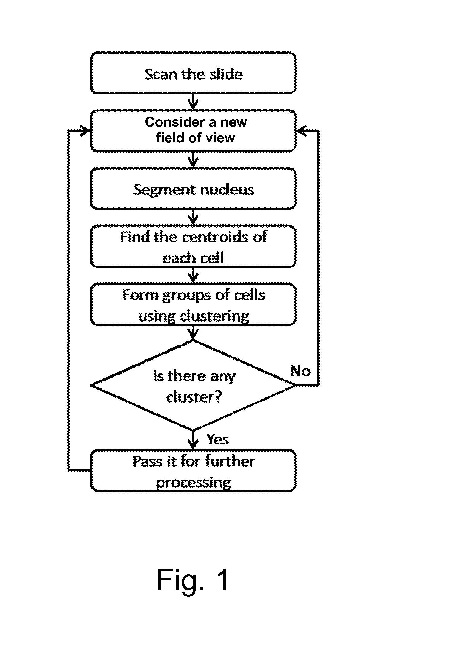
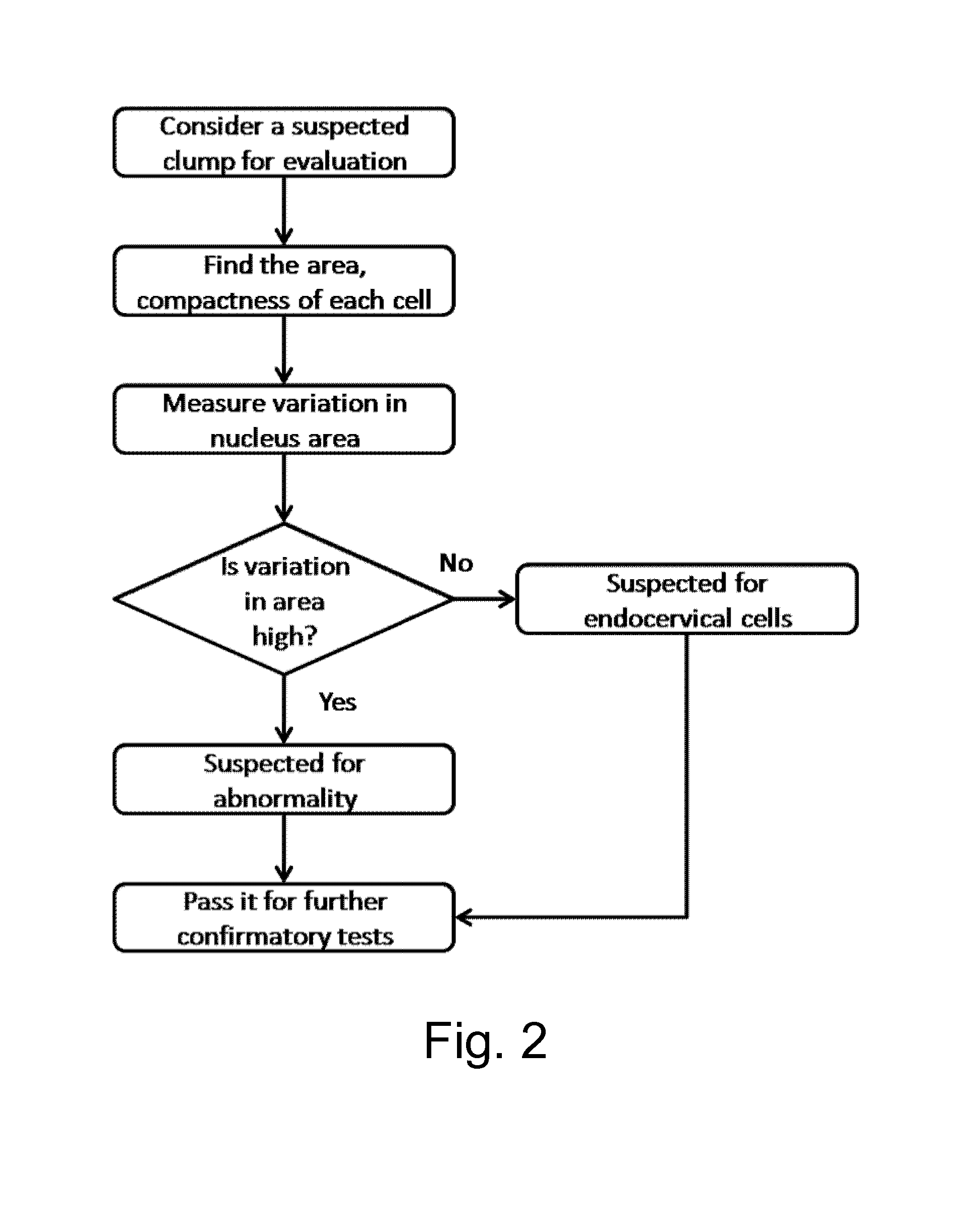
[0154]FIGS. 1 and 2 show flow charts which have been described in the text already.

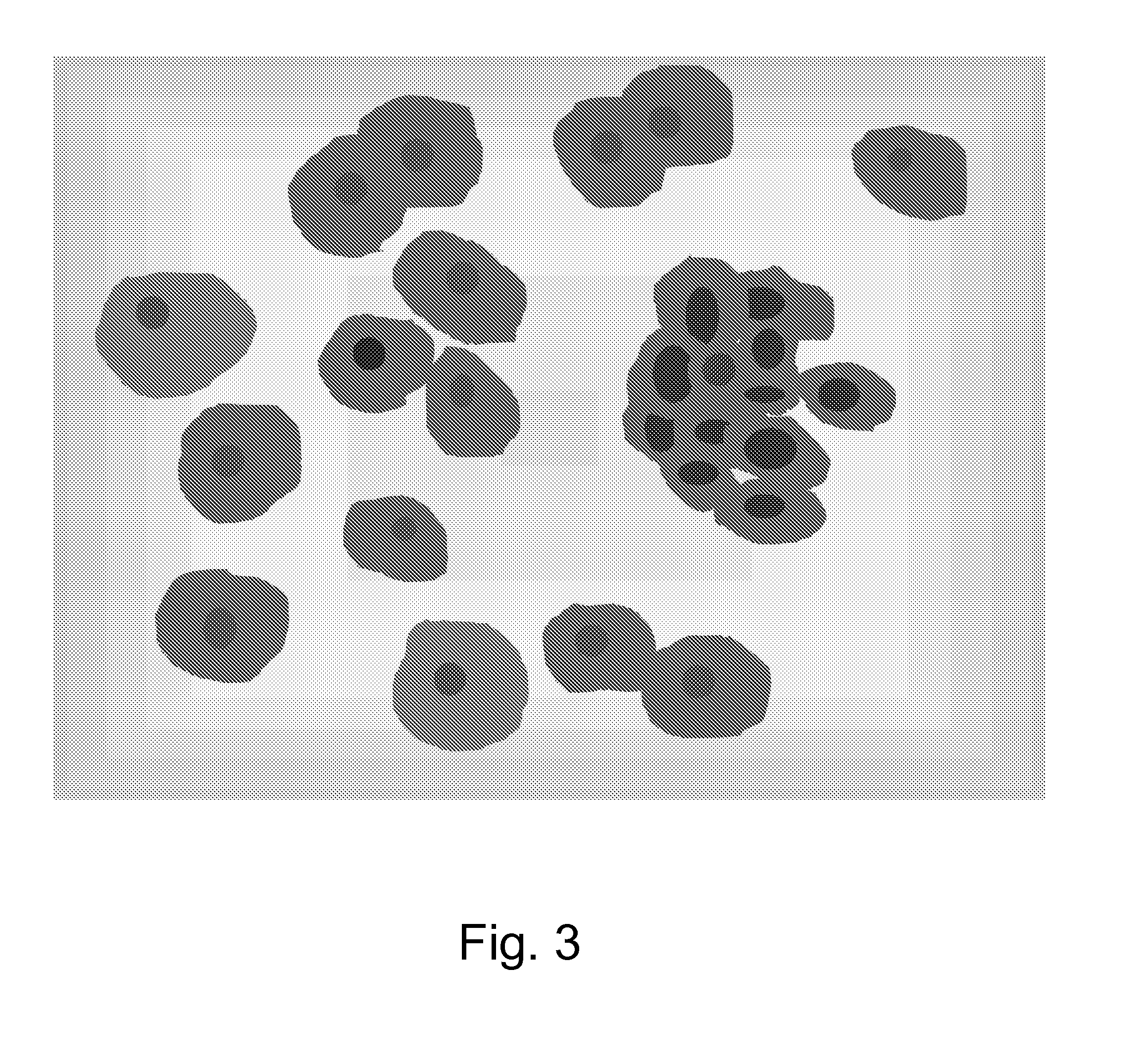
[0155]FIG. 3 shows an artif...


PUM

PropertyMeasurementUnit
digital image processingaaaaaaaaaa
sizeaaaaaaaaaa
size ratioaaaaaaaaaa

Abstract

The present invention is related to a method or system for the detection of abnormalities in a biological sample comprising cells. Said method or system comprises at least the following steps, or is capable to carry out, and/or comprises means capable to carry out, at least the following steps, respectively: a) acquiring an image of said sample by digital image acquisition, b) optionally, carrying out digital image processing, c) selecting a field of view, d) determining, by digital image processing, whether or not, in said field of view, cell aggregates exist, and e1) selecting a new field of view and carrying on with step c) if, in step d), it turns out that, in said field of view, the determination of cell aggregates is negative, or e2) carrying out further process steps if in step d), it turns out that, in said field of view, the determination of cell aggregates is affirmative.

Description

FIELD OF THE INVENTION[0001]The invention relates to the field microscopic detection of abnormalities in a biological sample comprising cells.BACKGROUND OF THE INVENTION[0002]Cancer arising from cervix is the number one cancer in women in many industrialized countries as well as emerging countries. About 30% of cancers in women are due to cervical cancer with more than 100,000 new cases diagnosed every year, e.g., in India. The estimated compounded annual growth rate (CAGR) for cervical cancer cases is 2.56% and at this growth rate approximately 175,000 new cases of cervical cancer will be detected in the year 2012.[0003]One of the recommended tools for screening of cervical cancer is to detect cytological precursors of cancer in Papanicolaou tests (also called Pap-smear, Pap-test, cervical smear, or smear test), which is a screening test used in gynecology to detect premalignant and malignant processes in the cervical canal especially in the transformation zone.[0004]In taking a Pa...

Claims


Application Information

Patent Timeline
Patent Type & Authority Applications(United States)
IPC IPC(8): C12Q1/70G06T7/00
CPCC12Q1/70G06T2207/30024G06T7/0012G06V20/695G06T7/0014G06V20/698
Inventor SETH, SUBHENDUNAIK, SARIF KUMARVENKATESAN, SHANKAR MOSURKUMAR, SUNILPAYAL, KESWARPUJAYAVANTH, SANJAY
Owner KONINKLIJKE PHILIPS ELECTRONICS NV
Why Eureka
Try Eureka