Image processing apparatus, image processing method, and image processing program

a technology which is applied in the field of image processing apparatus and image processing method, and image processing program, can solve the problem that the blood vessel structure cannot be accurately extracted, and achieve the effect of more accurate extraction

Inactive Publication Date: 2014-08-21
FUJIFILM CORP
View PDF3 Cites 4 Cited by
  • Summary
  • Abstract
  • Description
  • Claims
  • Application Information

AI Technical Summary

Benefits of technology

[0006]In view of the foregoing circumstances, it is an objective of the present invention to provide an image processing apparatus, an image processing method, and an image processing program which are capable of accurately specifying the position of a point of origin even in the case that the point of origin of a target structure is not sufficiently pictured in image data.
[0017]Further, the correlating means may employ an evaluation function, which evaluates the degree of similarity between graph parts, formed by a collection of a plurality of nodes that constitute the graph, correlated to the plurality of nodes that constitute the plurality of shape models, and the plurality of shape models, in an arbitrarily set correlative relationship, as the predetermined cost function. Thereby, the correlating means may correlate the plurality of shape models to the extracted graph, by determining a correlative relationship that achieves maximization of the degree of similarity.
[0022]Further, the entirety of the predetermined structure that includes the point of origin can be extracted more accurately than by the aforementioned conventional method, in the case that a graph that represents the predetermined structure is extracted, employing the obtained data that represents the position of the point of origin.

Problems solved by technology

Therefore, there is a problem that blood vessel structures cannot be accurately extracted by applying the aforementioned conventional method, which extracts a target blood vessel structure by fitting shape models corresponding to the entirety of the target blood vessel structure to a generated graph, under the presumption that the graph has been generated with respect to the entirety of the target blood vessel structure including the point of origin from image data.

Method used


Image

  • Image processing apparatus, image processing method, and image processing program
  • Image processing apparatus, image processing method, and image processing program
  • Image processing apparatus, image processing method, and image processing program

Examples


Embodiment Construction

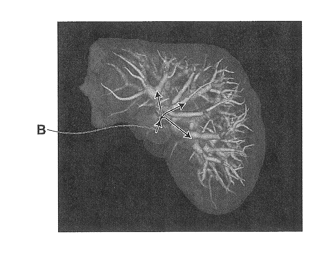
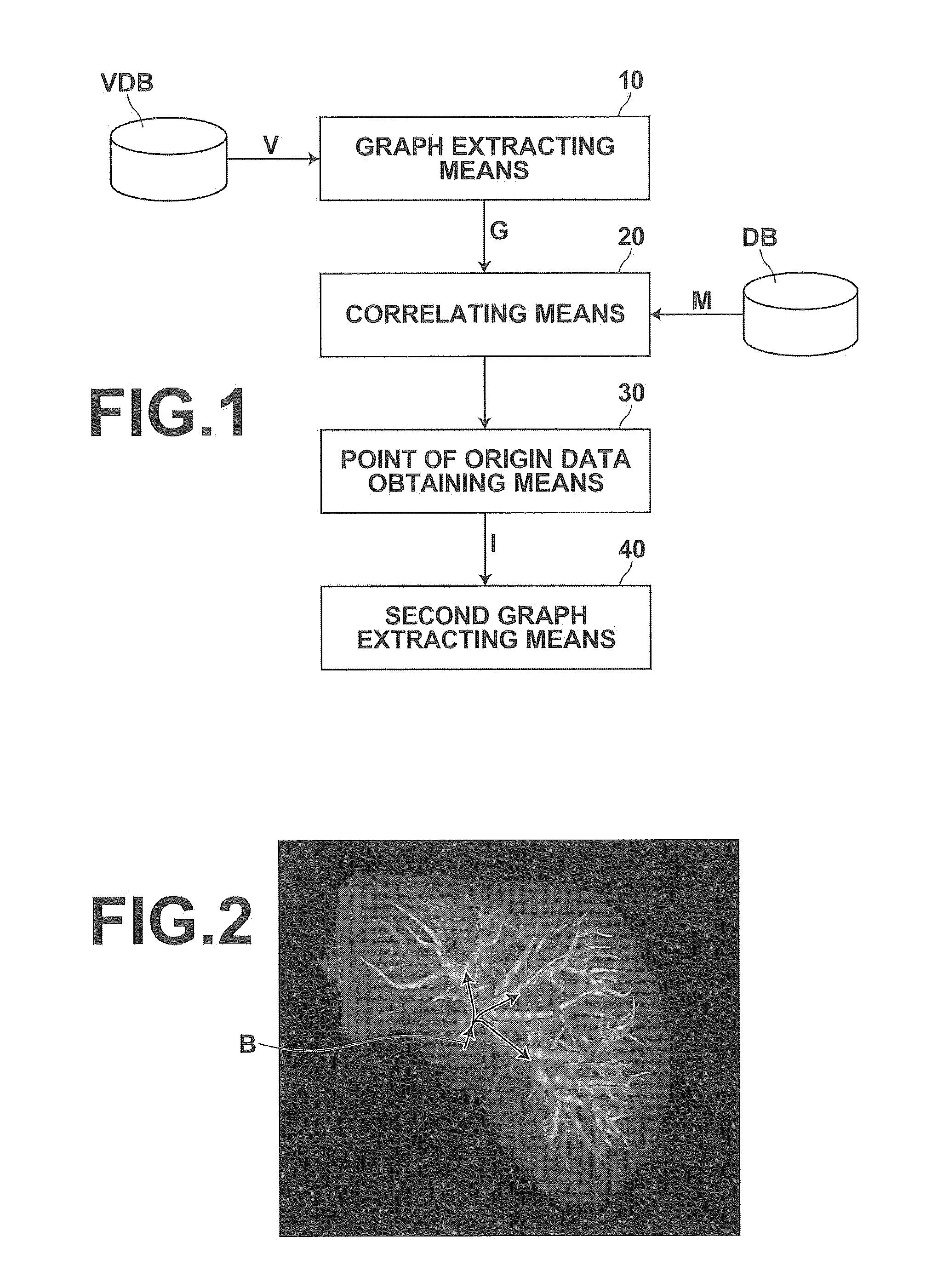
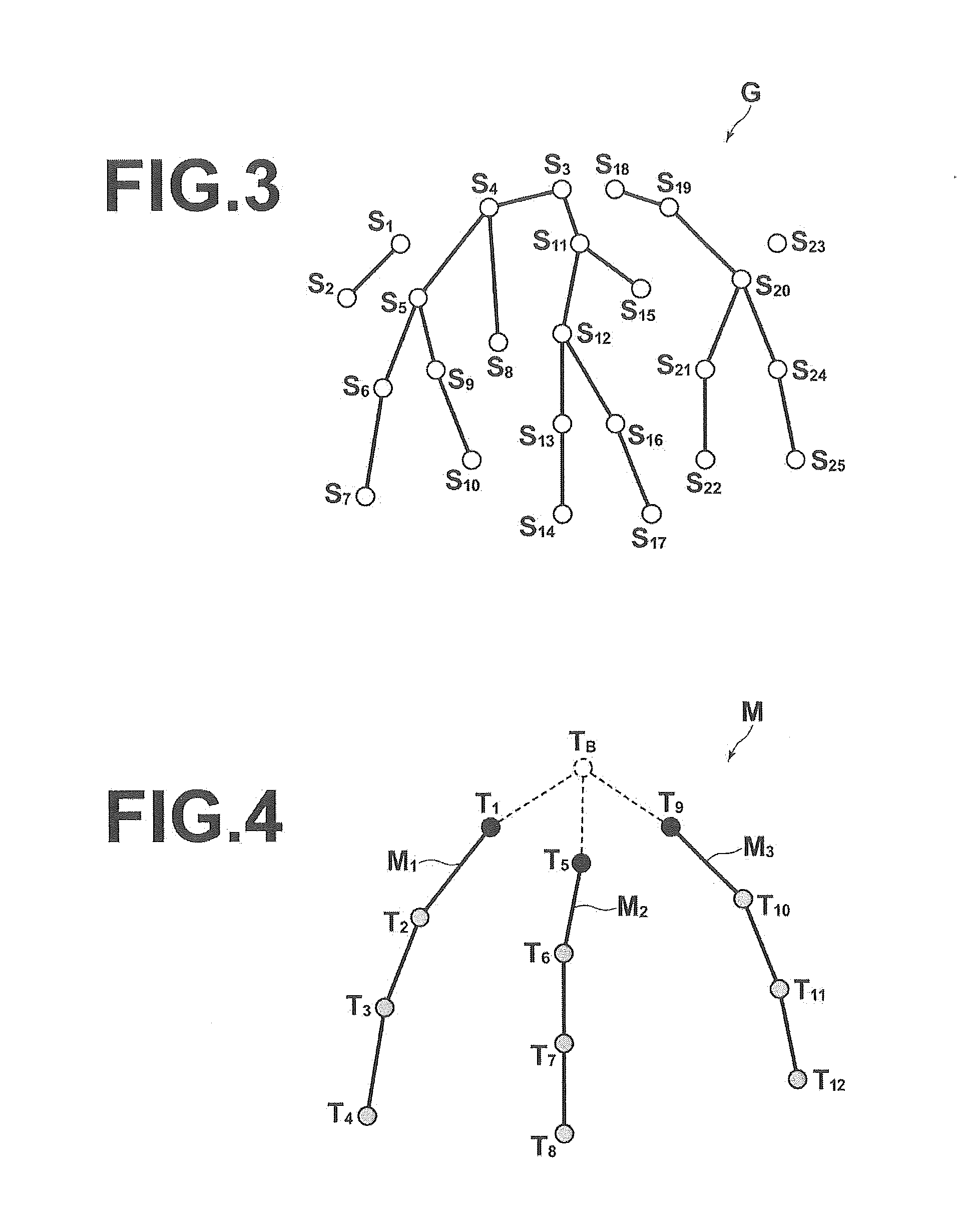
[0033]Hereinafter, embodiments of an image processing apparatus, an image processing method, and an image processing program of the present invention will be described with reference to the attached drawings. FIG. 1 is a diagram that schematically illustrates the configuration of an image processing apparatus 1 according to an embodiment of the present invention. Note that the configuration of the image processing apparatus 1 illustrated in FIG. 1 is realized by executing a medical image processing program loaded in an auxiliary memory device on a computer. At this time, the image processing program is recorded in a data recording medium such as a CD-ROM, or distributed via a network such as the Internet, and installed in the computer. The image processing apparatus 1 of FIG. 1 extracts a graph that represents a predetermined structure that spreads and extends by repeatedly branching from a single point of origin B, from image data that represents the predetermined structure. The im...


PUM


Abstract

A graph estimated to represent hepatic veins is extracted from image data. Shape models representing partial tree structures branched from a point of origin within a tree structure representing a common shape of hepatic veins are obtained. A cost function correlates the graph with the shape models. Whether parts extending from the peaks of correlated graph parts and not correlated with a shape model exist within the extracted graph is judged. Positional data of the peaks are obtained as data representing the position of the point of origin in the case that no such parts exist, or positional data of a node closest to an estimated position of the point of origin, specified by tracing along the nodes of such parts from the peaks to approach the estimated position, is obtained as data representing the position of the point of origin, in the case that such parts exist.

Description

TECHNICAL FIELD[0001]The present invention is related to an image processing apparatus, an image processing method, and an image processing program for obtaining data that represents the position of a point of origin from image data that represents a predetermined structure, which extends from the point of origin while repeatedly branching.BACKGROUND ART[0002]When performing ablative surgical procedures to remove diseased portions that include tumors from organs such as the liver and the lungs, it is necessary to specify blood vessels that are providing nutrients to the tumors and to appropriately determine the portions which are governed by such blood vessels as portions to be removed. This is because the portion of a portal vein that supplies nutrients to a tumor and a region governed by the portal vein, at which there is a possibility that substances to be noted such as cancer cells are being sent, are removed within a range that can maintain hepatic function, in ablative surgery...

Claims


Application Information

Patent Timeline
Patent Type & Authority Applications(United States)
IPC IPC(8): G06T7/00G06T11/20
CPCG06T11/206G06T7/0012A61B5/02007A61B2576/00G06T2207/10081G06T2207/10088G06T2207/10136G06T2207/20072G06T2207/30101G06T7/162G16H30/40
Inventor IHARA, SATOSHI
Owner FUJIFILM CORP
Why Eureka
Try Eureka