Medical X-Ray Photography Apparatus

a technology of x-ray photography and x-ray images, which is applied in the field of medical x-ray photography equipment, can solve the problems of troublesome operators and large burdens on patients, and achieve the effect of efficient operation

Active Publication Date: 2014-09-11
MORITA MFG CO LTD
View PDF8 Cites 18 Cited by
  • Summary
  • Abstract
  • Description
  • Claims
  • Application Information

AI Technical Summary

Benefits of technology

[0029]In accordance with an eleventh aspect of the present invention, in the medical X-ray photography apparatus of any of the second to tenth aspects, a first turning speed at which the moving mechanism moves the support to the photography starting position is faster than a second turning speed at...

Problems solved by technology

However, unfortunately it is necessary to previously dispose X-ray detection means in a mouth cavity of a patient, and a large burden is placed...

Method used


Image

  • Medical X-Ray Photography Apparatus
  • Medical X-Ray Photography Apparatus
  • Medical X-Ray Photography Apparatus

Examples


Embodiment Construction

[0067]Hereinafter, a preferred embodiment of the present invention will be described with reference to the accompanying drawings. In the following drawings, for the sake of convenience, sometimes a size or the number of pieces of each part is illustrated while magnified or simplified as needed basis.

1. Preferred Embodiment

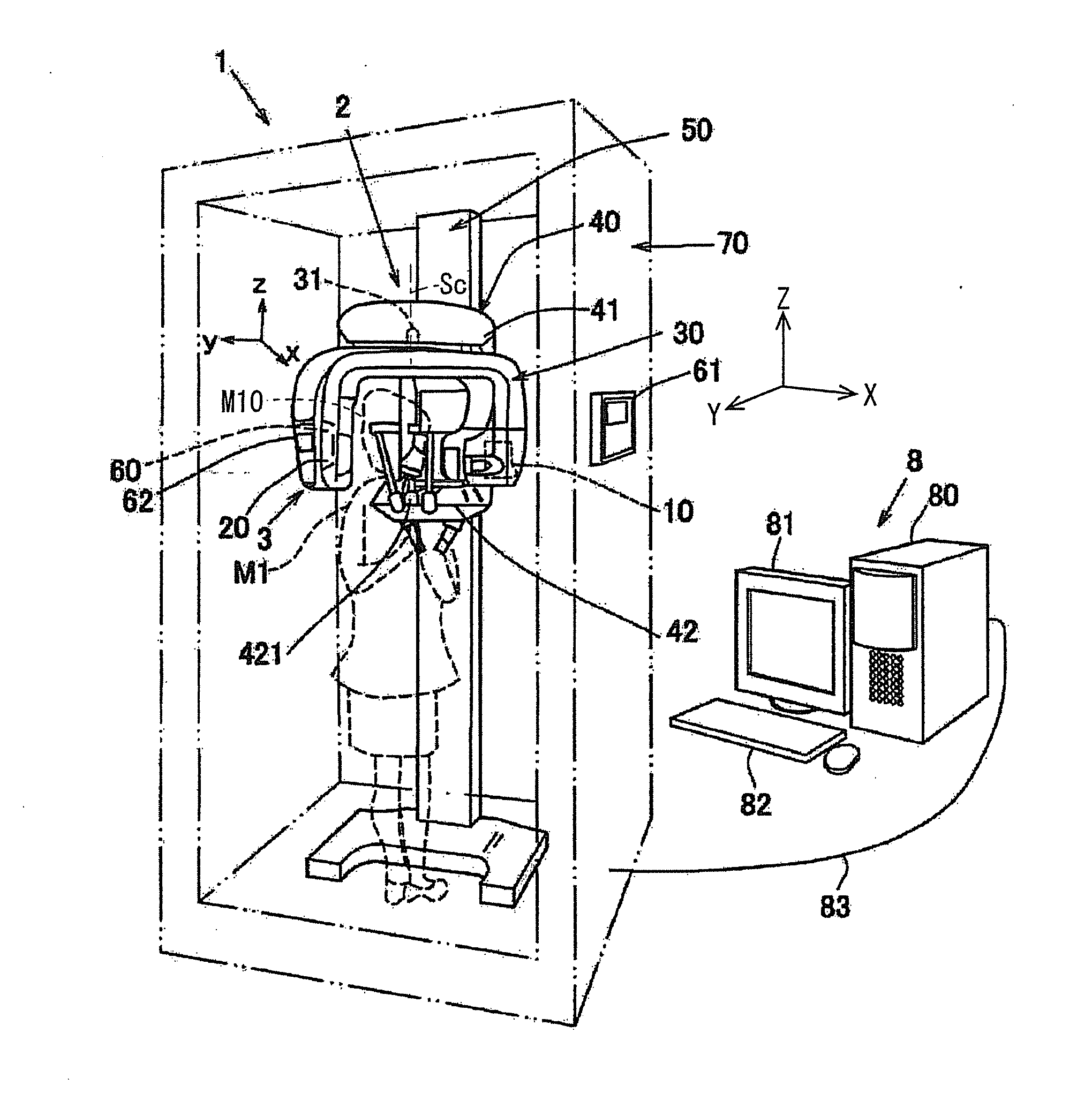
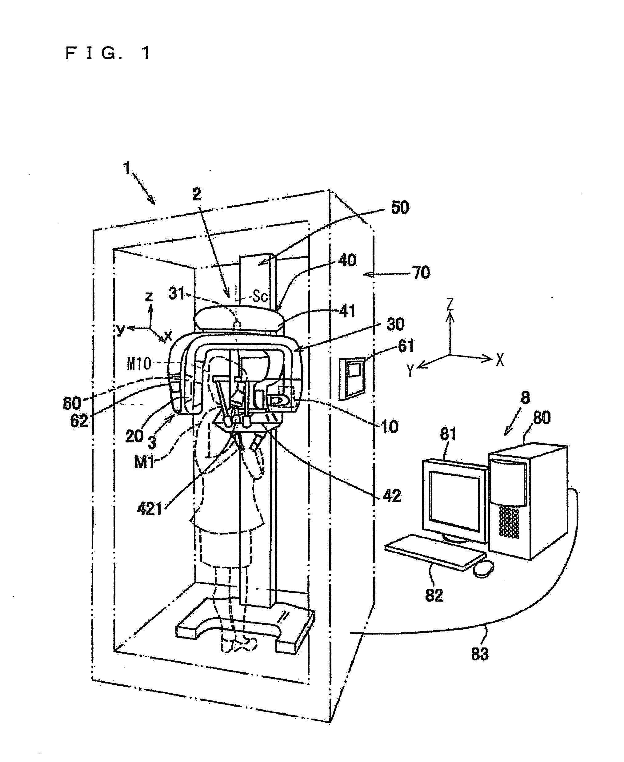
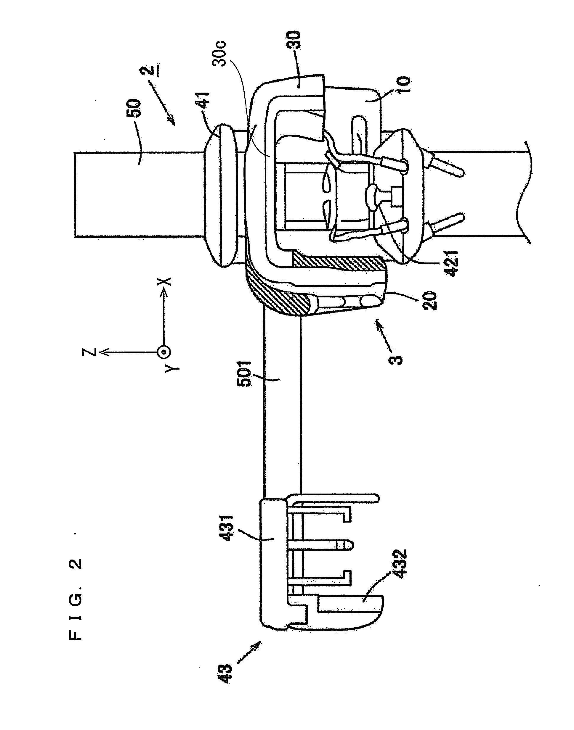
[0068]A medical X-ray photography apparatus 1 according to a first preferred embodiment will be described below. FIG. 1 is a schematic perspective view of the medical X-ray photography apparatus 1 of the first preferred embodiment. FIG. 2 is a partial front view of the medical X-ray photography apparatus 1 on which a cephalic part 43 is mounted. FIG. 3 is a block diagram illustrating a configuration of the medical X-ray photography apparatus 1. FIG. 4 is a front view of a signal output switch 71.

[0069]The medical X-ray photography apparatus 1 is roughly divided into operation display parts 61 and 62, a main body 2, and an information processing device 8. The operat...


PUM


Abstract

A medical X-ray photography apparatus includes the turning arm and a moving mechanism. The turning arm supports an X-ray generator and an X-ray detector. The moving mechanism includes a turning part that turns the turning arm and a moving part that moves the turning arm along a two-dimensional plane orthogonal to an axial direction of a turning shaft. The medical X-ray photography apparatus also includes a photographic region assignment receiving part that receives an operation to assign a part of a dental arch as a pseudo intraoral radiography region, a main-body control part that controls the moving mechanism based on a movement starting signal to move the turning arm to a predetermined photography starting position corresponding to the pseudo intraoral radiography region, and a signal output switch that includes a movement starting signal output part outputting the movement starting signal to the main body control part.

Description

BACKGROUND OF THE INVENTION[0001]1. Field of the Invention[0002]The present invention relates to a medical X-ray photography apparatus.[0003]2. Description of the Background Art[0004]There have been proposed some medical X-ray photography apparatuses that perform dental panoramic photography. In this kind of medical X-ray photography apparatus, a support, which supports an X-ray generator and an X-ray detector while the X-ray generator and the X-ray detector are opposed to each other with a subject (a head of a test subject) interposed therebetween, is turned about the subject to perform the panoramic photography (for example, see Japanese Patent Application Laid-Open No. 2011-152411).[0005]What is called intraoral radiography (dental radiography), in which X-ray detection means (such as an X-ray film and an X-ray sensor panel) is disposed in a mouth cavity of a patient to photograph part of a row of teeth or gums, is performed in X-ray photography of a dental field. In the intraora...

Claims


Application Information

Patent Timeline
IPC IPC(8): A61B6/14
CPCA61B6/14A61B6/145A61B6/4441A61B6/4452A61B6/462A61B6/463A61B6/466A61B6/469A61B6/5223A61B6/545A61B6/548A61B6/586A61B6/025
Inventor NAKAI, TERUJIYASUDA, KOUJI
Owner MORITA MFG CO LTD
Why Eureka
Try Eureka