
Methods for the non-invasive determination of heart and pulmonary pressures

a non-invasive and pulmonary pressure technology, applied in the field of non-invasive determination of heart and pulmonary pressure, can solve the problems of high invasiveness, time-consuming and potentially dangerous for patients, and high invasiveness of conventional monitoring and measuring techniques

Active Publication Date: 2014-09-18
KASSAB GHASSAN S +1
View PDF8 Cites 3 Cited by
  • Summary
  • Abstract
  • Description
  • Claims
  • Application Information

AI Technical Summary

Benefits of technology

The present patent describes a method for noninvasively determining the pressure of the heart using a patient-specific medical image. The method involves generating the image through magnetic resonance imaging or external ultrasound, obtaining at least one measurement related to the position of the left ventricular free wall and the interventricular septum, and determining the end-diastolic Young's modulus of the left ventricular free wall and patient-specific material parameters of the heart. The method can also involve measuring the blood flow velocity and determining the pressure gradient across the interventricular septum. The technical effects of this patent include the ability to noninvasively determine important cardiac pressure measurements, which can aid in the diagnosis and treatment of heart disease.

Problems solved by technology

Conventional monitoring and measuring techniques are typically invasive, often requiring sensors or other measurement devices to be placed within the heart or the vessels connected thereto.
Such invasive measurements are costly, time-consuming and potentially dangerous to the patient.
However, catheterizing the left ventricle is both highly invasive and risky, as it is associated with serious complications including stroke and death.
Accordingly, the conventional invasive techniques used to obtain direct pressure measurements from a heart are undesirable as routine diagnostic tests and are impractical in a non-clinical setting.
While some techniques exist for non-invasively measuring heart and pulmonary pressures during the cardiac cycle, such techniques are limited and, in some cases, only approximate the desired measurements.
However, the non-invasive estimation of LVEDP, right ventricle (RV) pressure, and pulmonary pressure is currently a great challenge using conventional techniques.
It is difficult, however, to non-invasively measure jugular vein pressure and to accurately compute the transtricuspid pressure gradient using a modified Bernoulli equation in patients.

Method used


Image

  • Methods for the non-invasive determination of heart and pulmonary pressures
  • Methods for the non-invasive determination of heart and pulmonary pressures
  • Methods for the non-invasive determination of heart and pulmonary pressures

Examples


Embodiment Construction

[0015]For the purposes of promoting an understanding of the principles of the present disclosure, reference will now be made to the embodiments illustrated in the drawings, and specific language will be used to describe the same. It will nevertheless be understood that no limitation of the scope of this disclosure is thereby intended.

[0016]At least one objective of the disclosure of the present application is to introduce new methods to determine LVEDP, RV and pulmonary pressures based on solid theoretical grounds. For the purpose of promoting understanding, the steps of the methods disclosed herein will first be generally described, followed by a more detailed description of the relevant computations associated with the various steps of the method embodiments.

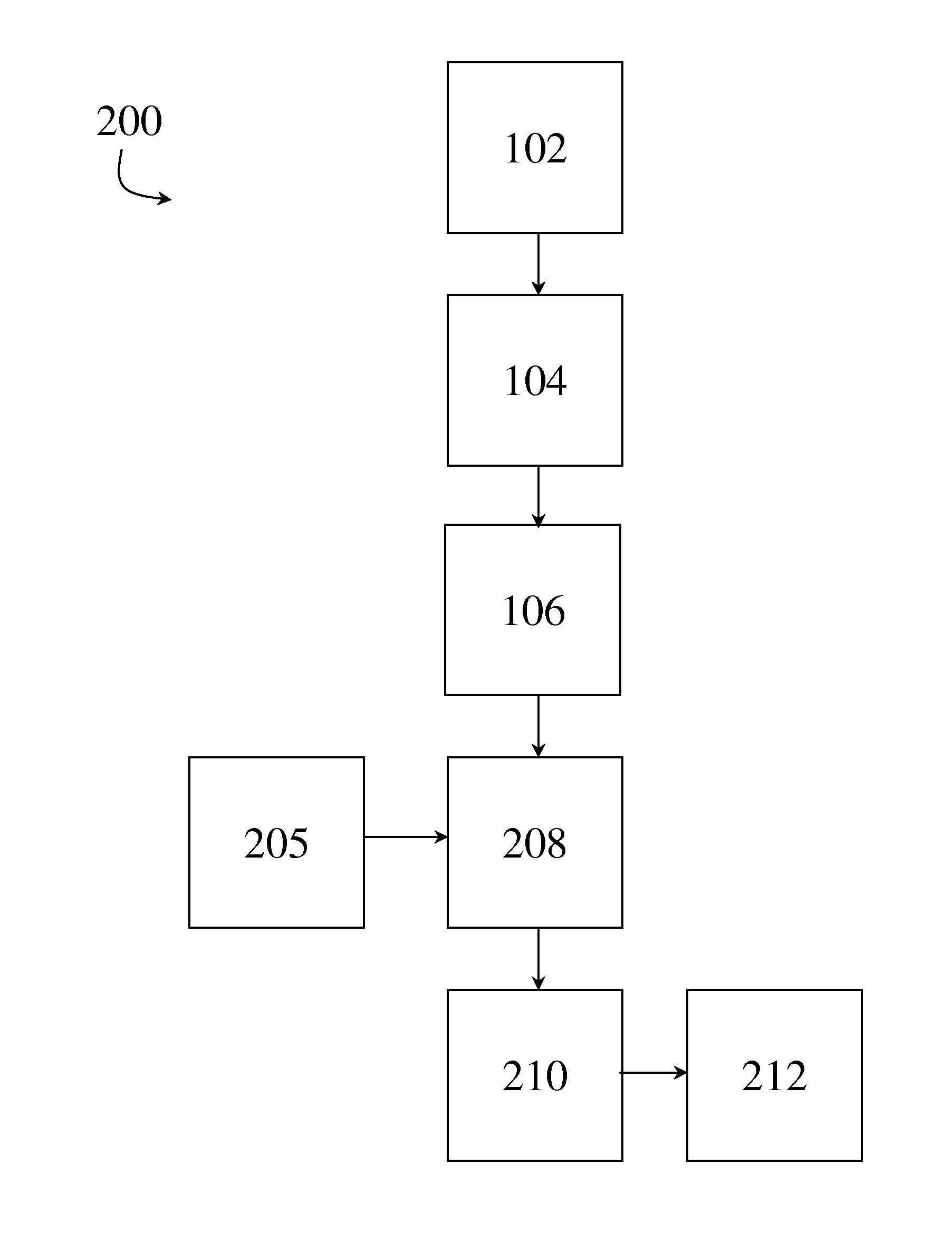
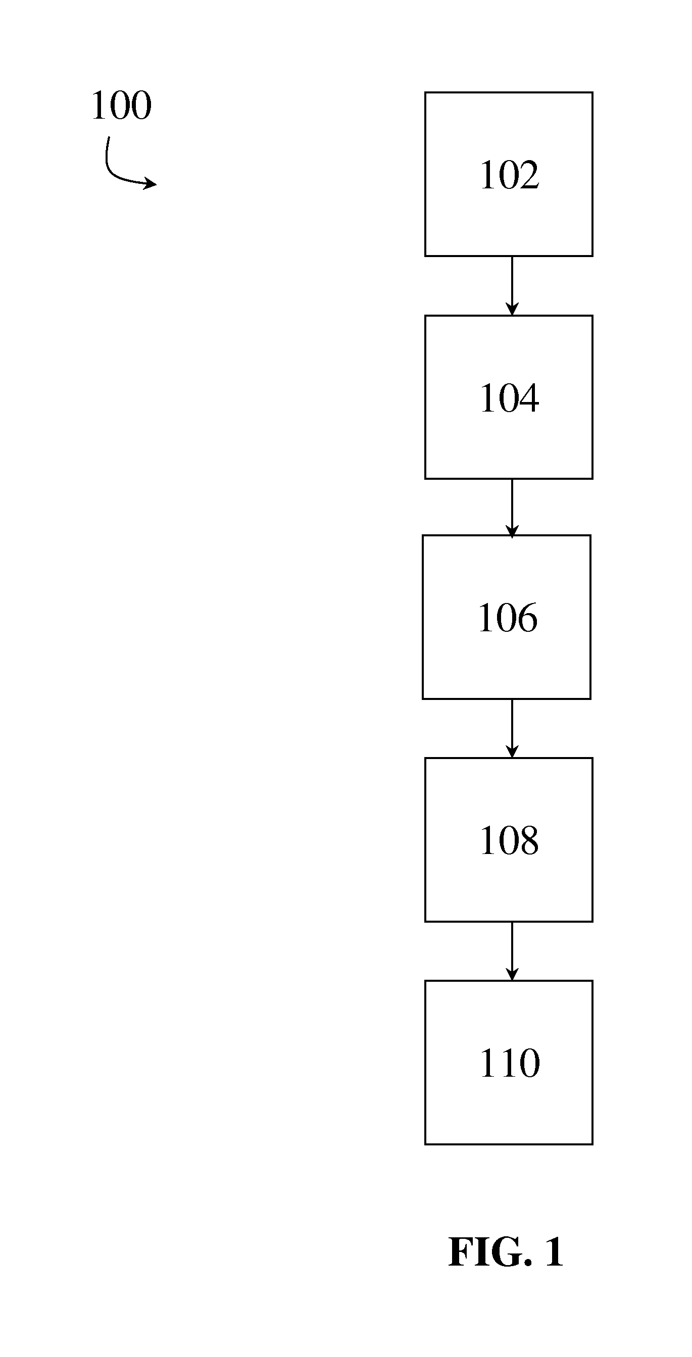
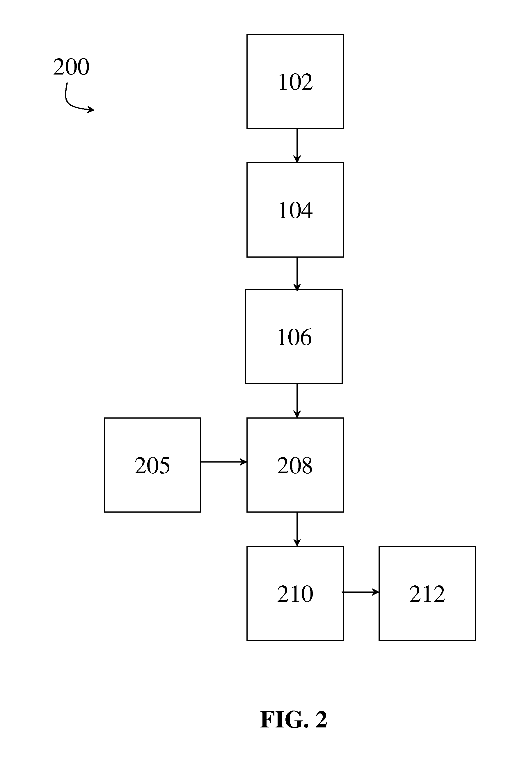
[0017]FIG. 1 shows a flow chart of at least one embodiment of a method 100 for noninvasively determining heart and / or pulmonary pressures in accordance with an exemplary embodiment of the present disclosure. For example, in at...


PUM


Abstract

The disclosure of the present application provides methods for the noninvasive determination of cardiac and pulmonary pressures based off of patient-specific medical images and one or more computations. In one exemplary embodiment, the method comprises noninvasively generating a patient-specific medical image of at least a portion of a heart, determining a free vibration measurement from the left ventricular free wall based on the patient-specific image, and performing at least one computation using the free vibration measurement to noninvasively determine a cardiac or pulmonary pressure.

Description

PRIORITY CLAIM[0001]This application is related to and claims the priority benefit of U.S. Provisional Patent Application Ser. No. 61 / 788,399 to Kassab et al., filed Mar. 15, 2013. The contents of the aforementioned application is hereby incorporated by reference in their entirety into this disclosure.BACKGROUND[0002]Measurements of heart and pulmonary pressures are very significant for diagnosis of systolic and diastolic heart failures, ventricular infarction, and other heart conditions. Specifically, such pressures are directly related to the mechanical properties of the cardiac muscle and, as such, different parameters of the cardiac cycle can be used to determine the status of a patient's heart function and / or to diagnose specific heart-related conditions.[0003]Conventional monitoring and measuring techniques are typically invasive, often requiring sensors or other measurement devices to be placed within the heart or the vessels connected thereto. Such invasive measurements are ...

Claims


Application Information

Patent Timeline
Patent Type & Authority Applications(United States)
IPC IPC(8): A61B5/021A61B8/04
CPCA61B8/04A61B5/02133A61B5/022A61B5/0263A61B5/0265A61B5/055A61B8/06A61B8/0883A61B8/485A61B8/5223G16H50/30G06T7/0012A61B6/503A61B6/504G01R33/20
Inventor KASSAB, GHASSAN S.HUO, YUNLONG
Owner KASSAB GHASSAN S
Learn More