Device for viewing a digital image

a digital image and device technology, applied in the field of medical and biological science, can solve the problems of not being able to concentrate easily, introducing a difference or an error into diagnosis and prognosis, and being difficult to study, etc., and achieve the effect of concentrating easily

Inactive Publication Date: 2014-12-18
UNIV PIERRE & MARIE CURIE +1
View PDF9 Cites 8 Cited by
  • Summary
  • Abstract
  • Description
  • Claims
  • Application Information

AI Technical Summary

Benefits of technology

[0012]One aim of the invention is to propose a device for viewing a digital image, such as a

Problems solved by technology

Since this real slide is very fragile, it may become damaged in transit, making it impossible to study.
Furthermore, the new real slide does not incorporate the same section as the initial slide, and therefore the expert colleague does not study the same section as the doctor who has requested the expert examination.
The differ

Method used


Image

  • Device for viewing a digital image
  • Device for viewing a digital image
  • Device for viewing a digital image

Examples


Embodiment Construction

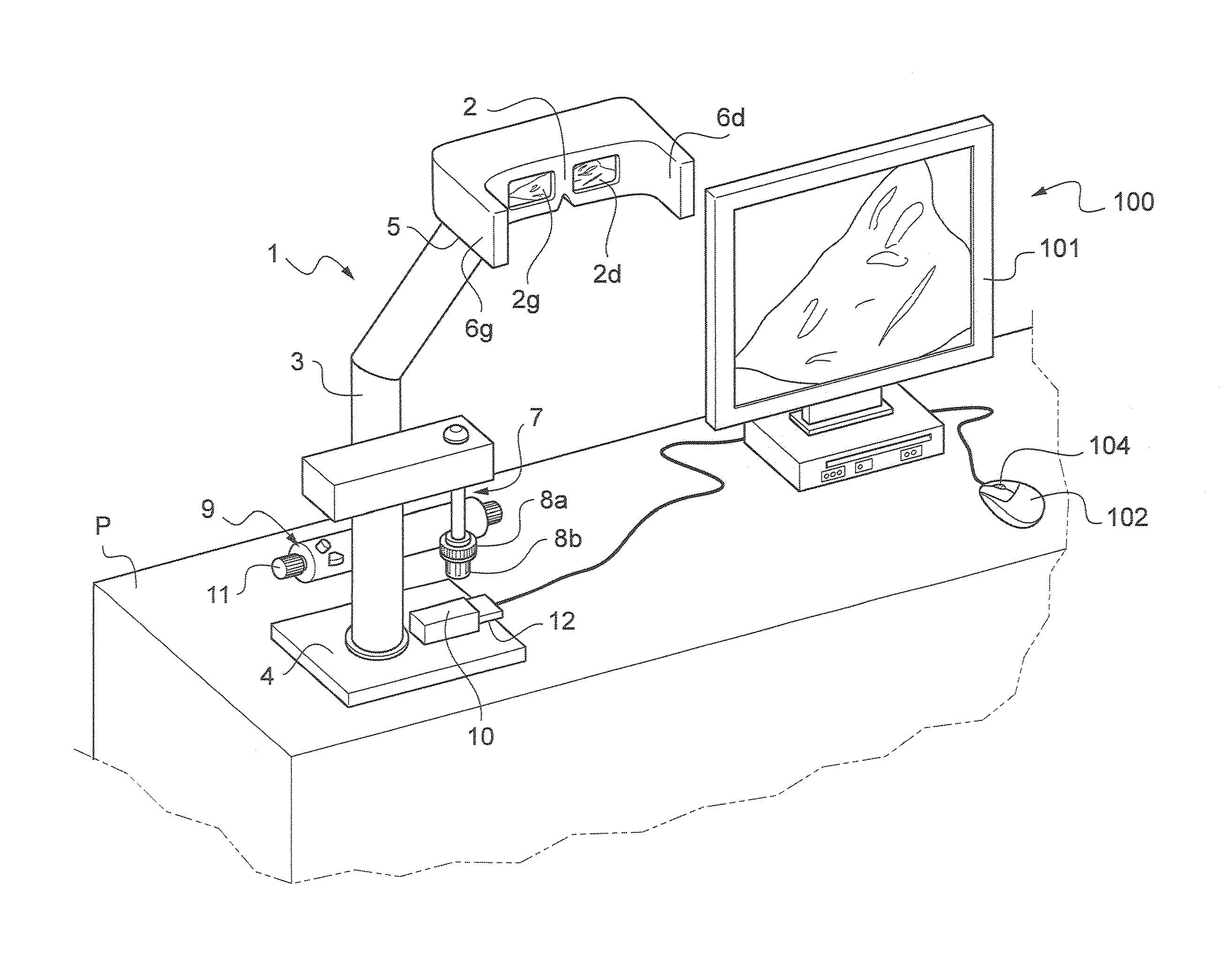
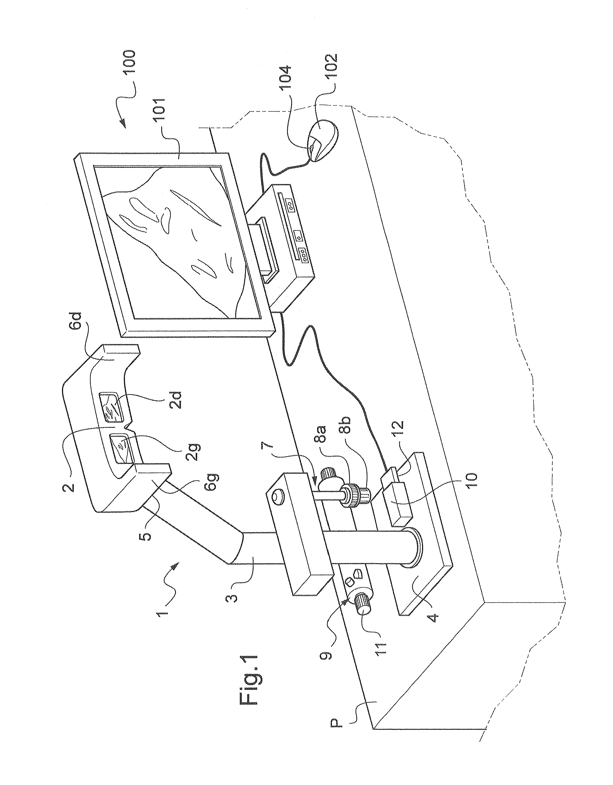
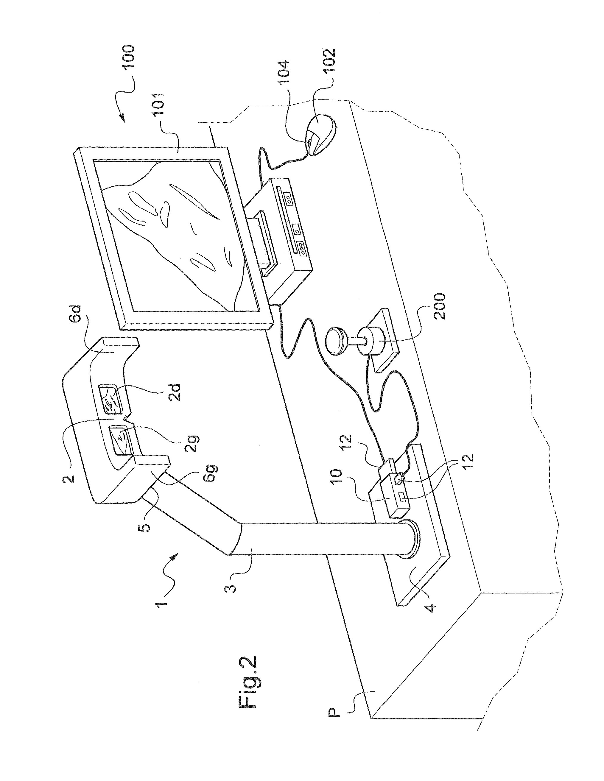
[0022]With reference to FIG. 1, the viewing device 1 according to the invention is, in this case, placed on a desk P. In this case, the viewing device 1 allows the viewing of a virtual slide where the user works in human or animal biology, plant biology or any other field in which microscopy, and therefore the study of virtual slides, is used.

[0023]The viewing device 1 includes means for viewing at least one part of a virtual slide.

[0024]Thus the viewing means include a viewing mask 2. The viewing device 1 further includes a support 3, a proximal end of which forms a base 4 resting on the desk P. A distal end 5 of the support 3 is shaped to hold the viewing mask 2 so that the viewing mask 2 is at the level of a user's eyes when, in operation, the user moves his eyes toward the viewing mask 2.

[0025]In this case, the support 3 includes a control unit 10 comprising a connection port 12. The viewing device 1 includes, in this case, a computer 100 near which the support 3 is placed, the ...


PUM


Abstract

The invention relates to a device for viewing a digital image comprising means (2) for viewing at least one portion of the digital image. According to the invention, the viewing means include a viewing mask (2) held by a stand (3) such that the viewing mask (2) is used at the eye level of a user such that same can bring his/her eyes closer to the viewing mask in order to examine the portion of the digital image displayed, the viewing mask comprising side extensions (6g, 6d) for protecting the user from outer visual interferences, and in that the viewing device further includes at least one movement handling device (7, 200) that can be controlled by the user in order to control virtual movement of the digital image such that the user is able to view another portion of the digital image.

Description

[0001]The invention relates to a device for viewing a digital image, and notably for viewing a virtual slide for the purpose of helping a user to analyze and study this digital image.TECHNICAL BACKGROUND OF THE INVENTION[0002]Histology is a medical and biological science concerned with the study of the structure of human, animal or plant tissues. Normally, a sample of the tissue to be studied is taken, treated by physical-chemical methods, and then cut with a microtome to provide a section with a thickness of about two micrometers, which is placed on a glass slide. The constituents specific to the tissue are detected in this section, using dyes or immunological markers. Finally, said section is covered with a second glass slide. The assembly then forms a physical glass slide, called a “real slide”, which is then conventionally observed with an optical microscope.[0003]For several years, it has also been possible to scan a real slide with a dedicated slide scanner. The real slide is ...

Claims


Application Information

Patent Timeline
IPC IPC(8): G02B21/36
CPCG02B21/365G02B27/028G02B27/02G02B27/026
Inventor LE NAOUR, GILLESDE TRAZEGNIES D'ITTRE, FRANCOISCAPRON, FREDERIQUE
Owner UNIV PIERRE & MARIE CURIE
Why Eureka
Try Eureka