
Method and System for Functional Assessment of Renal Artery Stenosis from Medical Images

a technology of functional assessment and renal artery stenosis, which is applied in the field of noninvasive functional assessment of renal artery stenosis, can solve the problems of atrophy of the affected kidney, ultimately renal failure, and the inability to assess the true hemodynamic significance of the ras

Active Publication Date: 2015-03-05
SIEMENS HEALTHCARE GMBH
View PDF1 Cites 67 Cited by
  • Summary
  • Abstract
  • Description
  • Claims
  • Application Information

AI Technical Summary

Benefits of technology

The present invention provides a non-invasive method for assessing the functional and anatomical severity of a renal artery stenosis using medical image data and flow simulations. This method allows for the computation of at least one hemodynamic quantity which can characterize the functional severity of the stenosis region. The technical effects of this invention are improved accuracy in the non-invasive assessment of renal artery stenosis and reduced risk of complications associated with conventional methods.

Problems solved by technology

RAS can impede blood flow to the target kidney and may result in hypertension, atrophy of the affected kidney, and ultimately renal failure if not treated.
Nevertheless, the true hemodynamic significance of RAS cannot typically be assessed by merely estimating the anatomical narrowing of the renal artery.
However, this procedure is not widely used for RAS, mostly due to the invasive nature of the procedure.
In recent years, large multi-center trials have demonstrated the drawbacks of using anatomical severity of the RAS of as the criterion for stenting.

Method used


Image

  • Method and System for Functional Assessment of Renal Artery Stenosis from Medical Images
  • Method and System for Functional Assessment of Renal Artery Stenosis from Medical Images
  • Method and System for Functional Assessment of Renal Artery Stenosis from Medical Images

Examples


Embodiment Construction

[0012]The present invention relates to a method and system for non-invasive functional assessment of renal artery stenosis using medical image data and blood flow simulations. Embodiments of the present invention are described herein to give a visual understanding of the methods for simulating blood flow and assessing renal artery stenosis. A digital image is often composed of digital representations of one or more objects (or shapes). The digital representation of an object is often described herein in terms of identifying and manipulating the objects. Such manipulations are virtual manipulations accomplished in the memory or other circuitry / hardware of a computer system. Accordingly, is to be understood that embodiments of the present invention may be performed within a computer system using data stored within the computer system.

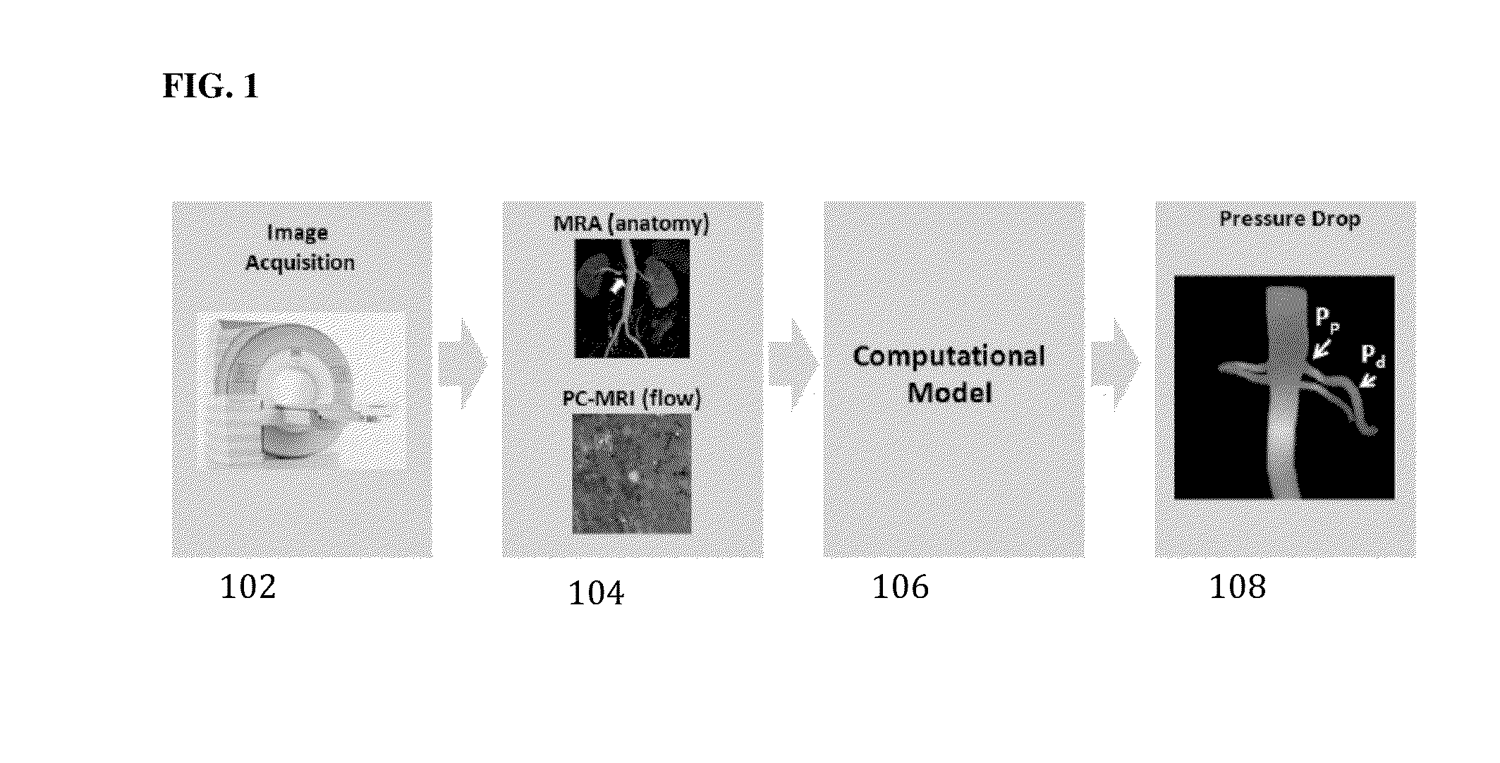
[0013]FIG. 1 illustrates a framework for non-invasive functional assessment of renal artery stenosis according to an embodiment of the present invention....


PUM


Abstract

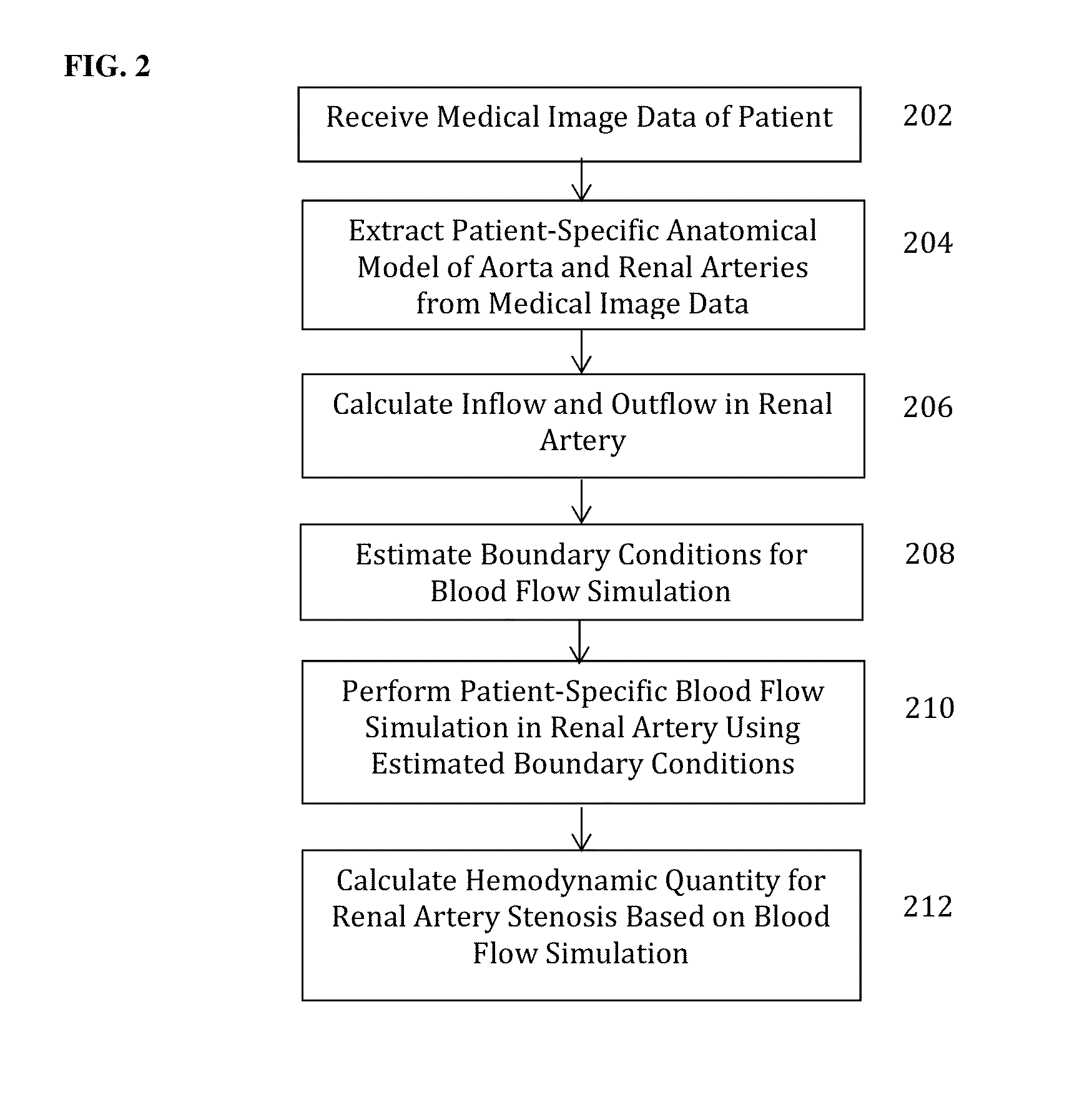
A method and system for non-invasive assessment of renal artery stenosis is disclosed. A patient-specific anatomical model of at least a portion of the renal arteries and aorta is generated from medical image data of a patient. Patient-specific boundary conditions of a computational model of blood flow in the portion of the renal arteries and aorta are estimated based on the patient-specific anatomical model. Blood flow and pressure are simulated in the portion of the renal arteries and aorta using the computational model based on the patient-specific boundary conditions. At least one hemodynamic quantity characterizing functional severity of a renal stenosis region is calculated based on the simulated blood flow and pressure in the portion of the renal arteries and aorta.

Description

[0001]This application claims the benefit of U.S. Provisional Application No. 61 / 873,399, filed Sep. 4, 2013, and U.S. Provisional Application No. 61 / 891,920, filed Oct. 17, 2013, the disclosures of which are herein incorporated by reference in their entirety.BACKGROUND OF THE INVENTION[0002]The present invention relates to functional assessment of renal artery stenosis, and more particularly, to non-invasive functional assessment of renal artery stenosis from medical image data and blood flow simulations.[0003]Renal artery stenosis (RAS) is the narrowing of the renal artery. RAS can impede blood flow to the target kidney and may result in hypertension, atrophy of the affected kidney, and ultimately renal failure if not treated. Medical image based anatomical assessment of RAS is routinely done in clinical practice. A number of imaging modalities, including magnetic resonance imaging (MRI), computed tomography (CT), x-ray angiography, and ultrasound, are used for this purpose, and e...

Claims


Application Information

Patent Timeline
Patent Type & Authority Applications(United States)
IPC IPC(8): A61B5/026G01R33/563A61B5/021
CPCA61B5/0263A61B2576/023G01R33/56316A61B5/021A61B5/055G01R33/5635G16H30/40
Inventor SHARMA, PUNEETRAPAKA, SAIKIRANMIHALEF, VIORELKAMEN, ALI
Owner SIEMENS HEALTHCARE GMBH
Learn More