Joint estimation of tissue types and linear attenuation coefficients for computed tomography

a computed tomography and tissue type technology, applied in the field of medical imaging, can solve the problems of difficult to accurately estimate linear attenuation coefficients and tissue types from photon counts, and use a priori information on tissue types to accurately estimate linear attenuation coefficients and tissue types

Active Publication Date: 2015-05-14
THE JOHN HOPKINS UNIV SCHOOL OF MEDICINE
View PDF5 Cites 12 Cited by
  • Summary
  • Abstract
  • Description
  • Claims
  • Application Information

AI Technical Summary

Problems solved by technology

This sequential method decouples the two steps and makes it difficult to use a priori information on t

Method used


Image

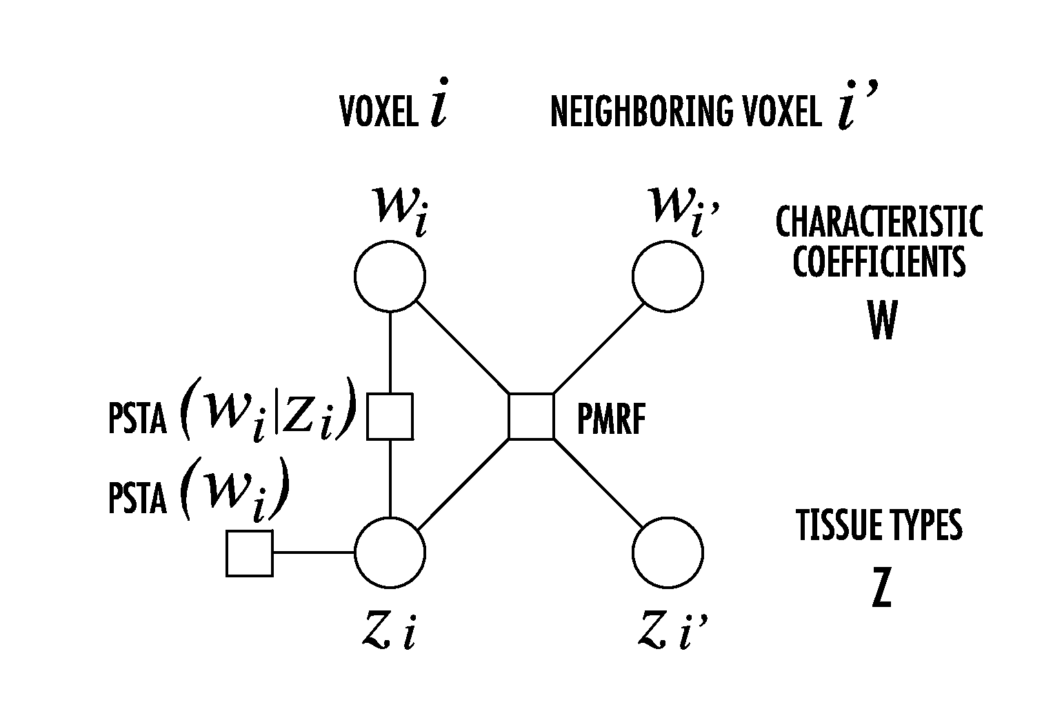
  • Joint estimation of tissue types and linear attenuation coefficients for computed tomography
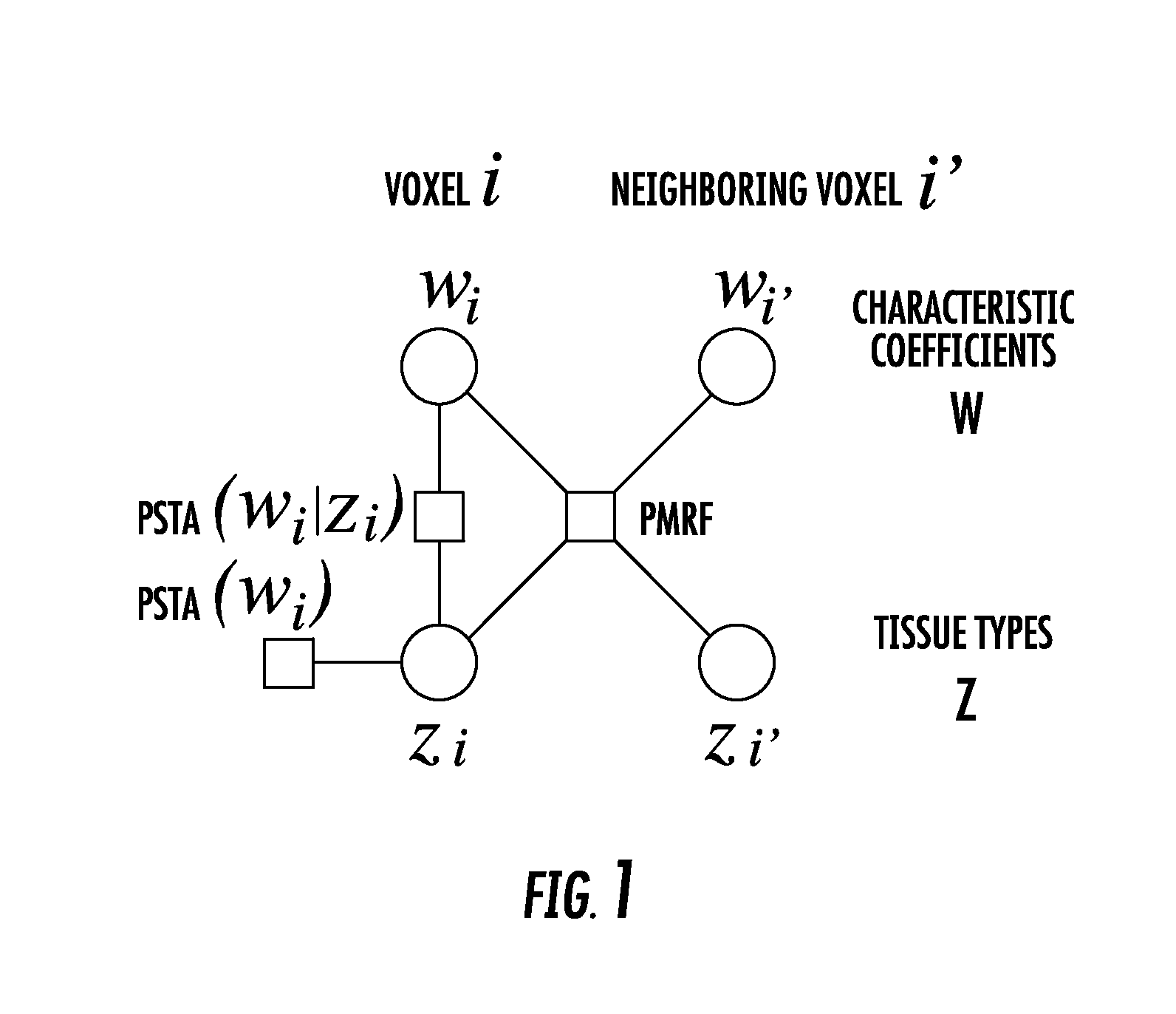
  • Joint estimation of tissue types and linear attenuation coefficients for computed tomography
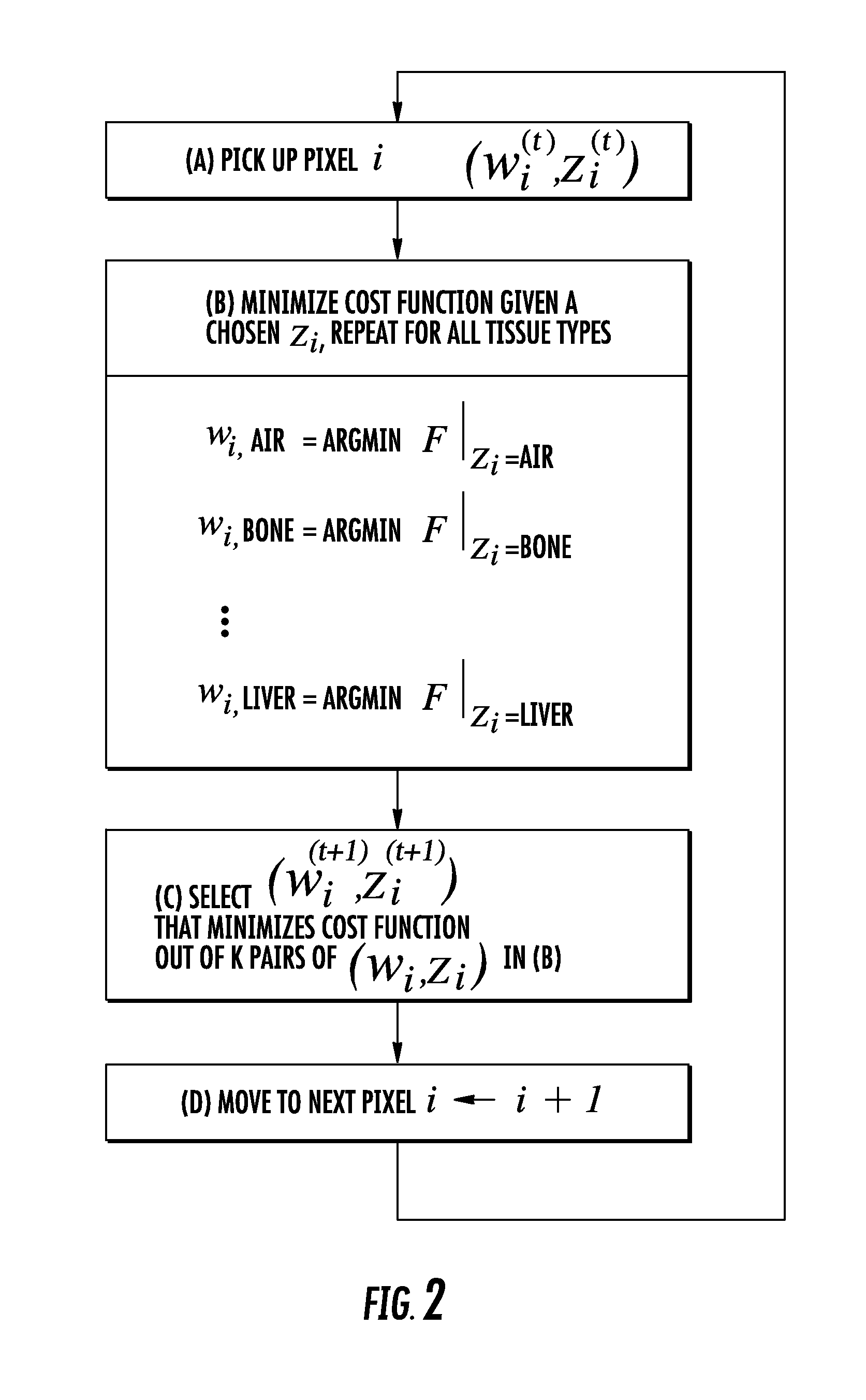
  • Joint estimation of tissue types and linear attenuation coefficients for computed tomography

Examples


Embodiment Construction

[0024]The presently disclosed subject matter now will be described more fully hereinafter with reference to the accompanying Drawings, in which some, but not all embodiments of the inventions are shown. Like numbers refer to like elements throughout. The presently disclosed subject matter may be embodied in many different forms and should not be construed as limited to the embodiments set forth herein; rather, these embodiments are provided so that this disclosure will satisfy applicable legal requirements. Indeed, many modifications and other embodiments of the presently disclosed subject matter set forth herein will come to mind to one skilled in the art to which the presently disclosed subject matter pertains having the benefit of the teachings presented in the foregoing descriptions and the associated Drawings. Therefore, it is to be understood that the presently disclosed subject matter is not to be limited to the specific embodiments disclosed and that modifications and other ...


PUM


Abstract

The present invention is directed to a new joint estimation framework employing MAP estimation based on pixel-based latent variables for tissue types. The method combines the geometrical information described by latent MRF, statistical relation between tissue types and P-C coefficients, and Poisson noise models of PCD data, and makes possible the continuous Baysian estimation from detected photon counts. The proposed method has better accuracy and RMSE than the method using FBP and thresholding. The joint estimation framework has the potential to further improve the accuracy by introducing more information about tissues in human body, e.g., the location, size, and number of tissues, or limited variation of neighboring tissues, which will be easily formulated by pixel-based latent variables.

Description

CROSS REFERENCE TO RELATED APPLICATION[0001]This application claims the benefit of U.S. Provisional Patent Application No. 61 / 895,542 filed on Oct. 25, 2013, which is incorporated by reference, herein, in its entirety.FIELD OF THE INVENTION[0002]The present invention relates generally to medical imaging. More particularly, the present invention relates to a method of tissue type estimation for use with computed tomography scanning.BACKGROUND OF THE INVENTION[0003]Energy sensitive photon counting detector-based, x-ray computed tomography (PCD-CT) has been one of the hottest research topics lately, as it is expected to provide various clinical benefits such as enhanced tissue contrast, decreased image noise, decreased radiation dose to patient, quantitative mono-energetic CT images, and more accurate material decomposition. Tissue types such as bones, fat, muscle, and iodine-enhanced blood can then be identified, allowing software applications to, e.g., separate blood vessels from bon...

Claims


Application Information

Patent Timeline
IPC IPC(8): G06T11/00A61B6/03G06T7/00A61B6/00
CPCG06T11/005A61B6/4241A61B6/032G06T2211/40G06T2207/10081G06T2207/20076G06T2207/30024G06T7/0012A61B6/5217G06T11/006G06T2211/408G16H50/30
Inventor TAGUCHI, KATSUYUKIAMAYA, KENJINAKADA, KENTO
Owner THE JOHN HOPKINS UNIV SCHOOL OF MEDICINE
Why Eureka
Try Eureka