Methods and systems for automatic location of optic structures in an image of an eye, and for automatic retina cup-to-disc ratio computation

a technology of automatic location and optic structure, applied in image generation, image enhancement, instruments, etc., can solve the problems of slowing down the progression of disease, loss of peripheral vision, and patients' complaints of vision loss, so as to reduce the workload of medical professionals and reduce the likelihood of occurren

Inactive Publication Date: 2015-07-02
AGENCY FOR SCI TECH & RES +1
View PDF5 Cites 24 Cited by
  • Summary
  • Abstract
  • Description
  • Claims
  • Application Information

AI Technical Summary

Benefits of technology

[0029]The invention may be used to perform optic cup and optic disc detection while reducing the workload of medical professionals. These can in turn be used to produce a cup-to disc ratio (CDR) which is indicative of the presence of medical conditions s

Problems solved by technology

Progression of the glaucoma results in loss of peripheral vision, so patients may complain of vision loss.
Although glaucoma cannot be cured, treatments can slow down the progression of the disease.
However, routine screening for glaucoma for the whole population is limited by poor sensitivity of current tests.
IOP measurement is neither specific nor sensitive enough to be an effective screening tool.
Moreover, visual field testing is not sensitive enough to detect early glaucoma damage as these patients do not have visual symptoms.
However, the technique involves manual assessment of the optic disc assessment, which is subjective, and the cost of training an ophthalmologist is high.
Deformable models are sensitive to poor initialization.
The

Method used


Image

  • Methods and systems for automatic location of optic structures in an image of an eye, and for automatic retina cup-to-disc ratio computation
  • Methods and systems for automatic location of optic structures in an image of an eye, and for automatic retina cup-to-disc ratio computation
  • Methods and systems for automatic location of optic structures in an image of an eye, and for automatic retina cup-to-disc ratio computation

Examples


first embodiment

ntation

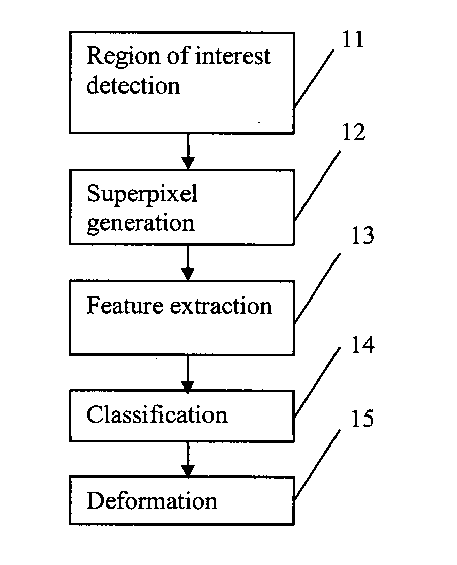
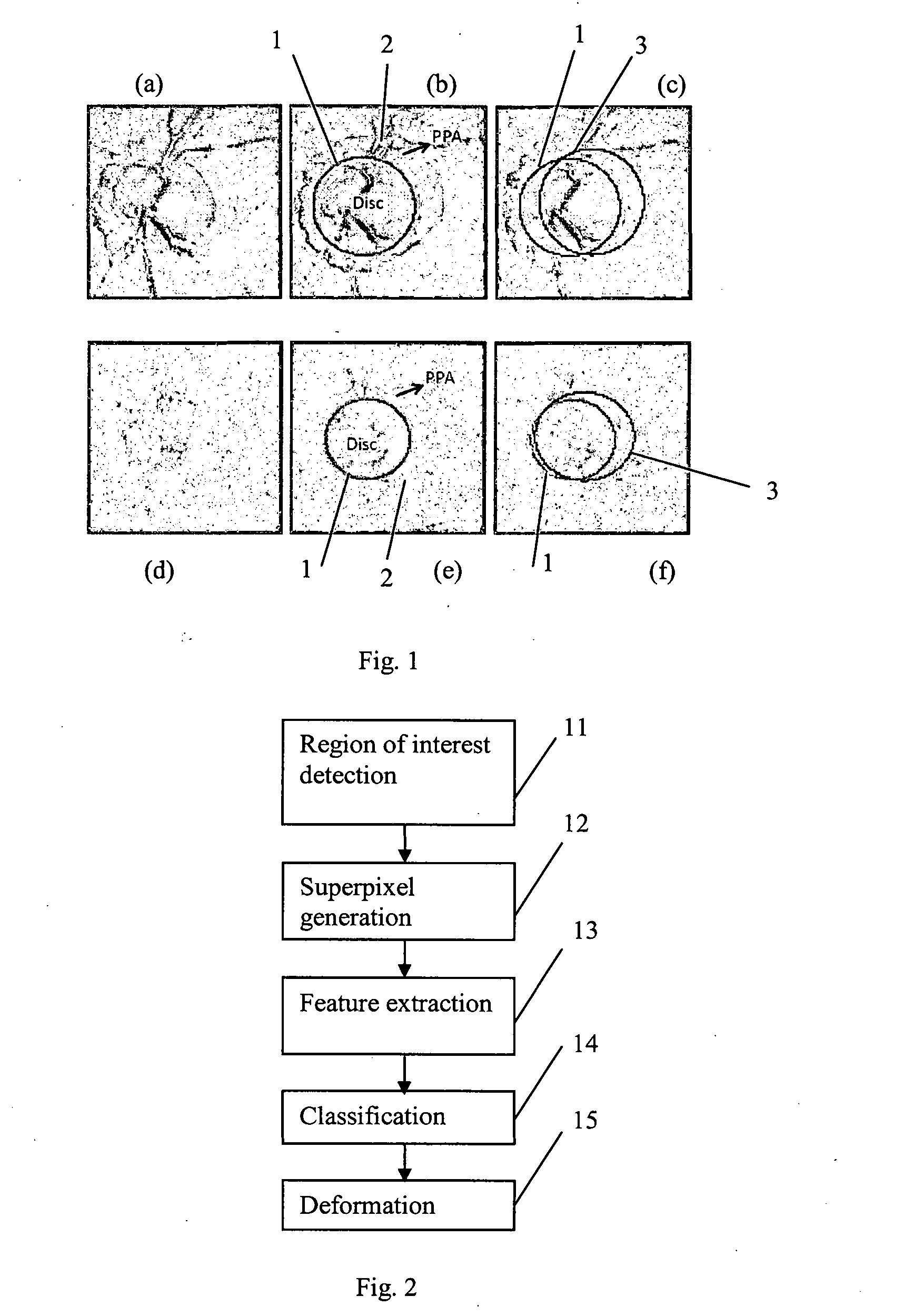
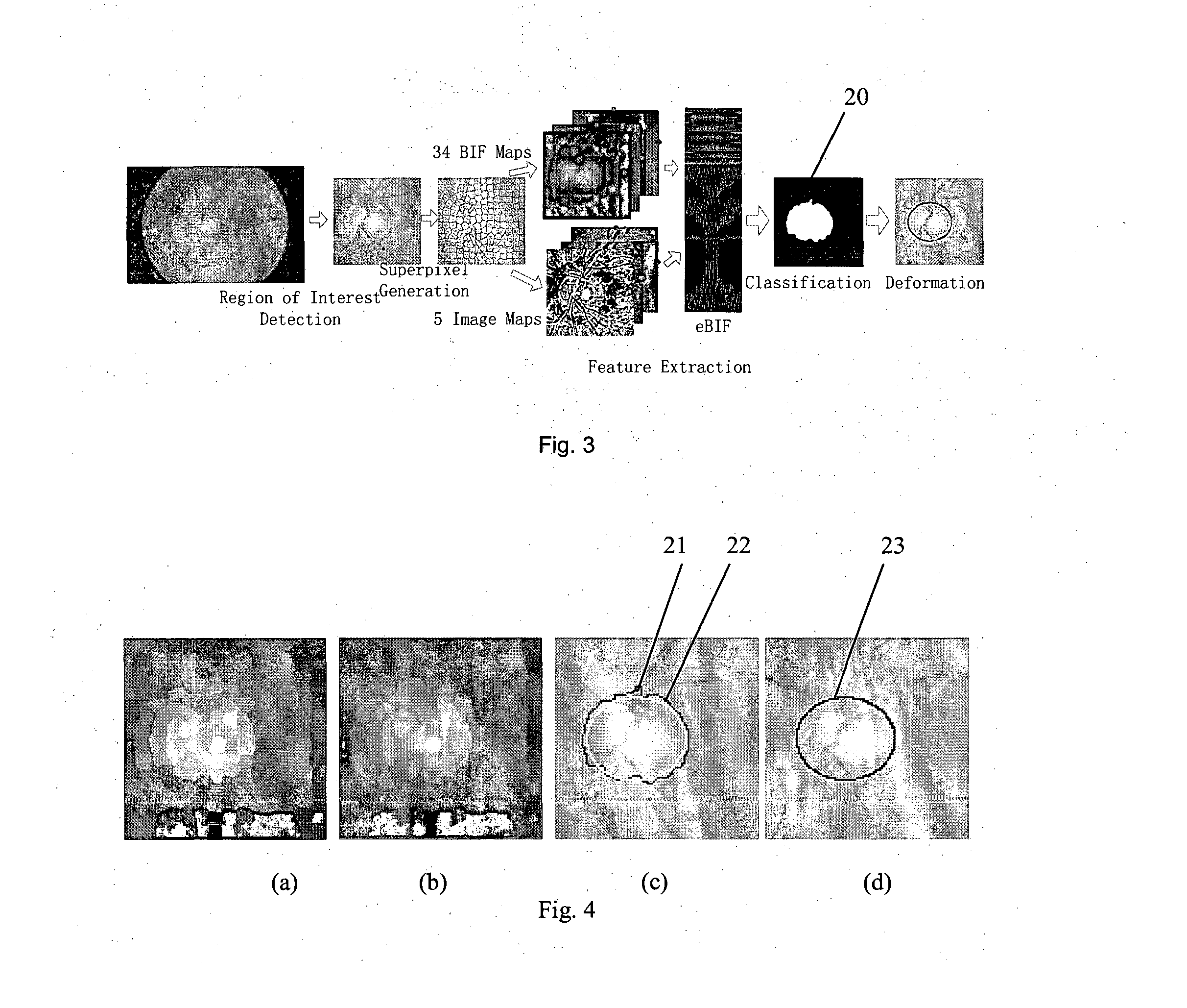
[0039]FIG. 2 shows the steps of a method which is a first embodiment of the invention, and which performs OD segmentation. The input to the method is a single two-dimensional retinal fundus image of the eye of a certain individual (or, in variations of the method, multiple fundus images of the eye). In a first step 11, a region of interest is detected by an optic disc localization step. In superpixel generation step 12, the region of interest is divided into sub-regions referred to as “superpixels”. In a feature extraction step 13, for each of the superpixels, extended biologically inspired features (eBIF) are computed from 34 biologically inspired feature maps and 5 image maps. In a classification step 14, they are used to classify the superpixels as disc or non-disc, so as to obtain a first partitioning of the region of interest into disc and non-disc sections. In a deformation step 15, deformation is applied to fine tune the disc.

[0040]The process is illustrated schematica...

second embodiment

tation

[0069]The optic cup is a central portion of the optic disc. Very few methods have been proposed for optic cup segmentation from 2-D images. Some of the present inventors have proposed the AGALI and AGLAIA techniques, which included thresholding followed by deformation methods. Because in many images there is no change of intensity at the optic cup boundary, additional information has been used, such as seeking small vessel bends (“kinks”) which anatomically mark the cup boundary. However, correct identification of kinks presents a challenge, and some retinal images do not exhibit kinks.

[0070]The second embodiment of the invention employs a superpixel-based approach for optic cup segmentation. The procedures for the superpixel-classification-based the optic cup segmentation are similar to those for optic disc segmentation and summarized in FIG. 5, and FIG. 6 shows schematically the data produced at each stage of the process. The cup segmentation may be done after the optic disc...


PUM


Abstract

A method is proposed for automatically locating the optic disc or the optic cup in an image of the rear of an eye. A portion of the image containing the optic disc or optic cup is divided into sub-regions using a clustering algorithm. Biologically inspired features, and optionally other features, are obtained for each of the sub-regions. An adaptive model uses the features to generate data indicative of whether each sub-region is within or outside the optic disc or optic cup. The result is then smoothed, to form an estimate of the position of the optic disc or optic cup.

Description

FIELD OF INVENTION[0001]The present invention relates to a system and a method for using an image of the fundus of an eye to located the optic cup and / or disc, and thereby compute the eye's retina cup-to-disc ratio (CDR).BACKGROUND OF THE INVENTION[0002]Glaucoma is a chronic eye condition in which the nerve that connects the eye to the brain (optic nerve) is progressively damaged. Patients with early stages of glaucoma do not have visual symptoms. Progression of the glaucoma results in loss of peripheral vision, so patients may complain of vision loss. Although glaucoma cannot be cured, treatments can slow down the progression of the disease. Thus, early detection of glaucoma is critical and beneficial to patients. However, routine screening for glaucoma for the whole population is limited by poor sensitivity of current tests. Screening may be useful for high risk individuals, such as first degree relatives of a glaucoma patient, older individuals (e.g. 65 years and older) and elder...

Claims


Application Information

Patent Timeline
IPC IPC(8): G06T7/00G06K9/46G06T11/20G06V10/764
CPCG06T7/0012G06T7/0042G06T11/203G06K9/4633G06K2009/4657G06T2207/30041G06T2207/30196G06T2210/41G06T2200/28G06T2207/10024G06T2207/20016G06T7/11G06T7/62G06T7/187G06V40/197G06V40/193G06V10/764G06F18/2411
Inventor CHENG, JUNLIU, JIANGXU, YANWUYIN, FENGSHOUTAN, NGAN MENGWONG, WING KEE DAMONLEE, BENG HAICHENG, XIANGANGGAO, XINTINGZHANG, ZHUOWONG, TIEN YINCHENG, CHING-YUCHEUNG, YIM-LUI CAROLMANI, BASKARANAUNG, TIN
Owner AGENCY FOR SCI TECH & RES
Why Eureka
Try Eureka