Radiation imaging system and operation method thereof, and radiation image detecting device and storage medium storing operation program therefor

a radiation imaging and operation method technology, applied in the field of radiation imaging system and operation method thereof, can solve the problems of long waiting time, inability to make the time taken from the completion of x-ray irradiation to the display of preview images shorter than the time taken for the above-described series of processing, etc., to achieve the effect of reducing the time required

Active Publication Date: 2015-07-02
FUJIFILM CORP
View PDF2 Cites 23 Cited by
  • Summary
  • Abstract
  • Description
  • Claims
  • Application Information

AI Technical Summary

Benefits of technology

[0011]In view of the foregoing, an object of the present invention is to provide a radiation imaging system capable of decreasing time required for displaying a preview image and an operation method thereof, and a radiation image detecting device and a storage medium storing an operation program therefor.
[0031]According to the present invention, radiation is irradiated twice in performing radiography once to acquire one sheet of radiation image for diagnosis, the preview image is produced based on the first X-ray irradiation, and output of the produced preview image to the monitor is started before the second X-ray irradiation is finished. Therefore, it is possible to display the preview image on the monitor before the second X-ray irradiation is finished and decrease the time required for displaying the preview image.

Problems solved by technology

However, since the above-described series of processing is started after the completion of the X-ray irradiation necessary for acquiring one sheet of X-ray image, it is impossible to make the time taken from the completion of the X-ray irradiation to the display of the preview image shorter than the time taken for the above-described series of processing.
Although the waiting time before the preview image is displayed is a small amount of time, an operator who is required to perform an operation speedily feels like that the waiting time is long.

Method used


Image

  • Radiation imaging system and operation method thereof, and radiation image detecting device and storage medium storing operation program therefor
  • Radiation imaging system and operation method thereof, and radiation image detecting device and storage medium storing operation program therefor
  • Radiation imaging system and operation method thereof, and radiation image detecting device and storage medium storing operation program therefor

Examples


first embodiment

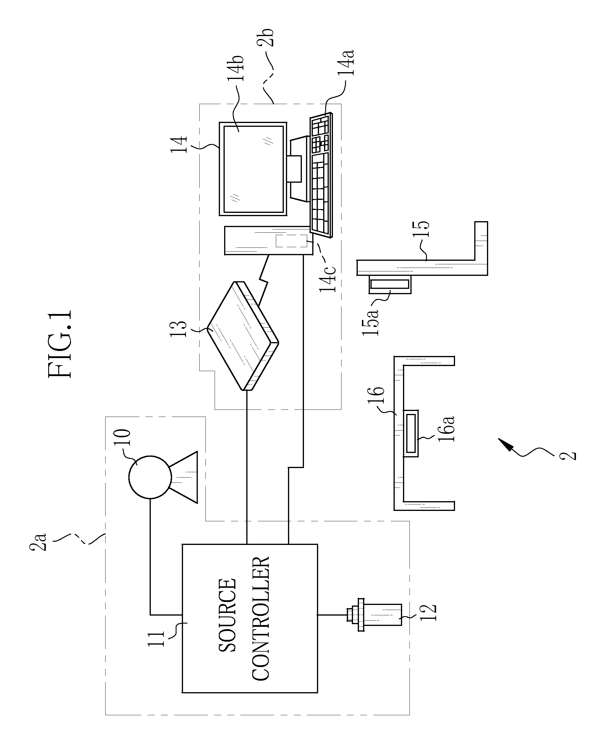
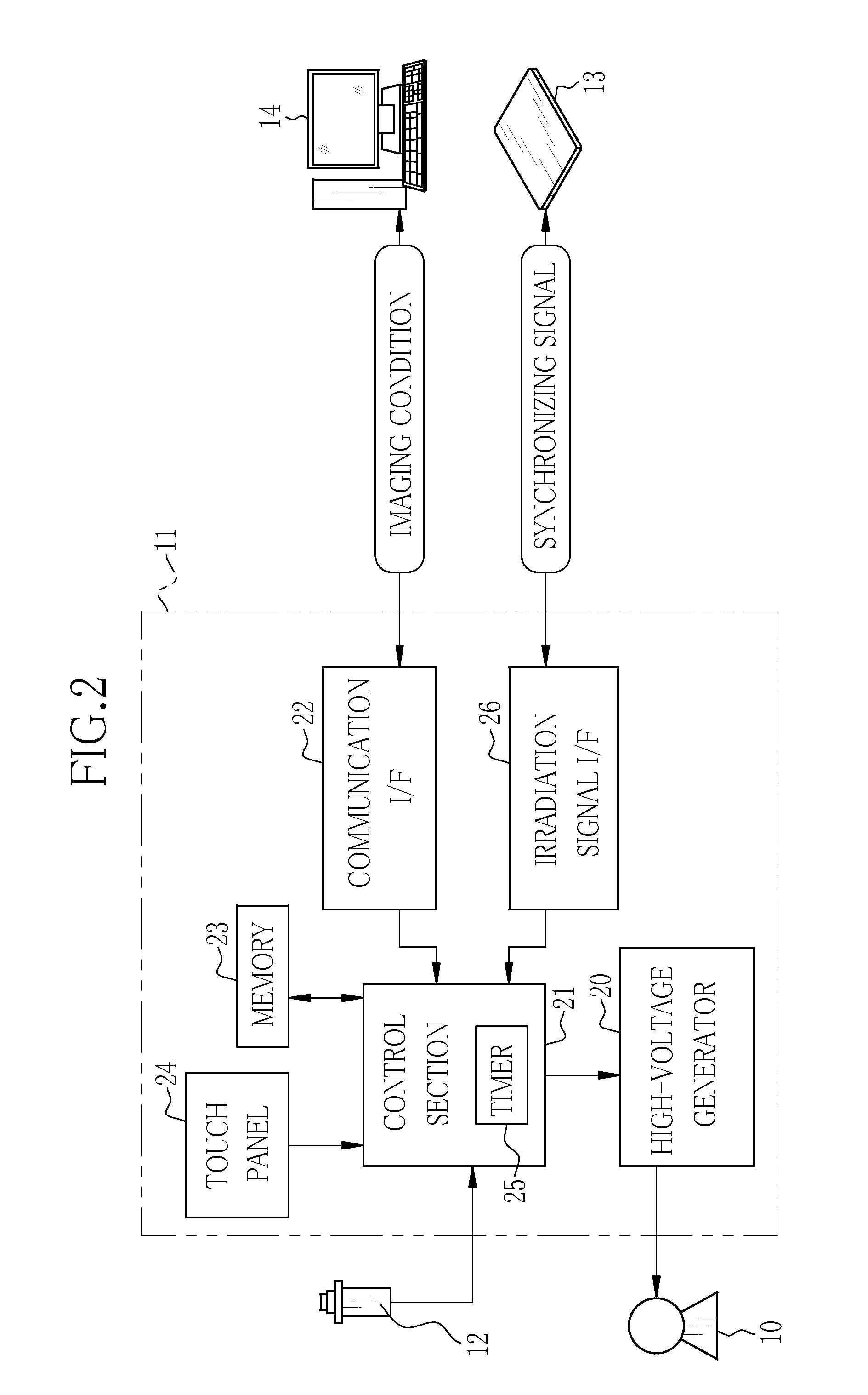
[0050]In FIG. 1, an X-ray imaging system 2 of the present invention includes an X-ray source 10 incorporating an X-ray tube for irradiating X-rays, a source controller 11 for controlling operation of the X-ray source 10, an irradiation switch 12 for giving a command to start warming-up and X-ray irradiation to the X-ray source 10, an electronic cassette 13 for detecting X-rays having passed through an object (i.e. patient) and outputting an X-ray image, a console 14 for performing operation control of the electronic cassette 13 and display processing of X-ray images, an upright-posture imaging table 15 for imaging the object in a standing posture, and a supine-posture imaging table 16 for imaging the object in a lying posture. The X-ray source 10, the source controller 11, and the irradiation switch 12 constitute an X-ray generating apparatus 2a. The electronic cassette 13 and the console 14 constitute an X-ray imaging apparatus 2b. Additionally, a source moving device (not shown in...

second embodiment

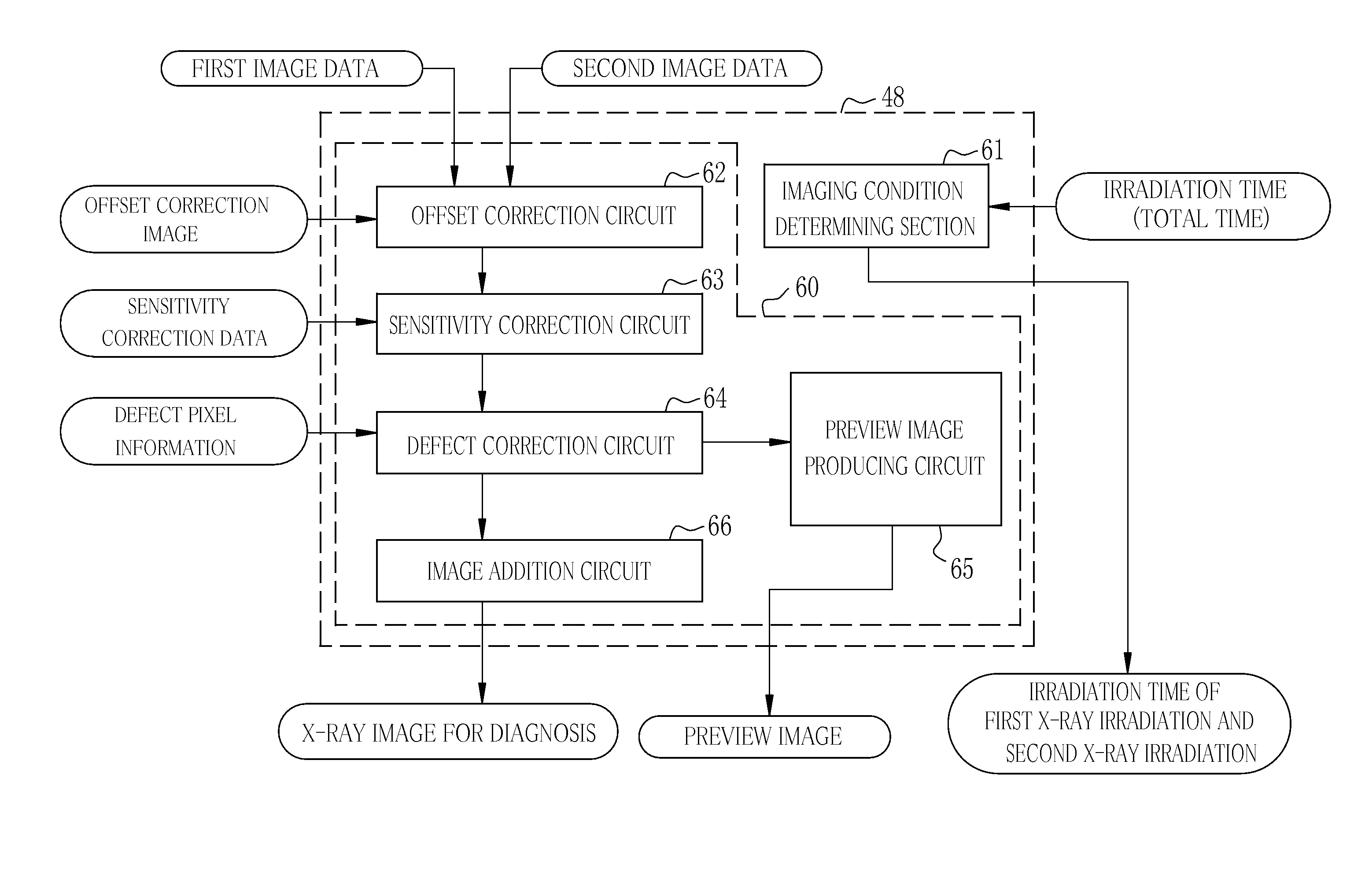
[0105]It is to be noted that body motion of an object may be detected based on the first and second image data respectively obtained by the first X-ray irradiation and second X-ray irradiation as described hereinbelow.

[0106]In FIG. 8, a control unit 70 is provided with a body motion detection circuit 71 (corresponding to a body motion detecting portion). Except the provision of the body motion detection circuit 71, the control unit 70 has the same structure as that of the control unit 48 in the first embodiment. After the second X-ray irradiation is finished, the body motion detection circuit 71 reads out the first image data obtained by the first X-ray irradiation and subjected to the image processing and the second image data obtained by the second X-ray irradiation and subjected to the image processing from the memory 54, and compares them. Then, the body motion detection circuit 71 adopts a well-known motion detection technique using object contour extraction, a motion vector, o...

third embodiment

[0110]Although the first image data is used to produce the preview image in the first embodiment, it is preferable that automatic exposure control (AEC) is performed with use of the first image data in order to improve the image quality of the X-ray image for diagnosis. As described in the first embodiment, although an approximate dose necessary for each part to be imaged is recognized, X-ray transmittance is varied depending on the body frame of the object such as thickness thereof. Therefore, even if the X-rays at the same dose are irradiated from the X-ray source 10, the dose of X-rays reaching the electronic cassette 13 is different for each object. Consequently, the AEC is performed in order to achieve more appropriate image quality. According to the AEC that is commonly performed, an accumulated dose of X-rays reaching the electronic cassette 13 during the X-ray irradiation is monitored, and the X-ray irradiation is stopped when the accumulated dose achieves a target dose. How...


PUM

PropertyMeasurementUnit
chargesaaaaaaaaaa
electric chargesaaaaaaaaaa
timeaaaaaaaaaa

Abstract

In an X-ray imaging system, first X-ray irradiation and second X-ray irradiation are performed in performing X-ray imaging once. A preview producing circuit subjects first image data outputted from a sensor panel after the first X-ray irradiation is finished to binning processing or thinning processing to produce a preview image. The produced image is transmitted through a communication I / F to a console while the sensor panel performs an accumulation operation in the second X-ray irradiation. The preview image is displayed on a monitor of the console in the second X-ray irradiation.

Description

CROSS-REFERENCE TO RELATED APPLICATIONS[0001]This application is a Continuation of PCT International Application No. PCT / JP2013 / 073286 filed on Aug. 30, 2013, which claims priority under 35 U.S.C §119(a) to Japanese Patent Application No. 2012-194582 filed Sep. 4, 2012. The above application is hereby expressly incorporated by reference, in its entirety, into the present application.BACKGROUND OF THE INVENTION[0002]1. Field of the Invention[0003]The present invention relates to a radiation imaging system for acquiring a radiation image and an operation method thereof, and a radiation image detecting device and a storage medium storing an operation program therefor.[0004]2. Description Related to the Prior Art[0005]In a medical field, an X-ray imaging system using radiation such as X-rays is known, for example. The X-ray imaging system includes an X-ray generating apparatus for generating X-rays and an X-ray imaging apparatus for acquiring an X-ray image of an object (i.e. patient) f...

Claims


Application Information

Patent Timeline
Patent Type & Authority Applications(United States)
IPC IPC(8): H04N5/32G01T1/17G01N23/04
CPCH04N5/32G01T1/17G01N23/04A61B6/4233A61B6/4283A61B6/488A61B6/52A61B6/5264A61B6/542A61B6/585G01T1/026
Inventor TAJIMA, TAKASHI
Owner FUJIFILM CORP
Why Eureka
Try Eureka