Image processing device and endoscope device

Active Publication Date: 2015-07-09
HOYA CORP
View PDF5 Cites 23 Cited by
  • Summary
  • Abstract
  • Description
  • Claims
  • Application Information

AI Technical Summary

Benefits of technology

[0021]As described above according to the embodiment of the invention, it becomes possib

Problems solved by technology

For this reason, even when the electronic endoscope device described in patent document 1 is used, the problem that identification of a lesion portion and the like is difficult is not resolved.
Furthermore, even when the electronic endoscope device described in the patent document 1 is used, judgm

Method used


Image

  • Image processing device and endoscope device
  • Image processing device and endoscope device
  • Image processing device and endoscope device

Examples


Embodiment Construction

[0033]In the following, an embodiment of the invention is explained in detail with reference to the accompanying drawings.

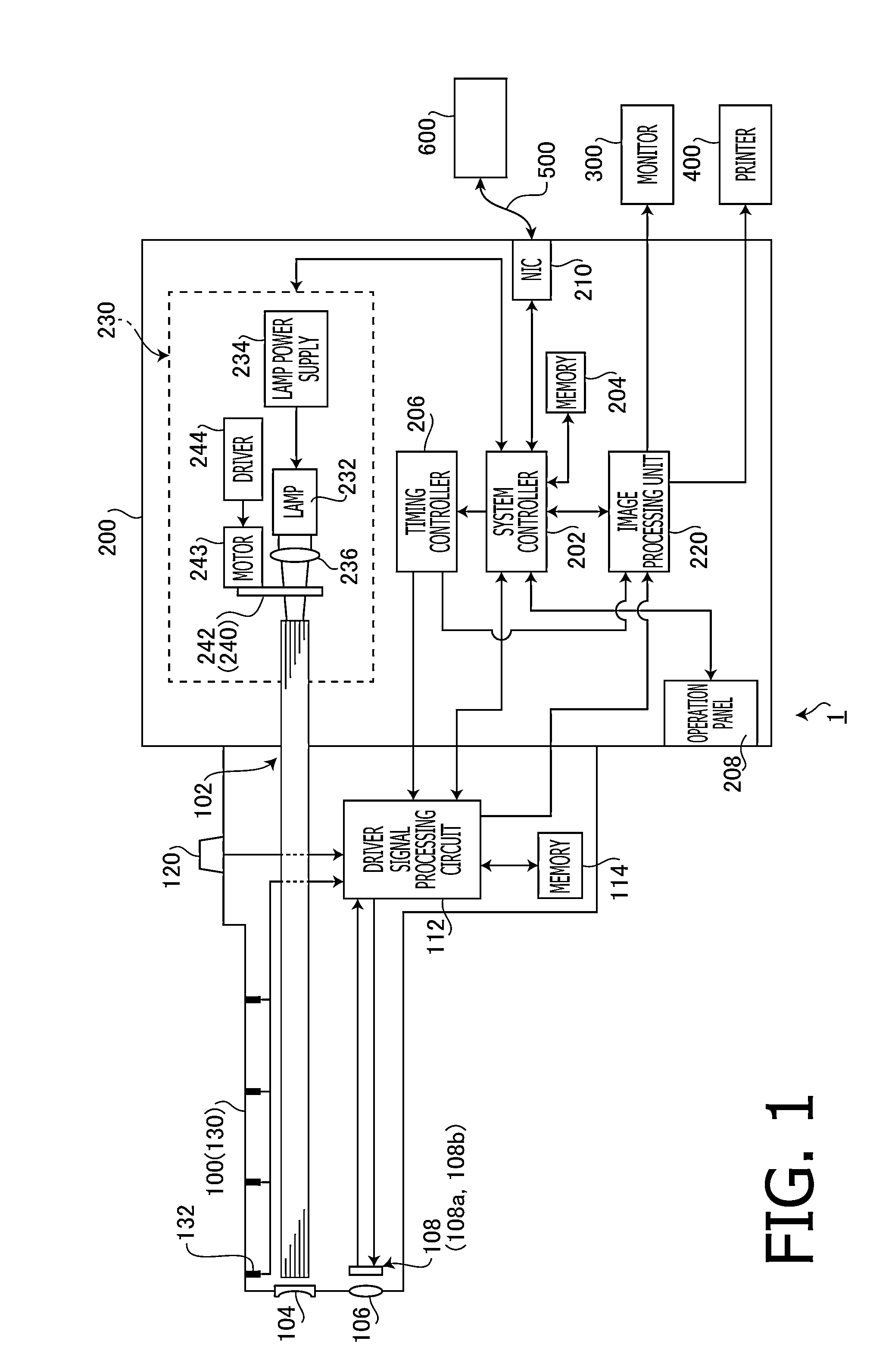
[0034]FIG. 1 is a block diagram illustrating a general configuration of an electronic endoscope device 1 according to the embodiment of the invention. As shown in FIG. 1, the electronic endoscope device 1 according to the embodiment includes an electronic scope 100, a processor 200 for an electronic endoscope, a monitor 300 and a printer 400.

[0035]The processor 200 for an electronic endoscope includes a system controller 202 and a timing controller 206. The system controller 202 executes various programs stored in a memory 204 and totally controls the entire electronic endoscope device 1. Further, the system controller 202 changes various settings of the electronic endoscope device 1 in accordance with an instruction from a user (an operator or an assistant) input to an operation panel 208. The timing controller 206 outputs clock pulses for adjusting operation ti...


PUM


Abstract

An image processing device, comprising an obtaining means that obtains color endoscopic image data, a color space conversion means that converts a color space of the color endoscopic image data to an HSI color space based on hue, saturation and intensity or an HSV color space based hue, saturation and a value, a lesion pixel judgment means that judges, for each of pixels constituting a color endoscopic image, whether or not each pixel is a lesion portion based on the hue and the saturation, an overlay processing means that executes an overlay process which changes a color of the pixel judged to be the lesion portion by the lesion pixel judgment means, and an overlay image display means that displays an overlay image obtained by subjecting the color endoscopic image to the overlay process.

Description

TECHNICAL FIELD[0001]The present invention relates an image processing device for endoscopic diagnosis and a medical endoscope device provided with the image processing device, and particularly to a device having the function of obtaining and displaying information on a lesion portion based on color information of a color endoscopic image.BACKGROUND ART[0002]In general, a lesion portion shows a color different from a color of a normal mucous membrane tissue. Thanks to enhancement of the performance of a color endoscope device, identification of a lesion portion of which color is slightly different from a color of a normal tissue has become possible. However, in order to become able to precisely identify a lesion portion from a normal tissue based on a slight difference in color on an endoscopic image, an operator needs to be trained under expert's instruction for a long period of time. Furthermore, even for a practiced operator, it is not easy to identify a lesion portion based on a...

Claims


Application Information

Patent Timeline
IPC IPC(8): G06T7/00G06T7/40
CPCG06T7/0014G06T7/408G06T2207/20172G06T2207/10024G06T2207/10068G06T2207/30096A61B1/00009A61B1/04G06T7/0012G06T2207/30028A61B5/061G06T5/007A61B1/000095
Inventor IKEMOTO, YOUSUKE
Owner HOYA CORP
Why Eureka
Try Eureka