Ultrasound imaging device operated by mobile display device and ultrasound imaging system

a technology of ultrasound imaging and mobile display, applied in tomography, applications, instruments, etc., can solve the problems of inconvenient movement in medical locations, large, heavy, and relatively expensive systems, and achieve the effect of improving the ultrasound imaging system

Inactive Publication Date: 2015-09-03
KONINKLJIJKE PHILIPS NV
View PDF11 Cites 13 Cited by
  • Summary
  • Abstract
  • Description
  • Claims
  • Application Information

AI Technical Summary

Benefits of technology

[0009]It is an object of the present invention to provide an improved ultr

Problems solved by technology

Of course, providing fully set up ultrasound imaging systems comprising every component as mentioned above make

Method used


Image

  • Ultrasound imaging device operated by mobile display device and ultrasound imaging system
  • Ultrasound imaging device operated by mobile display device and ultrasound imaging system
  • Ultrasound imaging device operated by mobile display device and ultrasound imaging system

Examples


Embodiment Construction

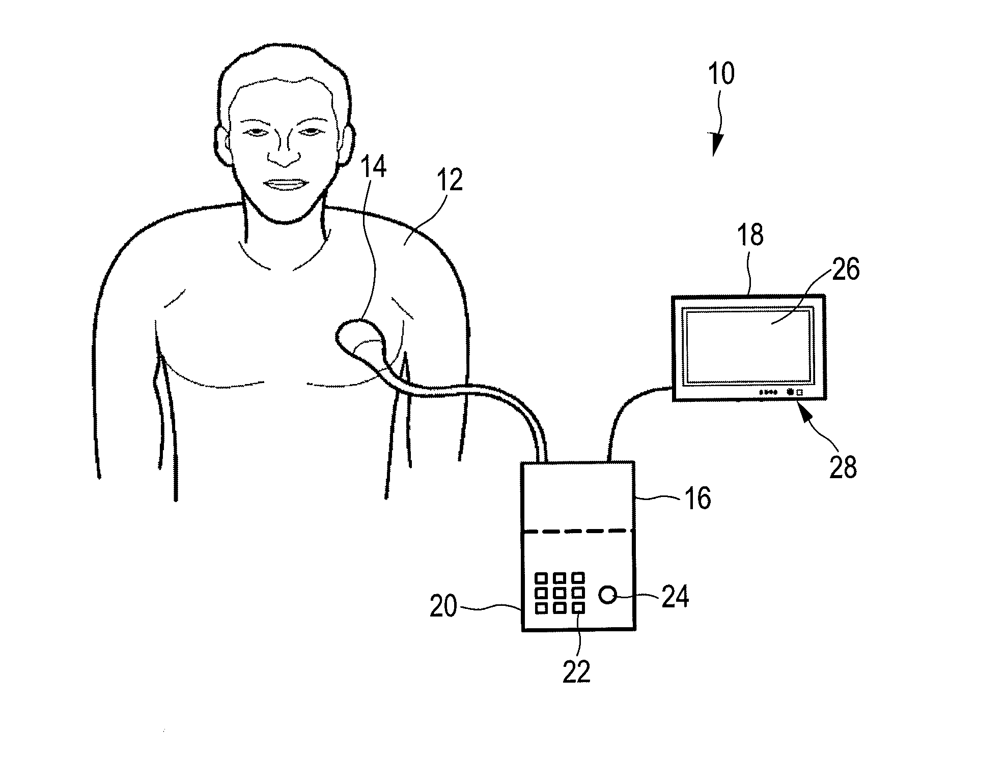
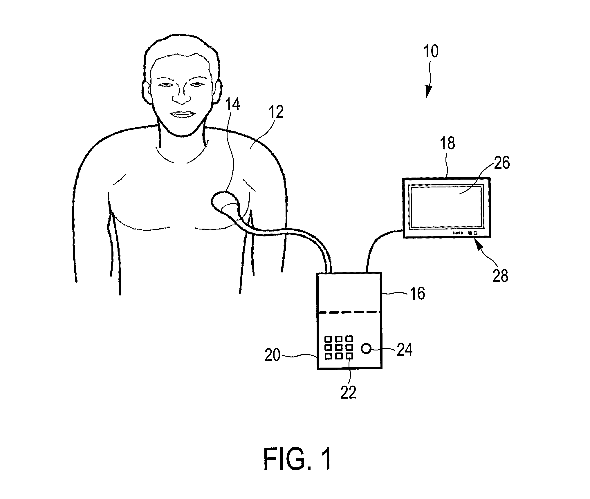
[0038]FIG. 1 shows an ultrasound imaging system 10. The ultrasound imaging system 10 is used for scanning an area or volume of the body of the patient 12.

[0039]For scanning the patient 12, a probe head 14 may be provided. The probe head 14 is connected to a docking unit 16 of the ultrasound imaging system 10. The docking unit 16 serves not only for connecting the probe head 14 but also for connecting a mobile display device 18. While the mobile display device 18 is connected to the docking unit 16 via a cable shown in FIG. 1, it is contemplated that the mobile display device 18 is connected to the docking unit 16 via a plug and socket connection, or may be easily connected in a wireless manner to the docking unit 16.

[0040]The docking unit 16 may comprise a first input device 20. The first input device 20 may have a keypad 22 and / or a trackball 24 to provide an input mechanism to a user of the ultrasound imaging system 10. Additionally or alternatively, other mechanisms may be presen...


PUM


Abstract

The present invention relates to an ultrasound imaging system comprising an ultrasound image acquisition station (46) and a mobile display device (18). When disconnected from the ultrasound image acquisition station (46), the mobile display device (18) may be operated by its own operating system providing general purpose applications to a user. When connected to the ultrasound image acquisition station (46), the hardware of the mobile display device (18) is operated by an ultrasound application dedicated operating system stored on the ultrasound image acquisition station (46). By this, a low weight and flexible to use ultra sound imaging system may be provided.

Description

FIELD OF THE INVENTION[0001]The present invention relates to an ultrasound image acquisition station for use together with a mobile display device to form an ultrasound imaging system. Further, the present invention relates to an ultrasound imaging system for providing an image of area tomographic plane or volume, for example an anatomical view within a body of a patient.BACKGROUND OF THE INVENTION[0002]Ultrasound imaging systems are widely known in the art. They are in particular used to provide anatomical imaging of patients. Both two-dimensional and three-dimensional imaging of bodies of patients is known to provide a reliable tool for medical practitioners to view parts of a body of a patient without the need for any surgical steps.[0003]Ultrasound imaging systems are complete stations that may be fixed to a certain location and are often movable on rollers to provide flexible use in different locations. The ultrasound imaging systems provide for every component needed to acquir...

Claims


Application Information

Patent Timeline
IPC IPC(8): A61B8/00G01S7/52A61B8/14G01S15/89
CPCA61B8/462G01S15/899G01S7/52055A61B8/4433G01S15/8915A61B8/14A61B8/54G01S7/52085A61B8/13A61B8/4411A61B8/4427A61B8/4438A61B8/4472A61B8/467A61B8/483
Inventor POLAND, MCKEE DUNN
Owner KONINKLJIJKE PHILIPS NV
Why Eureka
Try Eureka