Suppression of vascular structures in images

a vascular structure and image technology, applied in image enhancement, instruments, tomography, etc., can solve the problems of significant variability, difficult object detection in computer vision systems, and difficult object detection, so as to improve the rendering of selected anatomically suppressed or enhanced image data, and improve the detection of nodules

Active Publication Date: 2015-10-01
RIVERAIN TECH
View PDF12 Cites 11 Cited by
  • Summary
  • Abstract
  • Description
  • Claims
  • Application Information

AI Technical Summary

Benefits of technology

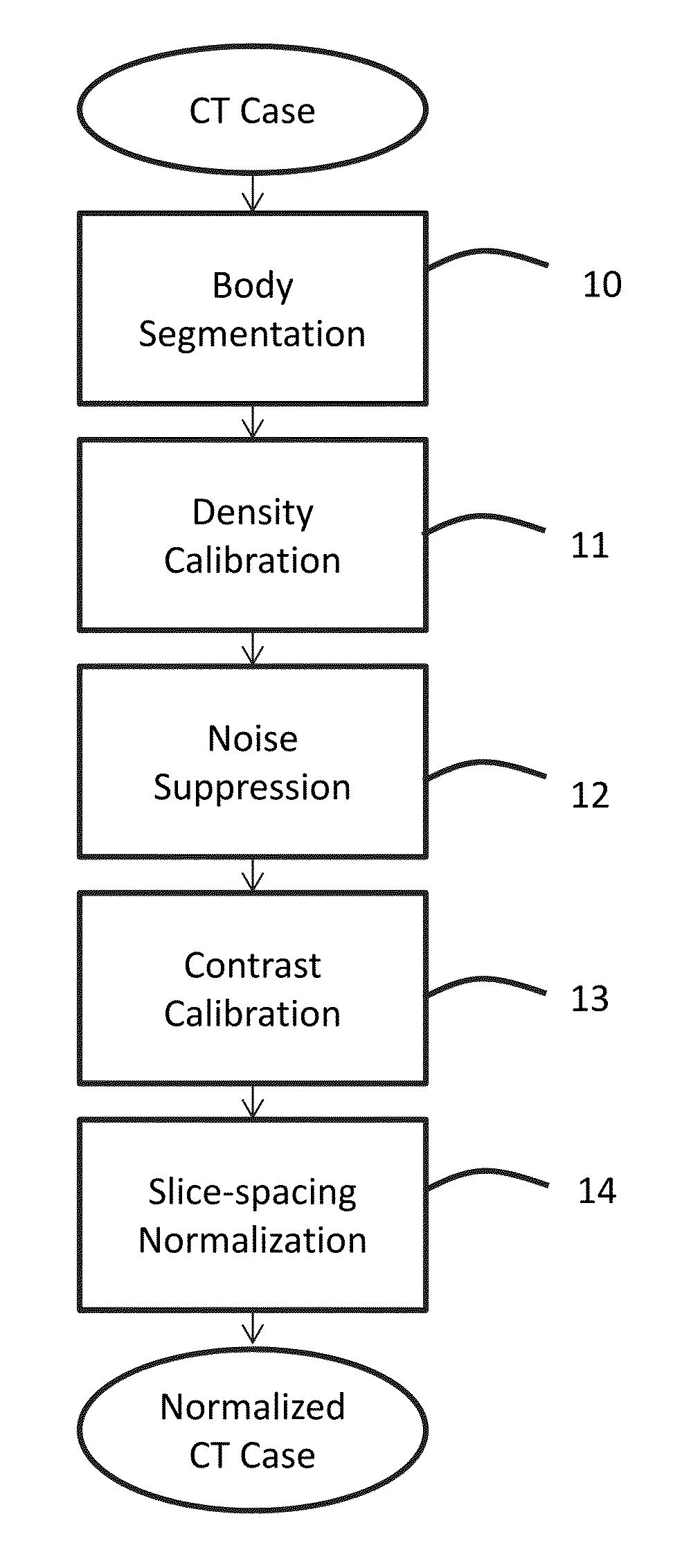
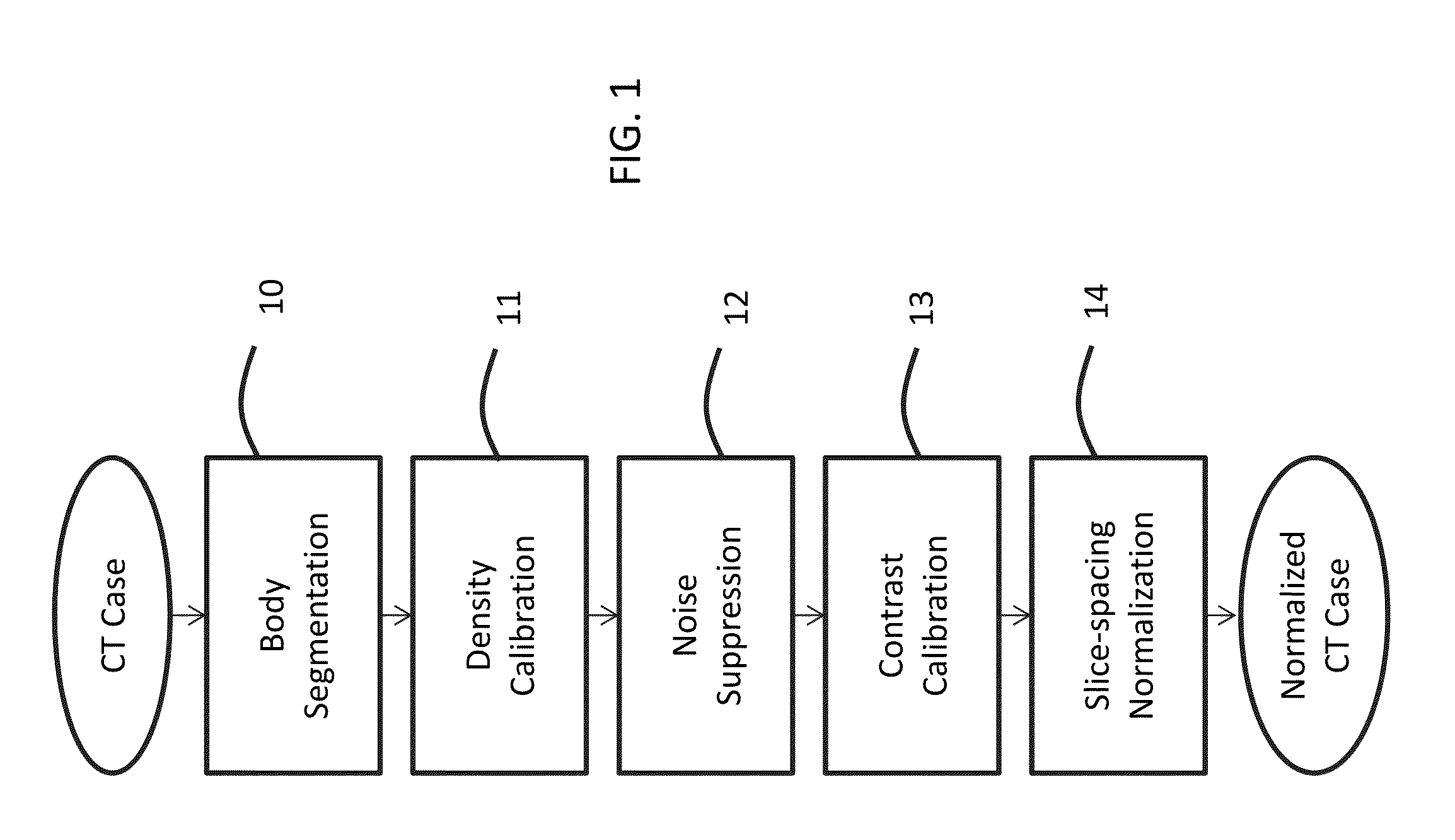
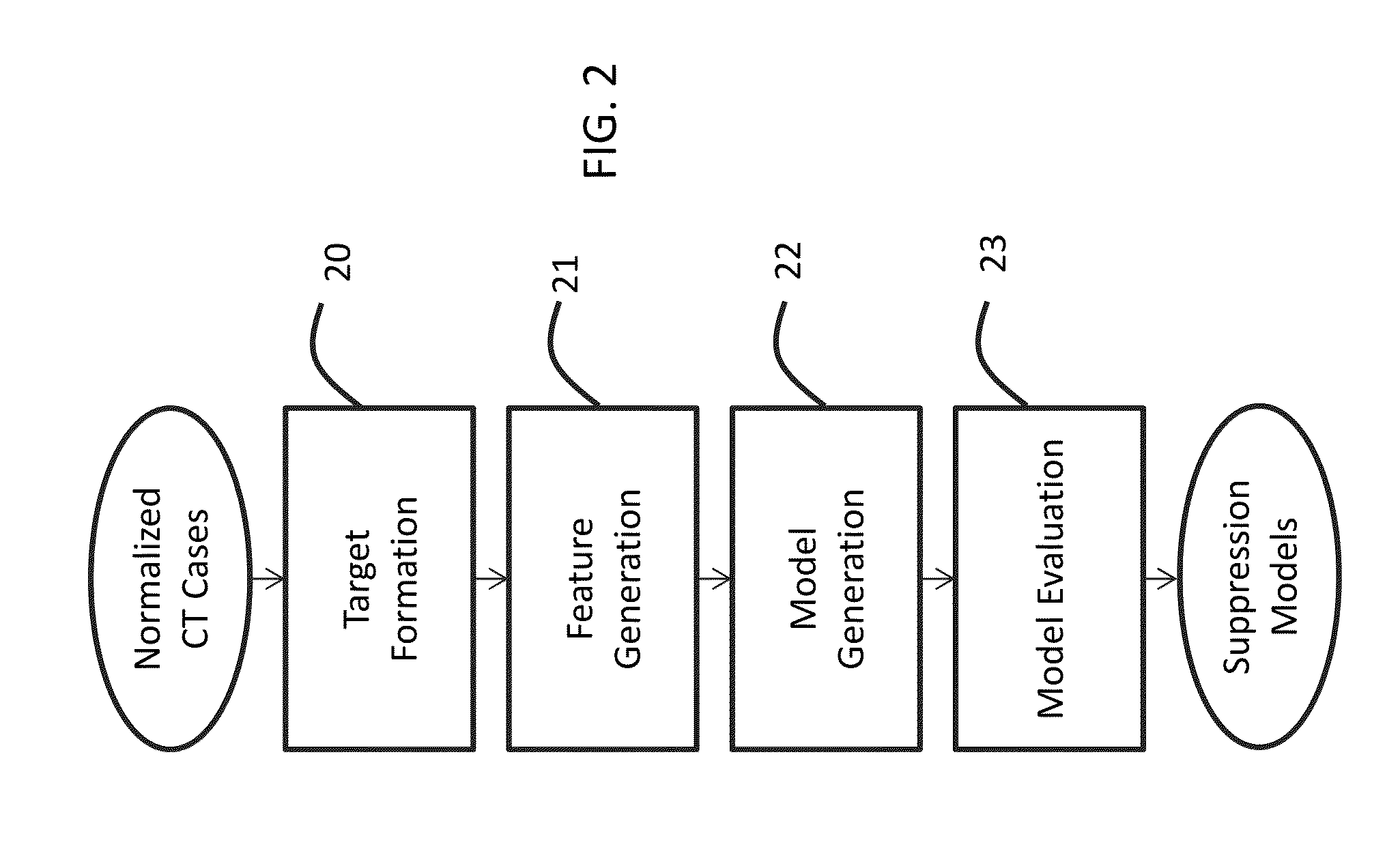
[0005]Various aspects of this disclosure may include an approach for normalizing medical image and/or voxel data captured under different acquisition protocols, and/or a method for suppressing selected non-nodule structures within medical image and/or voxel data of the chest. Most non-nodule structure is vascular content, and therefore, the term “vessel suppression” will be used in this disclosure as a general term for such non-nodule structure suppression. However, the disclosed techniques may also apply to structures other than “vessels”/vascular structures, e.g., bronchial walls and fissure lines in the thorax, a

Problems solved by technology

It is widely recognized that object detection is a challenging problem.
There are many aspects that make object detection difficult for computer vision systems, including factors such as variations in image acquisition, complexity of object appearance, and significant variability in object backgrounds (usually referred to as clutter), to name just a few.
Despite more than two d

Method used


Image

  • Suppression of vascular structures in images
  • Suppression of vascular structures in images
  • Suppression of vascular structures in images

Examples


Embodiment Construction

[0015]There are several approaches that may be taken to nodule detection, which may include template matching (e.g., Q. Li, S. Katsuragawa, and K. Doi, “Computer-aided diagnostic scheme for lung nodule detection in digital chest radiographs by use of a multiple-template matching technique,”Medical Physics, 2001, 28(10): 2070-2076; hereinafter “Li et al. 2001”), multi-level thresholding (e.g., S. Armato, M. Giger, and H. MacMahon, “Automated detection of lung nodules in CT scans: preliminary results,”Medical Physics, 2001, 28, 1552-1561; hereinafter “Armato et al. 2001”), enhancement filters (e.g., A. Frangi, W. Niessen, K. Vincken, and M. Viergever, “Multiscale vessel enhancement filtering,”MICCAI, 1998, 130-137; hereinafter “Frangi et al. 1998;” and Q. Li, S. Sone. and K. Doi, “Selective enhancement filters for nodules, vessels, and airway walls in two- and three-dimensional CT scans,”Medical Physics 2003, 30(8): 2040-2051; hereinafter “Li et al. 2003”), and voxel classification (e...


PUM


Abstract

Image processing techniques may include a methodology for normalizing medical image and/or voxel data captured under different acquisition protocols and a methodology for suppressing selected anatomical structures from medical image and/or voxel data, which may result in improved detection and/or improved rendering of other anatomical structures. The technology presented here may be used, e.g., for improved nodule detection within computed tomography (CT) scans. While presented here in the context of nodules within the lungs, these techniques may be applicable in other contexts with little modification, for example, the detection of masses and/or microcalcifications in full field mammography or breast tomosynthesis based on the suppression of glandular structures, parenchymal and vascular structures in the breast.

Description

CROSS-REFERENCE TO RELATED APPLICATION[0001]This application is a non-provisional patent application deriving priority from U.S. Provisional Patent Application No. 61 / 971,042, filed on Mar. 27, 2014, and incorporated by reference herein.FIELD OF ENDEAVOR[0002]The present disclosure may relate generally to suppressing unwanted structures within a medical image and / or voxel data using feature extraction and successive model-based prediction methods and may further relate to techniques that may improve detection of lung nodules within computed tomography (CT) scans. Model-based prediction in the context of this disclosure is defined as the use of an analytical model(s), empirical model(s) or combination thereof, e.g., neural network, to predict a value (e.g., a pixel or voxel) based on measures, either computed or derived from pixels / voxels.BACKGROUND[0003]It is widely recognized that object detection is a challenging problem. There are many aspects that make object detection difficult...

Claims


Application Information

Patent Timeline
IPC IPC(8): G06T7/00G06T11/00A61B6/03G06T5/30G06T3/00G06T1/00G06T5/00
CPCG06T7/0014G06T2210/41G06T7/0097G06T11/008G06T5/003G06T5/002G06T5/30G06T3/0068A61B6/032G06T2207/10081G06T2207/20141G06T2207/30068G06T2207/30064G06T2207/30101G06T2207/30196G06T1/0007A61B6/5217A61B6/5258G06T5/005G06T7/0012G06T2207/20016G06T2207/20036G06T2207/20076G06T7/136G16H50/30
Inventor KNAPP, JASON F.KAKUMANU, PRAVEENWORRELL, STEVE W.
Owner RIVERAIN TECH
Why Eureka
Try Eureka