

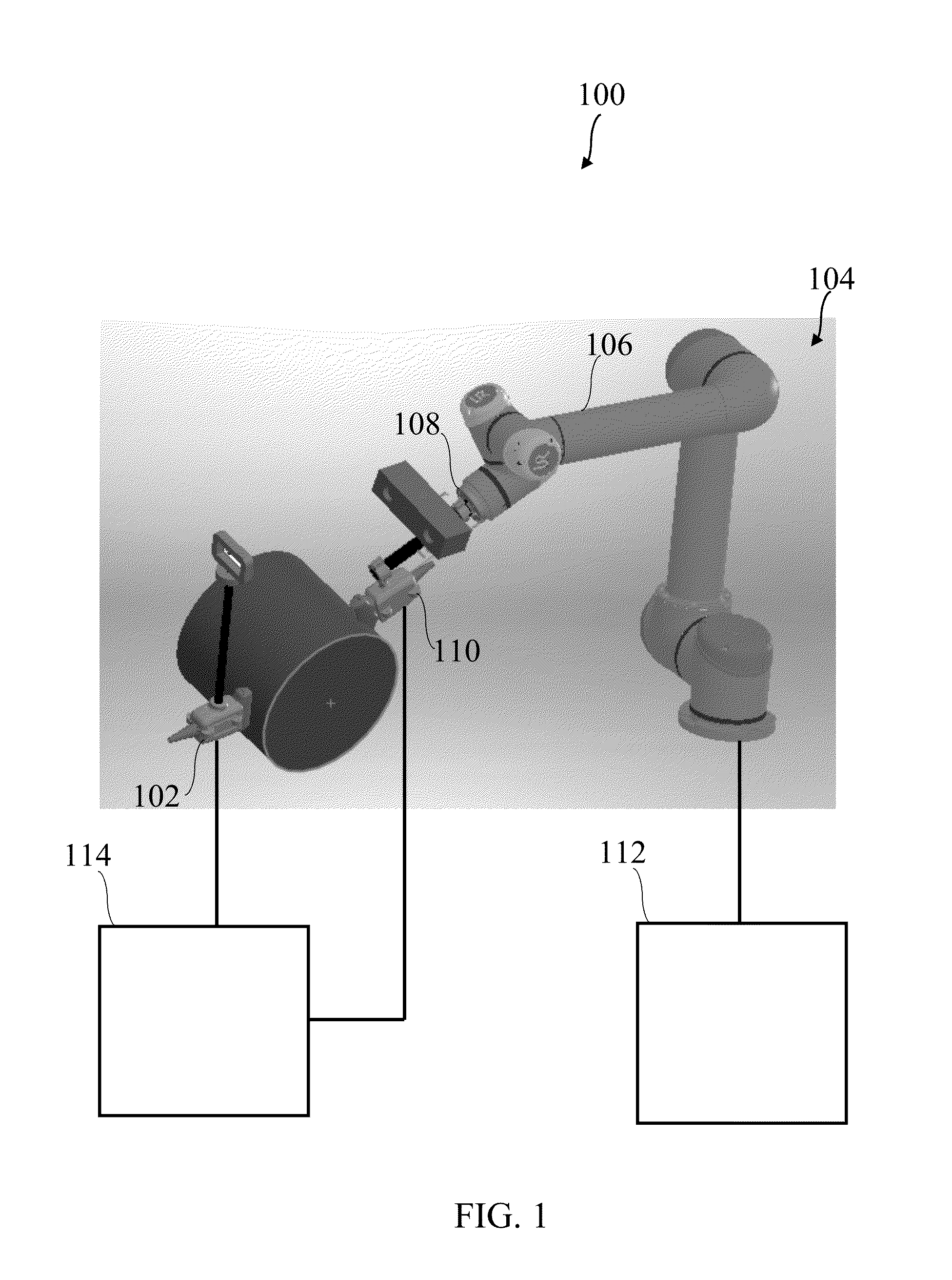
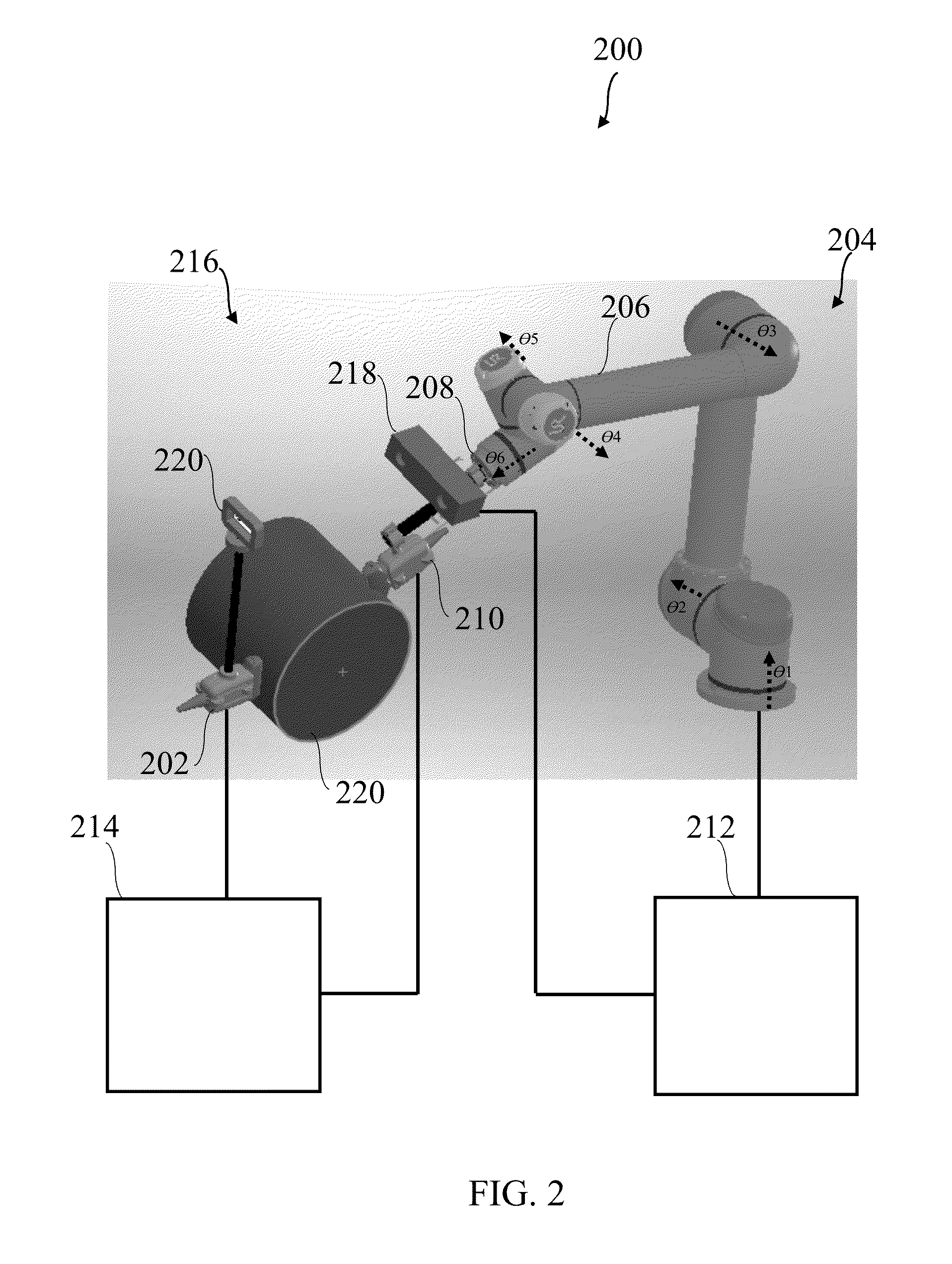
Robot assisted ultrasound system
a robotic assisted and ultrasound technology, applied in the field of ultrasound systems, can solve the problems of limiting the possible areas that can be scanned, limiting the use of ultrasonic imaging for thick tissues or obese patients, and relying on a large number of transducers instead of the existing ultrasonic imaging systems to enable this technology,
- Summary
- Abstract
- Description
- Claims
- Application Information
AI Technical Summary
Benefits of technology
Problems solved by technology
Method used
Image

Examples
example 1
References for Example 1
[0309][1] R. W. Prager, U. Z. Ijaz, A. H. Gee, G. M. Treece, “Three-dimensional US imaging,” Proc. of the Institution of Mechanical Engineers, Part H: J. of Engineering in Medicine 224(2), 193-223 (2010) [doi:10.1243 / 09544119JEIM586].[0310][2] A. Fenster, J. Bax, H. Neshat, N. Kakani, C. Romagnoli, “3D US imaging in image-guided intervention,” in Advancements and Breakthroughs in US Imaging, Ch. 1, INTECH (2013) [doi:10.5772 / 55230].[0311][3] R. W. Prager, R. N. Rohling, A. H. Gee, L. Berman, “Rapid calibration for 3-D freehand ultrasound,” Ultrasound in Medicine & Biology 24 (6), 855-869 (1998) [doi:10.1016 / 80301-5629(98)00044-1].[0312][4] J. Jago, A. Collet-Billon, C. Chenal, J. M. Jong, S. Makram-Ebeid, “XRES®: adaptive enhancement of ultrasound images,” Medicamundi 46(3), 36-41 (2002).[0313][5] C. Hansen, N. Huttebrauker, A. Schasse, W. Wilkening, H. Ermert, M. Hollenhorst, L. Heuser, G. Schulte-Altedorneburg, “Ultrasound breast imaging using full angle sp...
example 2
References for Example 2
[0342][1] Randall R. De Martino, Adam W. Beck, Matthew W. Edwards, “Impact of screening versus symptomatic measurement of deep vein thrombosis in a nationalquality improvement registry”, Journal of Vascular Surgery, 2012 (10), pp: 1045-1051.[0343][2]“DVT overview”, WebMD, available online: http: / / www.webmd.com / dvt / ss / slideshow-deep-vein-thrombosis-overview[0344][3] Akil P Patel, Michael T. Koltz, Charles A. Sansur, “An analysis of deep vein thrombosis in 1277 consecutive neurosurgical patients undergoing routine weekly ultrasonography”, Journal of Neurosurg, Vol. 118, March, 2013, pp: 505-509.[0345][4]“Deep Vein Thrombosis overview”, Society of Interventional, available noline: http: / / www.sirweb.org / patients / deep-vein-thrombosis / [0346][5] Talbot S, “Use of real-time imaging in identifying deep venous obstruction: a preliminary report”, Bruit, 1982, 7, pp: 41-42.[0347][6] Heather L. Gornik, Aditya M. Sharma, “Duplex ultrasound in the diagnosis of lower-extremi...
example 3
References for Example 3
[0363][1] Detmer P. R., Bashein G., Hodges T., Beach K. W., Filer E. P., Burns D. H., and Strandness Jr D. E., “3D ultrasonic image feature localization based on magnetic scanhead tracking: in vitro calibration and validation,” Ultrasound in Medicine and Biology, 20 (9), 923-936, 1994.[0364][2] Poon, T., Rohling, R., “Comparison of calibration methods for spatial tracking of a 3-D ultrasoundprobe.” Ultrasound in Medicine and Biology, 31(8), 1095-1108, 2005.[0365][3] Prager, R. W., Rohling, R. N., Gee, A. H., Berman, L., “Automatic Calibration for 3-D Free-Hand Ultrasound,” Dep. Eng., Cambridge Univ., 1997.[0366][4] Melvaer, E. L., Mørken, K., Samset, E., “A motion constrained cross-wire phantom for tracked 2D ultrasound calibration,” CARS, 7(4), 611-620, 2012.[0367][5] Cleary, K., Zhang, H., Glossop, N., Levy, E., Wood, B., Banovac, F., “Electromagnetic Tracking for Image-Guided Abdominal Procedures: Overall System and Technical Issues,” IEEE EMBS, 6748-6753,...
PUM
 Abstract
Description
Claims
Application Information