X-ray tomogram imaging device

a tomographic imaging and x-ray technology, applied in tomography, imaging enhancement, instruments, etc., can solve the problems of inability to use the panoramic imaging apparatus as an alternative to the x-ray intraoral imaging apparatus, still confronting difficulties, etc., and achieves a larger x-ray imaging room, high usability, and high resolution.

Inactive Publication Date: 2015-10-29
TAKARA TELESYST
View PDF0 Cites 66 Cited by
  • Summary
  • Abstract
  • Description
  • Claims
  • Application Information

AI Technical Summary

Benefits of technology

[0023]In the X-ray tomographic imaging apparatus according to the present invention, it is possible to provide tomographic images of higher resolution, while the apparatus can present high usability such that a radiation controlled area can be smaller and reconstructed tomographic images can be observed under treatment work of a dentist.
[0024]Particularly, applying this X-ray tomographic imaging apparatus to a dental X-ray extraoral imaging apparatus provides various advantages, including that a larger X-ray imaging room,...

Problems solved by technology

However, the panoramic imaging apparatus, which is provided as the foregoing radiation imaging apparatuses, is still confronted with a difficulty which is due to an issue concerning with an amount of X-ray exposure.
However, the panoramic imaging apparatus cannot be used as an alternative to the X-ray intraoral imaging apparatus, due to its lower resolution.
This movement is a bother for the patient as well as the dentist.
It is currently impossible to positionally confirm, ...

Method used


Image

  • X-ray tomogram imaging device
  • X-ray tomogram imaging device
  • X-ray tomogram imaging device

Examples


first embodiment

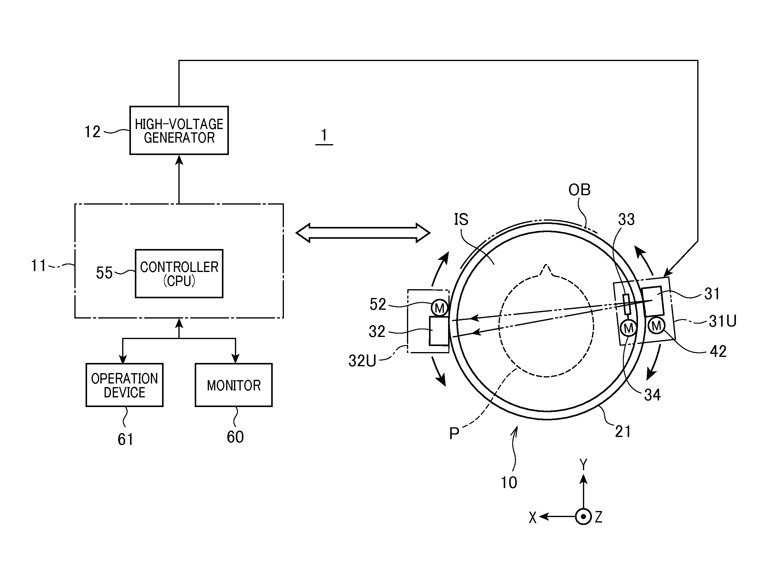
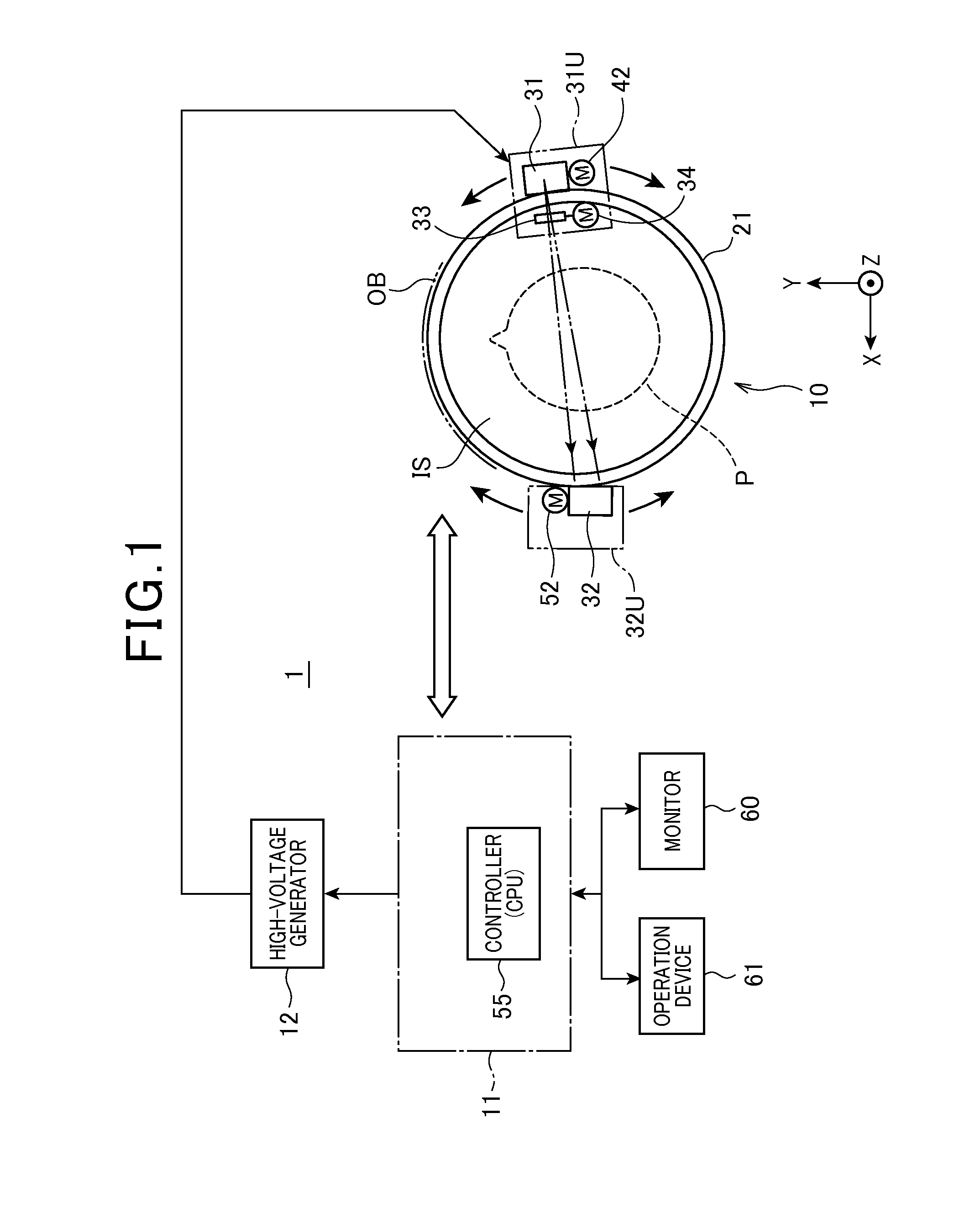
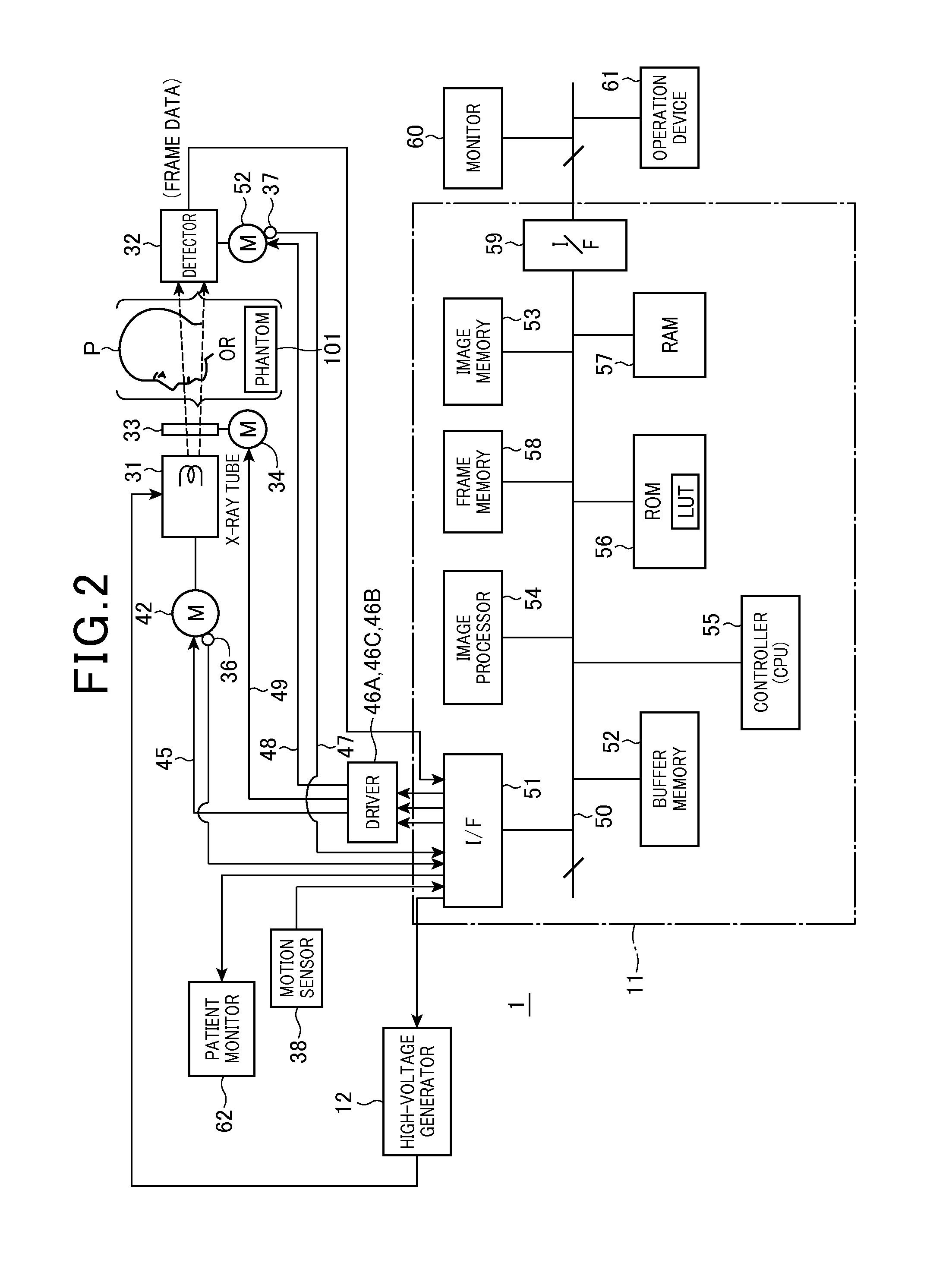
[0096]With reference to FIGS. 1-54, a first embodiment of a dental X-ray extraoral imaging apparatus, which is provided as an X-ray tomographic imaging apparatus according to the present invention, will now be described.

[0097]The present extraoral imaging apparatus is a modality capable of scanning an object (such as a tooth row) of the jaw of a patient P from outside the jaw with X-ray beams, processing data, acquired by scanning, with a tomosynthesis technique such that tomographic images of the object are produced. The X-ray extraoral imaging apparatus according to the present embodiment can provide images higher-resolution images which have not been provided by the conventional panoramic imaging apparatus, while the apparatus is still kept smaller in size and light in weight, in addition to performing the function of panoramic imaging apparatuses which are currently used for dental treatment. Further, employing this apparatus will improve an inconvenient workflow resulting from ...

second embodiment

[0404]With reference to FIGS. 58-68, the X-ray tomographic imaging apparatus of a second embodiment of the present embodiment will now be described.

[0405]In the present embodiment, the same or similar components to those in the apparatus described in the first embodiment will be given the same reference numerals for the sake of simplified description.

[0406]The X-ray tomographic imaging apparatus according to the present embodiment is provided with a construction for tomographic imaging (for panoramic imaging) based on the tomosynthesis technique and, without changes of its construction, a function for performing X-ray CT imaging by switching the apparatus to tomographic imaging based on a CT(Computed Tomography) technique, responsively to an operator's demand. In other words, there is provided a combined system capable of providing double role functions.

[0407]As shown in FIG. 58, an X-ray tomographic imaging apparatus 300 is equipped with a scan device 301 and a main cabinet 302. Th...


PUM


Abstract

An X-ray tomographic imaging apparatus includes an X-ray and a direct conversion type of detector. The X-ray tube and the detector are supported by the support means so as to be rotatable along curved orbits mutually independently. Under instructions from a computer, scans and image reconstruction are performed. The X-ray tube and the detector are moved along the orbits mutually independently so that X-ray beams are always transmitted through a desired tomographic plane of an object at desired angles. Acquired frame data are used to produce a panoramic image of the plane, while the frame data and the panoramic image are used to produce a tomographic image in which structural components of the object are optically focused and distortions caused due to differences in X-ray paths are suppressed. The apparatus can be used as devices for dental, medical diagnosis and nondestructive inspection, and can have a CT imaging function.

Description

FIELD OF THE INVENTION[0001]The present invention relates to an X-ray tomographic imaging apparatus that uses X-ray beams to image tomographic to images of an object being imaged, and in particular, to the X-ray tomographic imaging apparatus that images three-dimensional tomographic images of an object based on a tomosynthesis technique.BACKGROUND ART[0002]In recent years, tomographic imaging using a tomosynthesis technique has been used actively. The theory of this tomosynthesis technique has been known long before (for example, refer to patent literature 1), and recently, tomographic imaging that enjoys ease of image reconstruction performed using the tomosynthesis technique has been proposed (for example, refer to patent literatures 2 and 3). Especially, many such cases can be found in dental and mammographic fields (for example, refer to patent literatures 4, 5, 6, and 7).[0003]Conventionally, as one of radiation imaging apparatuses that employ the tomosynthesis technique by cho...

Claims


Application Information

Patent Timeline
IPC IPC(8): A61B6/14A61B6/03G06T11/00A61B6/00
CPCA61B6/14A61B6/466A61B6/035G06T2207/30196G06T2211/436G06T2207/10081G06T2207/30036G06T11/006A61B6/025A61B6/547A61B6/583
Inventor YAMAKAWA, TSUTOMUTSUJITA, MASAHIROKATSUMATA, AKITOSHIOGAWA, KOICHIAOKI, HISATOSHI
Owner TAKARA TELESYST
Why Eureka
Try Eureka