Ophthalmic elastography

a biomechanical and ophthalmic technology, applied in the field of ophthalmic elastography, can solve the problems of insufficient structural data alone to make informed clinical decisions, inability to in vivo methods that can image spatially resolved mechanical properties, and difficulty in prediction

Inactive Publication Date: 2015-11-05
OHIO STATE INNOVATION FOUND
View PDF2 Cites 7 Cited by
  • Summary
  • Abstract
  • Description
  • Claims
  • Application Information

AI Technical Summary

Benefits of technology

[0011]Also encompassed by the present invention are methods for correcting tonometric measurements of intraocular pressure. The method may comprise the steps of: (a) providing an ultrasound transducer in proximity to the eye or on the eye, wherein the ultrasound transducer has a center frequency ranging from about 50 Mite to about 100 MHz, or from about 50 MHz to about 80 MHz; (b) acquiring ultrasound data from at least one cross-section of at least one portion of the eye; (c) measuring ocular pulse and intraocular pressure; (d) processing the ultrasound data of step (b) with a speckle tracking algorithm to generate a strain profile and a stiffness index of the cross-section; and, (e) comparing the strain profile and the stiffness index to a standard, wherein the standard provides the strain profile and the stiffness index for normal subjects and / or patients having eye disease, and determining whether IOP is significantly under- or over-estimated by tonometric readings. The tonometric measurements may be from Goldmann Applanation Tonometry or Tonopen.

Problems solved by technology

However, it remains challenging to predict, for example, ectasia risk secondary to keratoconus and refractive surgery, even though detailed structural information has become available.
Thus, structural data alone are often not sufficient to make informed clinical decisions without also having functional information, such as would be available from measures of the biomechanical properties of the cornea in relationship to the status or changes in structure.
At the present there are no in vivo methods that can image spatially resolved mechanical properties of the cornea that are also independent of the intraocular pressure (IOP).

Method used


Image

  • Ophthalmic elastography
  • Ophthalmic elastography
  • Ophthalmic elastography

Examples


example 1

Analytical Model For Deriving the Biomechanical Index From Strain Measurements

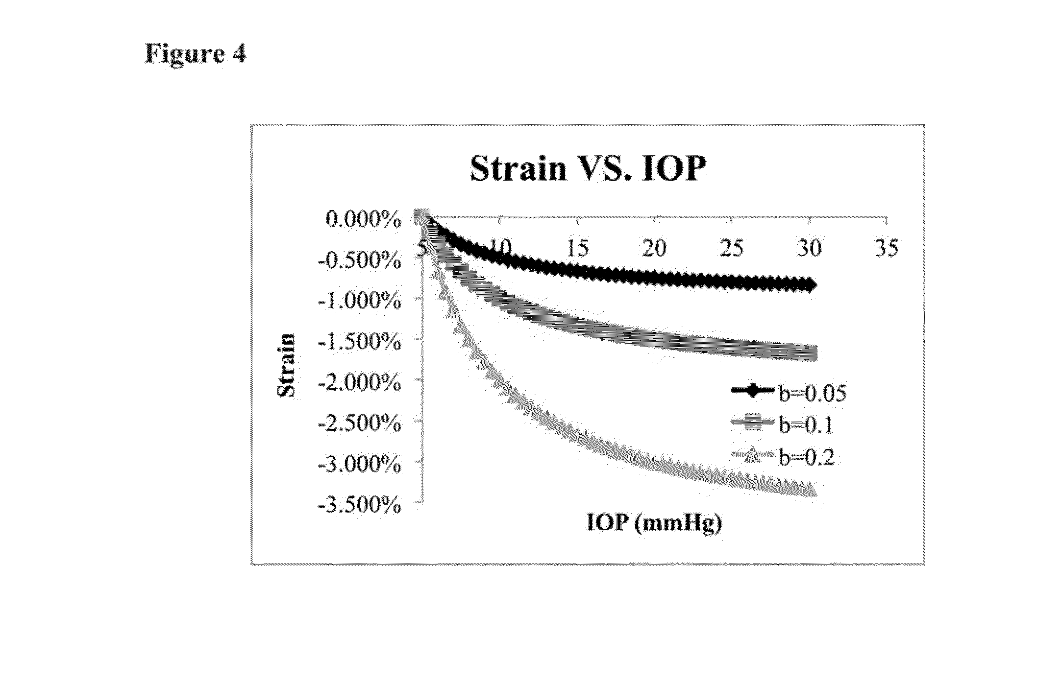
[0049]Based on the analysis of inflation tests of corneas, an analytical model is used to fit the strain-IOP curve. The strain-IOP curve passes through the point (5 mmHg, 0%) because an IOP of 5 mmHg is used as the reference pressure (i.e., 0% strain). Equation 2 (Eq. 2) shows the relationship between IOP and the radial strain, which can be accurately measured during ocular pulse.

strain=b*IOP−1−b*5−1   Eq. 2

[0050]In this model there is only one unknown parameter (“b”), which determines the overall shape of the strain-IOP curve, to predict the strain response of the cornea at a given IOP. As shown in FIG. 4, a smaller “b” value gives a shallower curve, indicating a stiffer cornea, and a larger “b” value gives a deeper curve, indicating a more compliant cornea. Therefore, “b” can be used as a stillness index obtained from the strain-IOP response.

[0051]FIG. 4 shows the plots of Eq. 2 when “b” values are 0.05,...


PUM


Abstract

This invention describes an ultrasound technique that maps out the mechanical properties of the cornea and the sclera to the intrinsic mechanical loadings in the eye. It helps identify the abnormally weaker or stiffer regions in the eye, to add functional information for early and definitive diagnosis of corneal diseases, surgical planning, prevention of surgical complications, as well as better interpretation of tonometric readings. This technique will allow a spatial mapping of the mechanical strains developed in the cornea or the sclera during ocular pulse or other intraocular pressure fluctuations. The envisioned use of this technique resembles the current clinical ophthalmic ultrasound in terms of the patient experience, but provides functional information about the eye tissue that is not available from current clinical ultrasound.

Description

CROSS REFERENCE TO RELATED APPLICATION[0001]This application claims priority to U.S. Provisional Application No. 61 / 987,684 filed on May 2, 2014, which is incorporated herein by reference in its entirety.FIELD OF THE INVENTION[0002]The present invention relates to methods and systems for determining the biomechanical properties of the eye. In particular, the present invention relates to in vivo mapping of the cornea and sclera biomechanical properties using high frequency ultrasound.BACKGROUND OF THE INVENTION[0003]Structural information of the cornea such as central corneal thickness and topography is critical for disease diagnosis and surgical planning. However, it remains challenging to predict, for example, ectasia risk secondary to keratoconus and refractive surgery, even though detailed structural information has become available. Thus, structural data alone are often not sufficient to make informed clinical decisions without also having functional information, such as would b...

Claims


Application Information

Patent Timeline
Patent Type & Authority Applications(United States)
IPC IPC(8): A61B8/10A61B3/16
CPCA61B3/16A61B8/10A61B8/485A61B8/5207
Inventor LIU, JUNCHEN, HONG
Owner OHIO STATE INNOVATION FOUND
Why Eureka
Try Eureka