Blue collagenase assay

a blue collagenase and assay technology, applied in the field of blue collagenase, can solve the problems of inability to measure the activity of soluble or insoluble cell or tissue-associated collagenase, individually or in combination, and achieve the effect of improving the detection accuracy and reproducibility

Inactive Publication Date: 2016-01-07
UNIV OF SOUTH FLORIDA
View PDF0 Cites 0 Cited by
  • Summary
  • Abstract
  • Description
  • Claims
  • Application Information

AI Technical Summary

Benefits of technology

[0011]The present invention may address one or more of the problems and deficiencies of the prior art discussed above. However, it is contemplated that the invention may prove useful in addressing other proble

Problems solved by technology

However, each of these references has their own respective drawbacks and would be incapable, individually or in combination, of measuring soluble or insoluble cell or tissue-associated collagenase activity using Coomassie dye.
Furthermore, where a definition or us

Method used


Image

  • Blue collagenase assay
  • Blue collagenase assay

Examples


example 1

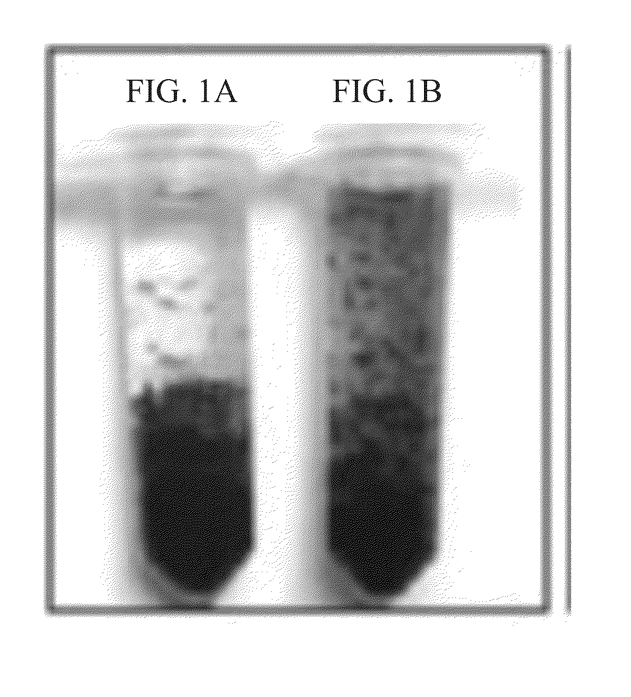
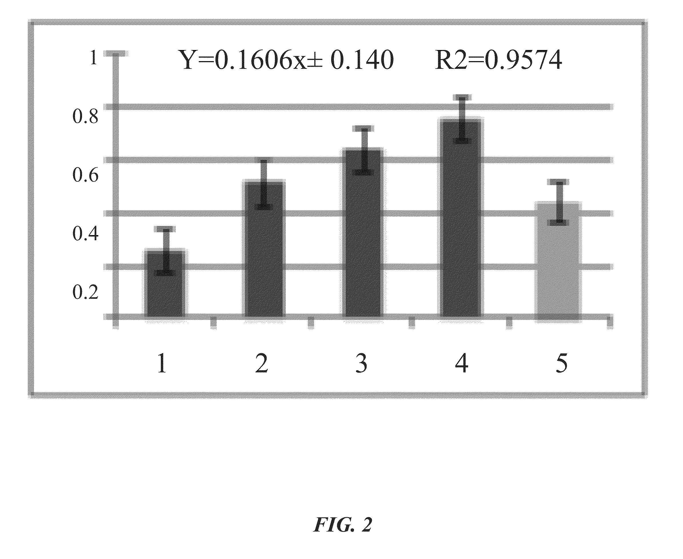
[0024]As an example, application of the blue collagenase assay to soluble Clostridium histolyticum collagenase (see FIGS. 1A-IB) and cell-associated Streptococcus mutans collagenase (see FIG. 2) is described herein.

[0025]The method generally includes the following steps:

[0026]1. The collagen fibrils are stained using Coomassie Brilliant Blue R-250 (C46-H44-N3-07-S2-Na).

[0027]2. The Blue collagen fibrils are suspended in collagenase substrate buffer (50 mM Tris, 50 mM NaCl, 10 mM CaCl2, pH7.5).

[0028]3. The blue collagen fibrils in suspension are incubated with the test sample at 37° C. on a rotator.

[0029]4. The collagenase activity results in the production of blue collagen particulates that can be readily observed.

[0030]5. After the incubation time of three (3) hours or longer, the mixture is filtered through glass wool / fibers to which the blue collagen fibrils are retained, and the small blue fragments are collected in the filtrate,

[0031]6, To determine the amount of digested colla...

example 2

1. Staining of Collagen

[0037]Type I fibrillar Collagen from Bovine Achilles Tendon (SIGMA-ALDRICH, product no. C9879) was cut with a pair of sharp scissors into shorter fibrils. The fibrils were passed through a 1 mm sieve and collected as dry collagen.

[0038]The dry collagen was mixed with 0.2% Coomassie Brilliant Blue in a solution of 10% acetic acid, 40% methanol in deionized water (standard method for staining protein bands in polyacrylamide gel), at a ratio of 500 mg collagen in 30 mL acetic acid-methanol-water solution, in a 50 mL conical centrifuge tube on a rotor at room temperature for 30 mins to 1 hour until all the collagen was saturated with the blue dye, after which time acetic acid-methanol-water (10:40:50) was added up to 50 mL.

[0039]The blue collagen suspension was centrifuged at 200×g for 2 minutes, and the supernatant was discarded. The excess dye was removed by rinsing with the solution of 10% Acetic acid, 40% methanol in deionized water.

[0040]With repeated mixing...


PUM


Abstract

A method of measuring soluble or insoluble cell or tissue-associated collagenase activity. The substrate includes native (fibrillar) collagen fragments that were stained with Coomassie Brilliant Blue R-250. Incubation with collagenase can be observed in real-time by the generation of digested smaller fragments. The degraded blue fragments are obtained by filtration through class fibers, onto which intact collagen fibrils are retained. The filtrate containing the blue collagen fragments is incubated with a detergent in order to extract the blue dye, and the mixture is centrifuged in order to separate the dye in the supernatant from the pellet, which contains de-stained collagen fragments and other insoluble materials contained in the test samples (such as bacterial cells or tissues). The amount of dye extracted is quantified by measuring the amount of dye extracted from these fragments, i.e., the absorbance at 600 nm using a spectrophotometer or an ELISA reader.

Description

CROSS-REFERENCE TO RELATED APPLICATIONS[0001]This nonprovisional application is a continuation of and claims priority to U.S. Provisional Patent Application No. 62 / 020,179, entitled “Blue Collagenase Assay”, filed Jul. 2, 2014, by the same inventor, the entirety of which is incorporated herein by reference.BACKGROUND OF THE INVENTION[0002]1. Field of the Invention[0003]This invention relates, generally, to collagenases. More specifically, it relates to blue collagenase assays.[0004]2. Brief Description of the Prior Art[0005]Collagenases are defined as proteases that can digest native collagen in the triple helix region. There is no spectrophotometric assay that uses native collagen fibrils [“Collagenase”, Worthington Biochemical Company, I.U.B, 3.4.24.3, C.A.S. 9001-12-1]. Commonly used substrates in collagenase assay kits include synthetic peptides, or denatured collagen such as acid soluble collagen and heat-gelling collagen or gelatin. Since denatured collagen can also be degrade...

Claims


Application Information

Patent Timeline
IPC IPC(8): C12Q1/37
CPCC12Q1/37G01N2333/78G01N2333/96494
Inventor DAO, MY, LIEN
Owner UNIV OF SOUTH FLORIDA
Why Eureka
Try Eureka