Ambulatory Electrocardiography Monitor Recorder Optimized For Capturing Low Amplitude Cardiac Action Potential Propagation

a low-amplitude, electrocardiography technology, applied in bioelectric signal measurement, medical science, diagnostics, etc., can solve the problems of unworkable ecg monitoring systems, significant challenge in recording sufficient ecg and related physiological data over an extended period, and realizing a 30-day observation period, so as to improve the ability of wearable monitors

Active Publication Date: 2016-01-14
BARDY DIAGNOSTICS
View PDF8 Cites 18 Cited by
  • Summary
  • Abstract
  • Description
  • Claims
  • Application Information

AI Technical Summary

Benefits of technology

[0016]Physiological monitoring can be provided through a lightweight wearable monitor that includes two components, a flexible extended wear electrode patch and a reusable monitor recorder that removably snaps into a receptacle on the electrode patch. The wearable monitor sits centrally (in the midline) on the patient's chest along the sternum oriented top-to-bottom. The ECG electrodes on the electrode patch are tailored to be positioned axially along the midline of the sternum for capturing action potential propagation in an orientation that corresponds to the aVF lead used in a conventional 12-lead ECG that is used to sense positive or upright P-waves. The placement of the wearable monitor in a location at the sternal midline (or immediately to either side of the sternum), with its unique narrow “hourglass”-like shape, significantly improves the ability of the wearable monitor to cutaneously sense cardiac electrical potential signals, particularly the P-wave (or atrial activity) and, to a lesser extent, the QRS interval signals indicating ventricular activity in the ECG waveforms. In addition, the monitor recorder includes an ECG sensing circuit that measures raw cutaneous electrical signals using a driven reference containing power supply noise and system noise to the reference lead, which is critical to preserving the characteristics of low amplitude cardiac action potentials, particularly P-waves.

Problems solved by technology

However, recording sufficient ECG and related physiological data over an extended period remains a significant challenge, despite an over 40-year history of ambulatory ECG monitoring efforts combined with no appreciable improvement in P-wave acquisition techniques since Dr.
Realizing a 30-day observation period has proven unworkable with existing ECG monitoring systems, which are arduous to employ; cumbersome, uncomfortable and not user-friendly to the patient; and costly to manufacture and deploy.
The location of the atria and their low amplitude, low frequency content electrical signals make P-waves difficult to sense, particularly through ambulatory ECG monitoring.
The atria are located posteriorly within the chest, and their physical distance from the skin surface adversely affects current strength and signal fidelity.
The distance between the heart and ECG electrodes reduces the magnitude of electrical potentials in proportion to the square of change in distance, which compounds the problem of sensing low amplitude P-waves.
Both approaches are unsatisfactory with respect to the P-wave and the accurate, medically actionable diagnosis of the myriad cardiac rhythm disorders that exist.
Additionally, maintaining continual contact between ECG electrodes and the skin after a day or two of ambulatory ECG monitoring has been a problem.
These factors adversely affect electrode adhesion and the quality of cardiac signal recordings.
Furthermore, the physical movements of the patient and their clothing impart various compressional, tensile, bending, and torsional forces on the contact point of an ECG electrode, especially over long recording times, and an inflexibly fastened ECG electrode will be prone to becoming dislodged.
Moreover, dislodgment may occur unbeknownst to the patient, making the ECG recordings worthless.
Further, some patients may have skin that is susceptible to itching or irritation, and the wearing of ECG electrodes can aggravate such skin conditions.
However, like all forms of implantable medical device (IMD), use of this monitor requires invasive surgical implantation, which significantly increases costs; requires ongoing follow up by a physician throughout the period of implantation; requires specialized equipment to retrieve monitoring data; and carries complications attendant to all surgery, including risks of infection, injury or death.
A “looping” Holter (or event) monitor can operate for a longer period of time by overwriting older ECG tracings, thence “recycling” storage in favor of extended operation, yet at the risk of losing event data.
Although capable of extended ECG monitoring, Holter monitors are cumbersome, expensive and typically only available by medical prescription, which limits their usability.
Further, the skill required to properly place the electrodes on the patient's chest precludes a patient from replacing or removing the sensing leads and usually involves moving the patient from the physician office to a specialized center within the hospital or clinic.
The location is used to simulate surgically implanted monitors, but without specifically enhancing P-wave capture.
Both devices represent compromises between length of wear and quality of ECG monitoring, especially with respect to ease of long term use, female-friendly fit, and quality of cardiac electrical potential signals, especially atrial (P-wave) signals.

Method used


Image

  • Ambulatory Electrocardiography Monitor Recorder Optimized For Capturing Low Amplitude Cardiac Action Potential Propagation
  • Ambulatory Electrocardiography Monitor Recorder Optimized For Capturing Low Amplitude Cardiac Action Potential Propagation
  • Ambulatory Electrocardiography Monitor Recorder Optimized For Capturing Low Amplitude Cardiac Action Potential Propagation

Examples


Embodiment Construction

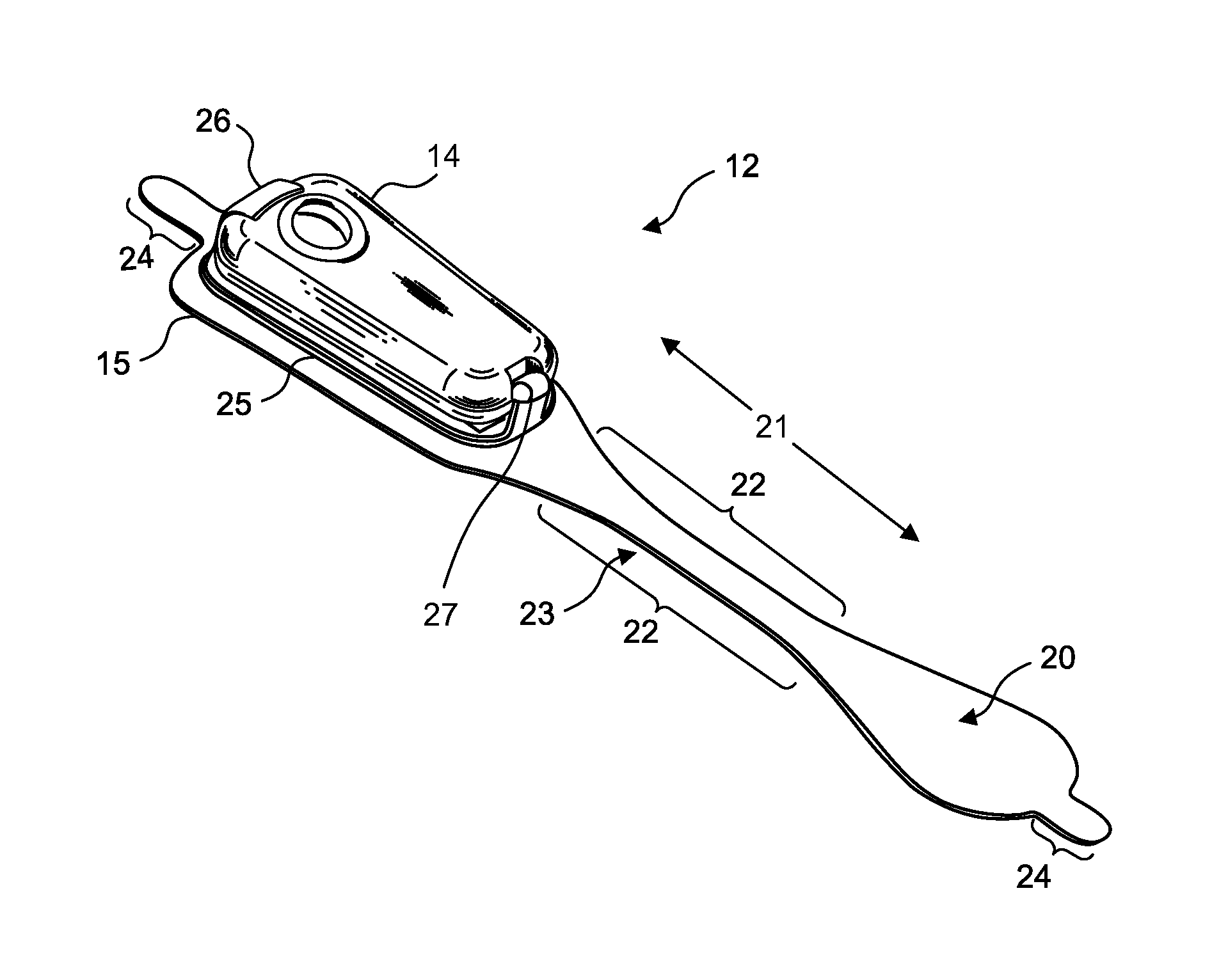
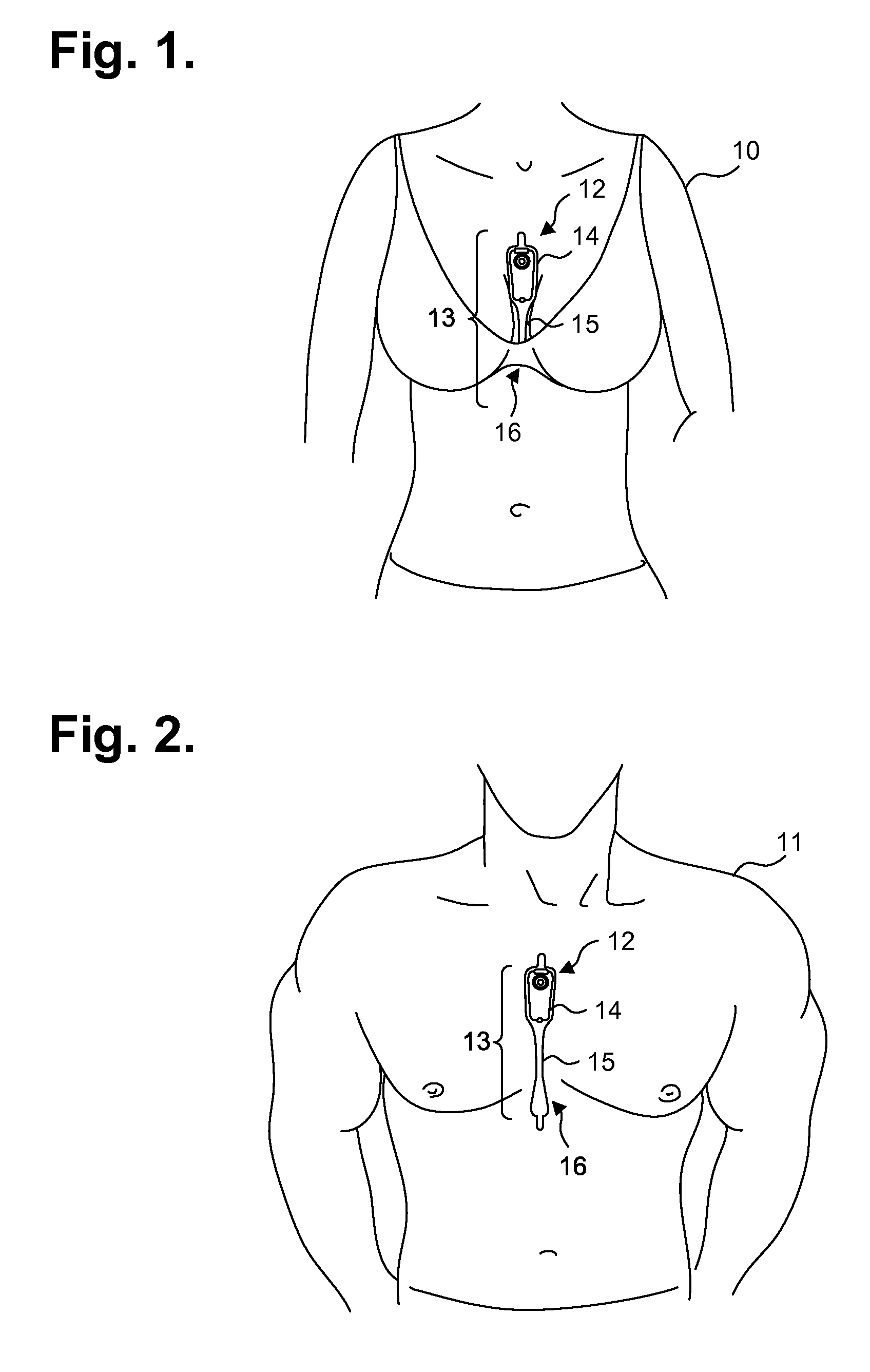
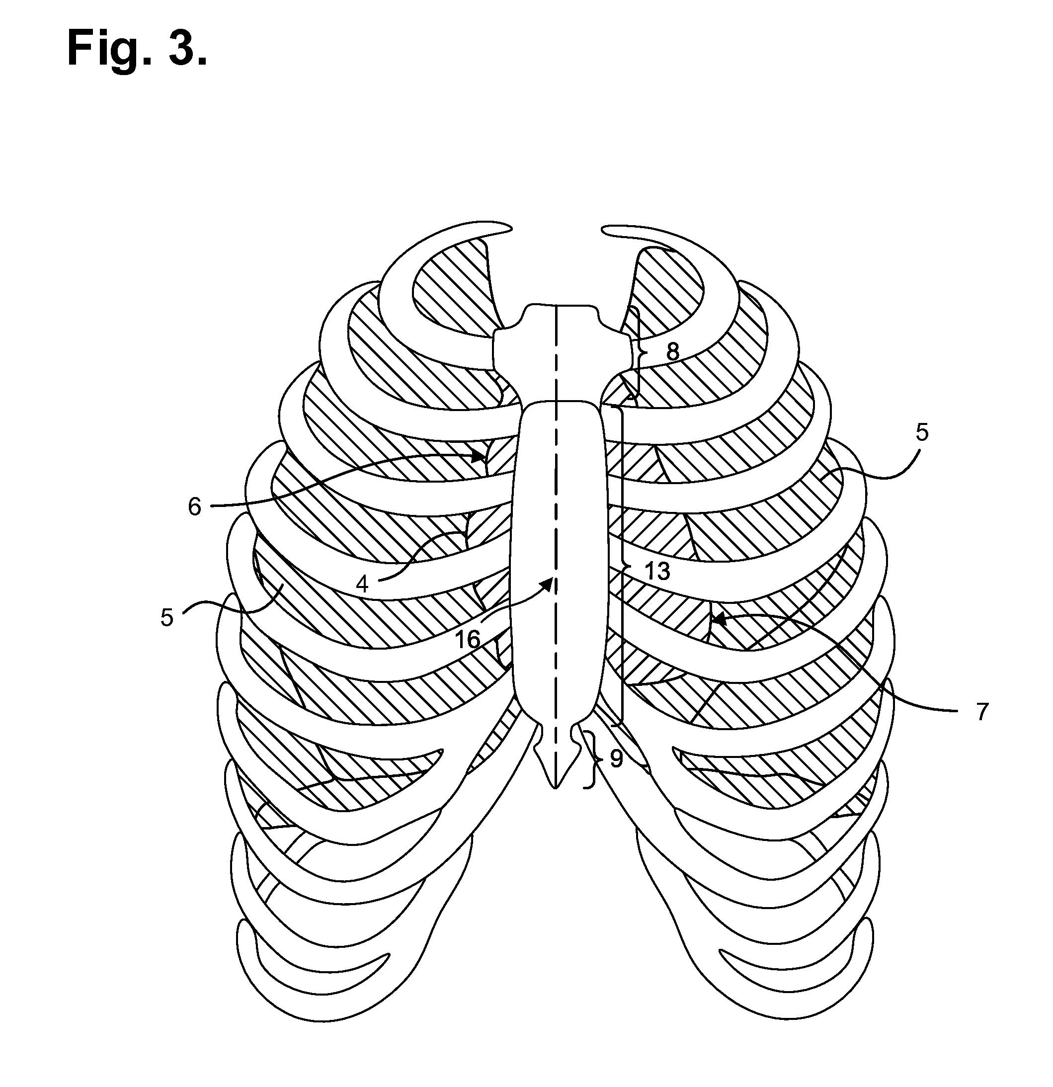
[0043]ECG and physiological monitoring can be provided through a wearable ambulatory monitor that includes two components, a flexible extended wear electrode patch and a removable reusable (or single use) monitor recorder. Both the electrode patch and the monitor recorder are optimized to capture electrical signals from the propagation of low amplitude, relatively low frequency content cardiac action potentials, particularly the P-waves generated during atrial activation. FIGS. 1 and 2 are diagrams showing, by way of examples, an extended wear electrocardiography monitor 12, including a monitor recorder 14, in accordance with one embodiment, respectively fitted to the sternal region of a female patient 10 and a male patient 11. The wearable monitor 12 sits centrally, positioned axially along the sternal midline 16, on the patient's chest along the sternum 13 and oriented top-to-bottom with the monitor recorder 14 preferably situated towards the patient's head. In a further embodimen...


PUM


Abstract

Physiological monitoring can be provided through a lightweight wearable monitor that includes two components, a flexible extended wear electrode patch and a reusable monitor recorder that removably snaps into a receptacle on the electrode patch. The wearable monitor sits centrally (in the midline) on the patient's chest along the sternum oriented top-to-bottom. The placement of the wearable monitor in a location at the sternal midline, with its unique narrow “hourglass”-like shape, significantly improves the ability of the wearable monitor to cutaneously sense cardiac electrical potential signals, particularly the P-wave and, to a lesser extent, the QRS interval signals indicating ventricular activity in the ECG waveforms. Additionally, the monitor recorder includes an ECG sensing circuit that measures raw cutaneous electrical signals using a driven reference containing power supply noise and system noise to the reference lead, which is critical to preserving the characteristics of low amplitude cardiac action potentials, particularly P-waves.

Description

CROSS-REFERENCE TO RELATED APPLICATION[0001]This non-provisional patent application is a continuation-in-part of U.S. patent application Ser. No. 14 / 080,725, filed Nov. 14, 2013, pending, and further claims priority under 35 U.S.C. §119(e) to U.S. Provisional Patent application, Ser. No. 61 / 882,403, filed Sep. 25, 2013, the disclosures of which are incorporated by reference.FIELD[0002]This application relates in general to electrocardiographic monitoring and, in particular, to an ambulatory electrocardiography monitor recorder optimized for capturing low amplitude cardiac action potential propagation from the atria.BACKGROUND[0003]The first electrocardiogram (ECG) was invented by a Dutch physiologist, Willem Einthoven, in 1903, who used a string galvanometer to measure the electrical activity of the heart. Generations of physicians around the world have since used ECGs, in various forms, to diagnose heart problems and other potential medical concerns. Although the basic principles u...

Claims


Application Information

Patent Timeline
Patent Type & Authority Applications(United States)
IPC IPC(8): A61B5/00A61B5/308A61B5/332
CPCA61B5/04325A61B5/0452A61B5/0404A61B5/7232A61B5/6823A61B5/725A61B5/04085A61B5/332A61B5/259A61B5/282A61B5/335A61B5/349
Inventor FELIX, JASONBISHAY, JON MIKALSONBARDY, GUST H.
Owner BARDY DIAGNOSTICS
Why Eureka
Try Eureka