Medical image dectection system and method

a technology of medical image and detection system, applied in the field of medical image detection software, can solve the problems of shortening the operation, reducing and increasing the anesthesia time, so as to reduce the incidence of rsis, reduce the cost, and improve the quality of patient car

Active Publication Date: 2016-03-10
RAPID MEDICAL TECH
View PDF1 Cites 23 Cited by
  • Summary
  • Abstract
  • Description
  • Claims
  • Application Information

AI Technical Summary

Benefits of technology

[0023]From a technical point of view, the contribution of our methods is in several areas. First, algorithms for enhancing images and increasing the contrast in them were developed for the specific purpose of foreign object detection. Focusing the algorithms on foreign object detection increases their effectiveness compared with general enhancement algorithms. Second, algorithms were developed to effectively cope with deformable and small objects. Third, algorithms were developed distinguishing between clutter and relevant objects. Fourth, algorithms for classifying the context of images such as anatomical regions or exposure level were developed. Finally, algorithms for producing large amounts of training data were developed to overcome the problem of low RSI incidence in actual images. The combination of two or more of these algorithms in a new methodology provides significant improvements in detection.
[0024]The limitations of the prior art motivated an automated image analysis solution for RSI detection in X-ray images based on computer vision machine learning techniques. Computerized algorithms may examine the image methodically, are fast and cost effective, and are more effective in the detection of objects with small dimensions. The core algorithms are based on image enhancement, candidate detection, feature extraction, and recognition.
[0025]Further, if a RSI is identified, additional projections / portable X-rays are usually necessary to approximate its position in the surgical field. This is particularly relevant in cases of emergent abdominal or chest surgeries with relatively large operative fields. By rapidly identifying the RFSO in the different projections, software according to embodiments of the present invention also significantly shorten time needed to locate the RSI within the surgical wound. This shortens the operation, anesthesia time, and associated risks.
[0027]The new method for RSI detection that works in tandem with existing protocols, and serve as an additional safeguard. Our system, routinely scan X-ray images taken in the OR for RSIs even when the focus of the image is a different issue. By using embodiments of the inventive system it is possible to reduce the incidence of RSIs and so reduce the cost and increase the quality of patient care.
[0028]From a technical point of view, the contribution of embodiments of the invention occur in several areas. First, algorithms for enhancing images and increasing the contrast we developed and tested for the purpose of foreign object detection. Focusing the algorithms on foreign object detection increases their effectiveness compared with general enhancement algorithms. Second, algorithms were developed and tested that effectively cope with deformable and small objects. Third, algorithms that distinguish between clutter and relevant objects were developed and tested. Fourth, algorithms for classifying the context of images such as anatomical regions or exposure level were developed and tested. Finally, algorithms for marking the location of RSIs in actual images were developed and tested. Combinations of such algorithms create an effective and useful computer aided decision tool for use by physicians in relation to hospital operations.
[0029]The costs related to RSIs are significant and justify investments in embodiments of the present invention. The methods and software provided increase the efficacy of RSI identification X-ray protocols, improve patient OR safety, prevent costly medical and consequent legal expenses, shorten the time needed for image analysis, and improve utilization of the OR time. These objectives are particularly important given patient outcome intensives in the Affordable Care Act. Integration of the core software into a PACS and / or portable X-ray machine software environment through their application programming interfaces (API) makes embodiments of the invention cost effective and widely applicable in everyday clinical practice.

Problems solved by technology

This shortens the operation, anesthesia time, and associated risks.

Method used


Image

  • Medical image dectection system and method
  • Medical image dectection system and method
  • Medical image dectection system and method

Examples


Embodiment Construction

[0042]The embodiment disclosed below is not intended to be exhaustive or limit the invention to the precise form disclosed in the following detailed description. Rather, the embodiment is chosen and described so that others skilled in the art may utilize its teachings.

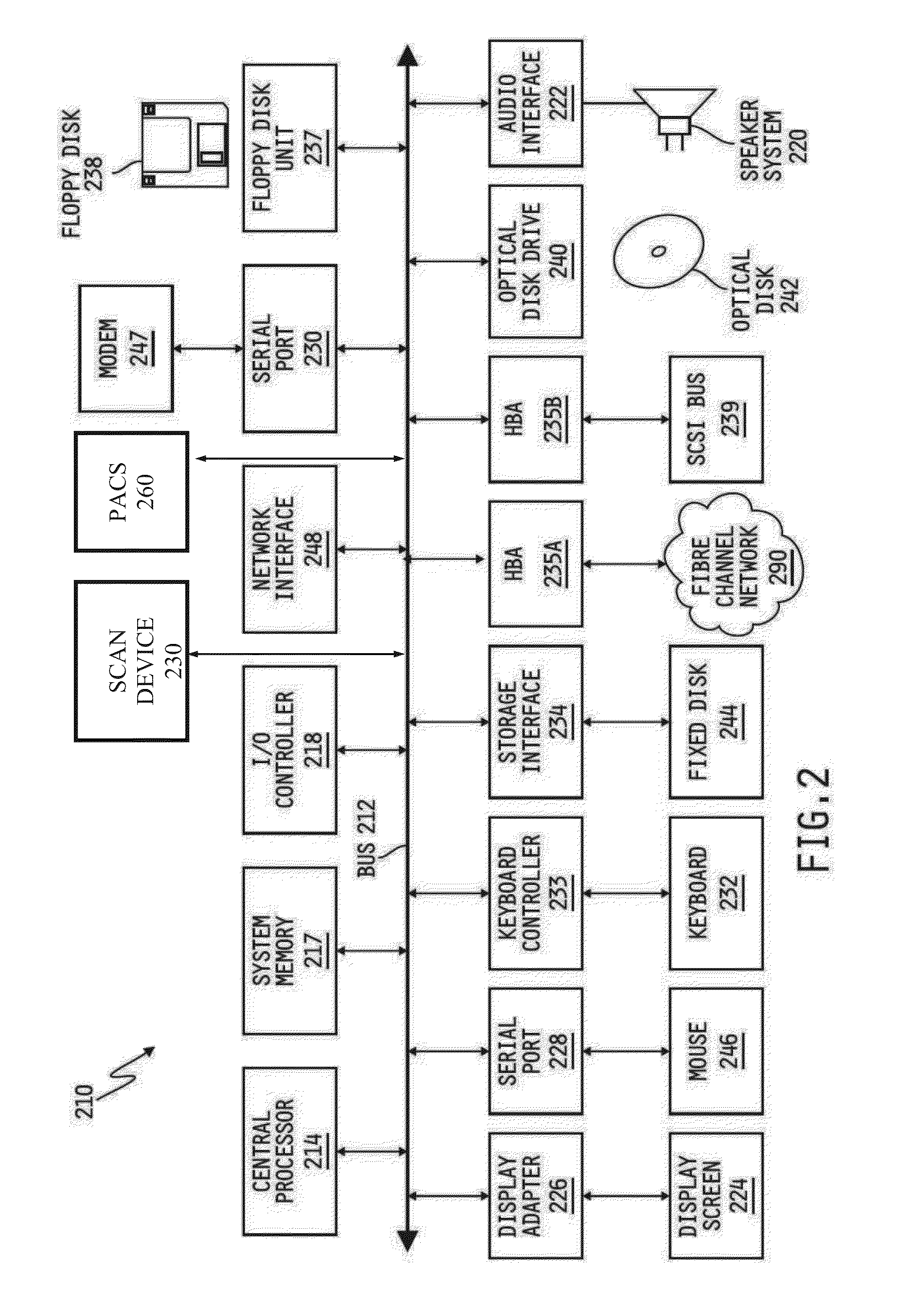
[0043]The detailed descriptions which follow are presented in part in terms of algorithms and symbolic representations of operations on data bits within a computer memory representing alphanumeric characters or other information. A computer generally includes a processor for executing instructions and memory for storing instructions and data. When a general purpose computer has a series of machine encoded instructions stored in its memory, the computer operating on such encoded instructions may become a specific type of machine, namely a computer particularly configured to perform the operations embodied by the series of instructions. Some of the instructions may be adapted to produce signals that control operation of ...


PUM


Abstract

The present invention involves a foreign object detection system and method which involves combining several algorithms specifically enhanced for foreign object detection to provide an improved detection system.

Description

BACKGROUND OF THE INVENTION[0001]1. Field of the Invention[0002]The invention relates to medical imaging software. More specifically, but not exclusively, the field of the invention is that of medical image detection software for detecting non-body objects.[0003]2. Description of the Related Art[0004]Techniques are know for the identification of implanted medical devices (“IMDs”) and retained foreign objects (“RFOs”) in medical images.[0005]Approximately 25 million patients in the United States have or have had an implanted medical device (“IMD”). Driven by a rapidly increasing aged population and supported by new technologies, the demand for IMDs and their further proliferation can only be expected to increase.[0006]An IMD is a medical device that is partly or totally surgically inserted into the human body or a natural orifice and is expected to remain implanted for an extended period or may be permanent. IMDs can further be classified as either active, those that use electricity,...

Claims


Application Information

Patent Timeline
Patent Type & Authority Applications(United States)
IPC IPC(8): G06T7/00G06T11/60G06V10/774
CPCG06T7/0012G06T11/60G06K9/46G06T2207/20081G06K9/6218G06T2207/10116G06T7/0081G06T2200/24G06T2207/20036G06T2207/30052G06V10/255G06V2201/034G06V10/774G06F18/214
Inventor AGAM, GADYLIN, GAN
Owner RAPID MEDICAL TECH
Why Eureka
Try Eureka