Medical Imaging System Providing Disease Prognosis

a medical imaging and disease technology, applied in the field of medical imaging systems, can solve problems such as mismatch between the number of dimensions of clinical data (the total number of clinical data, the number of samples (for example, patients) in clinical trials) and the number of dimensions of clinical data

Active Publication Date: 2016-03-17
WISCONSIN ALUMNI RES FOUND
View PDF5 Cites 13 Cited by
  • Summary
  • Abstract
  • Description
  • Claims
  • Application Information

AI Technical Summary

Benefits of technology

[0008]The present inventors have developed a medical imaging system that provides a machine learning architecture that can learn from clinical data with high dimensionality but a low number of samples incident to a small clinical study. Generally, the system divides up the data (splits the high dimensional data into multiple sets of smaller dimensions) for independent training of different “smaller” machine learning modules so that significant input data will be exposed and emphasized during the training, without forcing the system to be lost in the high dimensionality of the data. Once exposed and emphasized, the outputs of multiple such smaller machine learning modules are combined, for example, by a second machine learning module, to provide an overall result.

Problems solved by technology

A serious obstacle to the training of machine learning systems using clinical data is the mismatch between the number of dimensions of the clinical data (the total number of measurements obtained from all types of data collecting instruments, for example MRI and PET etc.) and the number of samples (for example, patients) in the clinical trial.
In these cases, where the dimensionality of the data far exceeds the number of samples, the machine learning system will almost surely not be able to learn the underlying concept (the distinction between disease and no disease) due to the few number of samples to learn from.
This problem of unnecessary over fitting is fundamentally inevitable given the cost and difficulty of performing clinical studies with larger numbers of patients and the increasingly powerful medical imaging devices that provide increasing dimensions of measurement.

Method used


Image

  • Medical Imaging System Providing Disease Prognosis
  • Medical Imaging System Providing Disease Prognosis
  • Medical Imaging System Providing Disease Prognosis

Examples


Embodiment Construction

Hardware Elements

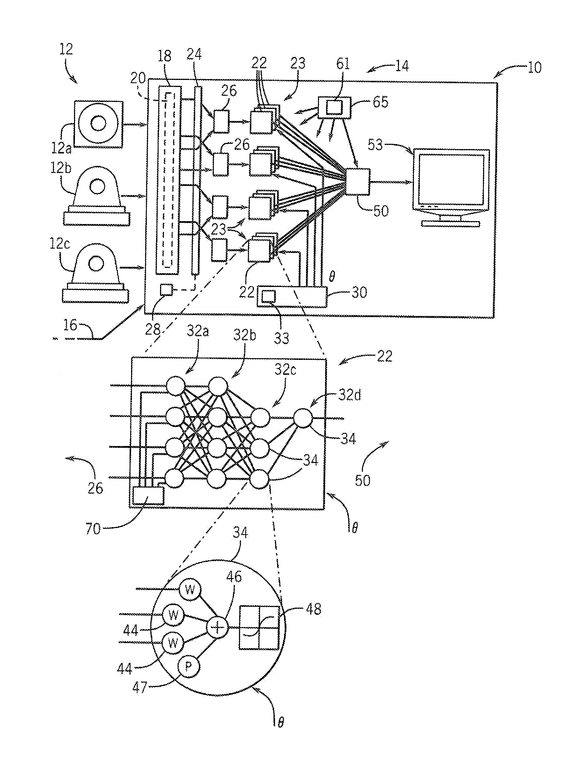
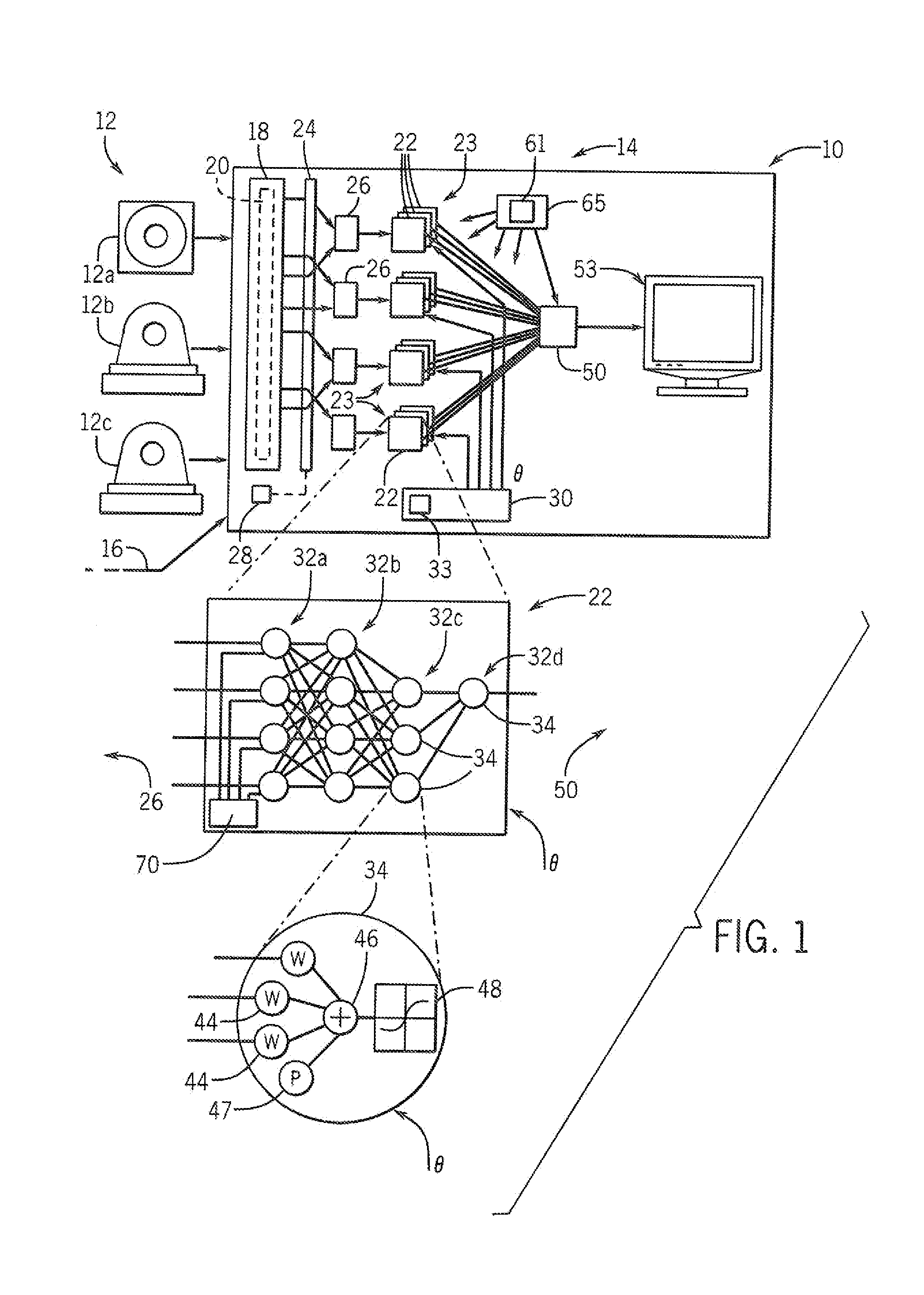
[0038]Referring now to FIG. 1, a medical imaging system 10 may employ one or more medical imaging sources 12 providing data to a processing engine 14. In the example of a medical imaging system 10 for diagnosing Alzheimer's disease, three medical imaging sources 12 may be employed including: an magnetic resonance imaging (MRI) machine 12a, a positron emission tomography (PET) machine 12b for providing amyloid data and a PET machine 12c for providing Fludeoxyglucose (FDG) data. The medical imaging system 10 may also accept non-image data sources 16 such as degree of cognitive impairment and parental family history.

[0039]A set of data 20 collected from these medical imaging sources 12 and non-image data sources 16 may be provided to the processing engine 14 to be stored in electronic memory 18. Generally this set of data 20 has dimensions ν much greater than the number of samples n.

[0040]The set of data 20 is mapped to a set of stacked de-noising autoencoders 22 accor...


PUM


Abstract

A medical imaging system that processes input data (imaging and/or non imaging) having high dimensionality and few samples to learn from, by using multiple ranks of machine learning modules each dealing with a separate portion of the clinical data. The outputs of the individual machine learning modules are the combined to provide a result reflective of the complete image data set.

Description

CROSS REFERENCE TO RELATED APPLICATION[0001]This application claims the benefit of U.S. provisional 62 / 050,592 filed Sep. 15, 2015 and hereby incorporated by reference.STATEMENT REGARDING FEDERALLY SPONSORED RESEARCH OR DEVELOPMENT[0002]This invention was made with government support under AG040396 awarded by the National Institutes of Health and 1252725 awarded by the National Science Foundation. The government has certain rights in the invention.BACKGROUND OF THE INVENTION[0003]The present invention provides a medical imaging system that can process medical imaging data to present a disease prognosis based on limited clinical data.[0004]Modern medical diagnostic tools such as magnetic resonance imaging (MRI) and positron emission tomography (PET) provide clinicians with a wealth of data that promises to greatly advance our ability to measure and predict disease progression. In one example, there is strong evidence that Alzheimer's disease manifest in such brain imaging data years ...

Claims


Application Information

Patent Timeline
Patent Type & Authority Applications(United States)
IPC IPC(8): A61B5/00A61B5/055A61B6/00A61B6/03G06N99/00G06V10/70
CPCA61B5/7267G06N99/005G06K9/6217A61B5/7271A61B5/742A61B2560/0475A61B5/055A61B6/037A61B6/5211A61B6/461A61B5/0042A61B5/7203A61B6/4417A61B6/501A61B6/5247A61B5/4088G16H50/70G06V10/70G06F18/21G06F18/256
Inventor ITHAPU, VAMSI K.SINGH, VIKASJOHNSON, STERLING C.OKONKWO, OZIOMA C.
Owner WISCONSIN ALUMNI RES FOUND
Why Eureka
Try Eureka