Linking breast lesion locations across imaging studies

a breast lesion and imaging study technology, applied in the field of breast imaging technology, can solve the problems of time-consuming, difficult, error-prone, etc., and achieve the effect of convenient comparability, fast and effective linking breast lesion locations, and less prone to errors

Active Publication Date: 2016-04-14
KONINKLJIJKE PHILIPS NV
View PDF8 Cites 6 Cited by
  • Summary
  • Abstract
  • Description
  • Claims
  • Application Information

AI Technical Summary

Benefits of technology

[0017]In a supplemented embodiment, the provided coordinates of the lesion in the first image are transformed into corresponding coordinates of the generic coordinate system of the breast model. Said coordinates of the generic coordinate system can be stored for a subsequent comparison with a second image. This will be elaborated in more detail with respect coordinates within standard clinical terminology including “upper outer quadrant and o'clock positions”. In other words, the provided coordinates may describe the lesion location in terms of “upper outer quadrant and o'clock positions” and can then be transferred into the normalized breast coordinates used by the breast model. This facilitates comparability. More aspects and technical details of such a coordination transformation will be explained hereinafter.
[0018]The presented method advantageously allows for fast and effective linking breast lesion locations across different imaging studies. By this approach, a lesion marked by the radiologist in the first image can for example be assigned either a unique three-dimensional coordinate (if the lesion was marked in a three-dimensional image, or a pair of two-dimensional images), or

Problems solved by technology

Consequently, when combining information from multiple reports, corresponding lesions need to be identified by the reader, which can be a challenging task.
When reviewing a foll

Method used


Image

  • Linking breast lesion locations across imaging studies
  • Linking breast lesion locations across imaging studies
  • Linking breast lesion locations across imaging studies

Examples


Embodiment Construction

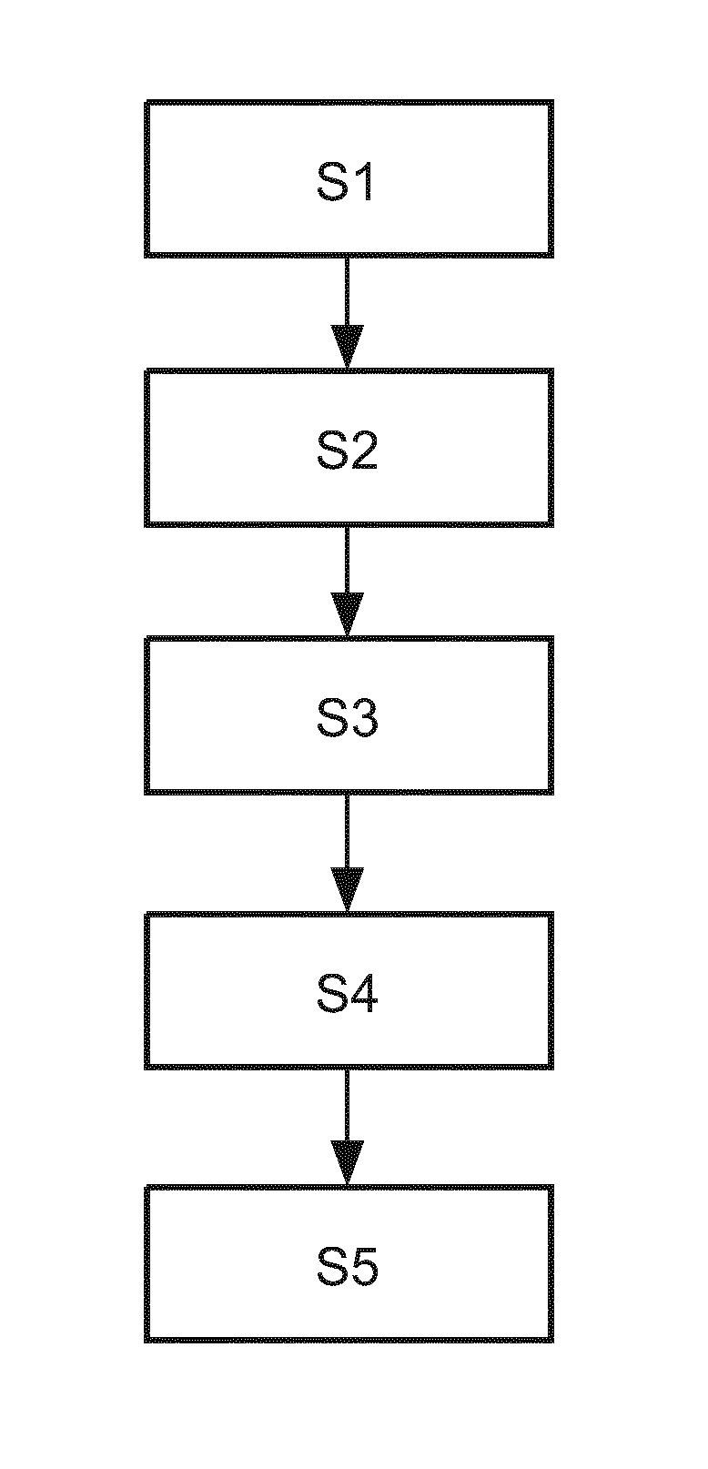
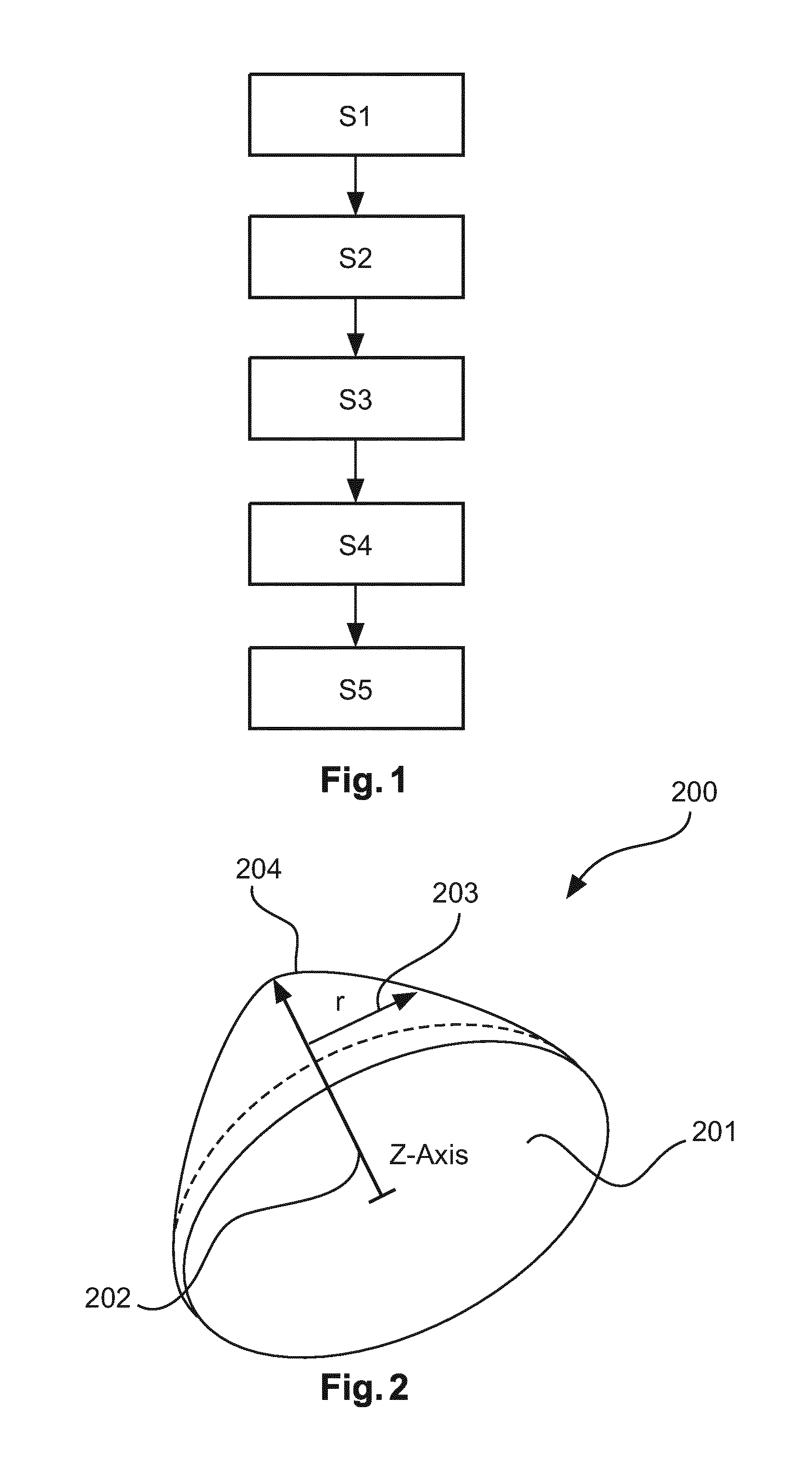
[0045]In the following, aspects about the breast model and the used coordinate systems will be described in detail. The following aspects are applicable to all embodiments depicted in FIGS. 1 to 4. It should be noted that all steps described for the first image are equally applicable and disclosed herewith for the second image and any further image.

[0046]In a first embodiment, the breast model is a generic breast model and is represented by a parametric function. For example, a two-dimensional Gaussian function (G(x,y)), restricted to a circular supporting domain D, parameterized either by Cartesian coordinates (x,y) or polar coordinates (r,φ) is a possible embodiment. Moreover, the anatomic structures can be presented by the apex, i.e. the nipple position, the flat circular domain with the z-coordinate=0 (posterior end of breast; pectoral muscle), the surface defined by the Gaussian function values G(x,y) (i.e. the skin surface), and by aligning the axis orientations with patient c...


PUM


Abstract

The present invention provides for means for linking breast lesion locations across imaging studies. In particular, a generic three-dimensional representation of the female breast is used. Automatic translation of the lesion location into standard clinical terminology and aligning the breast model with individual patient images is comprised. Moreover, a mechanism for linking image locations showing a lesion to a location in the breast model is presented. If desired, a region of interest can be calculated by a region of interest definition module that predicts a region of interest of a known lesion in terms of the breast model representation in a new imaging study.

Description

FIELD OF THE INVENTION[0001]The present invention relates to breast imaging technology and the respective image data analysis. In particular, the present invention relates to a breast model based method for linking breast lesion locations, an imaging system for imaging a patient's breast, a computer program element and a computer-readable medium.BACKGROUND OF THE INVENTION[0002]Breast imaging for breast cancer screening and diagnostic work-up follows a multi-modality approach, including X-ray mammography and tomosynthesis, ultrasound, and Magnetic Resonance (MR) imaging. In order to completely characterize a lesion, it is essential for the radiologist to track individual lesions across multiple imaging studies and to structure the report on the level of individual lesions. In current practice, the radiologist has to visually identify corresponding lesions across imaging studies, since the patient positioning differs significantly between the different modalities, e.g., two projectio...

Claims


Application Information

Patent Timeline
IPC IPC(8): G06T7/00G06T7/60
CPCG06T7/0012G06T7/60G06T2207/30068G06T2207/10088G06T2207/30096G06T7/0032G06T2207/10116G06T2207/10132G06T7/33G06T7/344
Inventor BUELOW, THOMASBERGTHOLDT, MARTINWIEMKER, RAFAELMEETZ, KIRSTEN REGINASABCZYNSKI, JOERGKUTRA, DOMINIK BERJAMIN
Owner KONINKLJIJKE PHILIPS NV
Why Eureka
Try Eureka