Metal resistant mr imaging

a mr imaging and metal technology, applied in the field of mr imaging, can solve the problems signal voids and other geometric distortions, mr imaging near metal is typically compromised, etc., and achieves the effects of reducing the probability of mr signal pile-up

Active Publication Date: 2016-08-04
KONINKLJIJKE PHILIPS NV
View PDF1 Cites 7 Cited by
  • Summary
  • Abstract
  • Description
  • Claims
  • Application Information

AI Technical Summary

Benefits of technology

[0027]a spin echo sequence or a stimulated echo sequence, if a metal implant is present. As mentioned above, a gradient echo sequence, such as a FFE acquisition protocol, is the standard for the SENSE reference scan. This sequence allows a fast acquisition of the spatial sensitivity profiles of the used RF coils, but it is very sensitive to susceptibility effects. When the system determines that a metal resistant SENSE reference scan is required it may select a spin echo sequence, such as, for example, a TSE SENSE reference scan which is more robust against susceptibility effects.
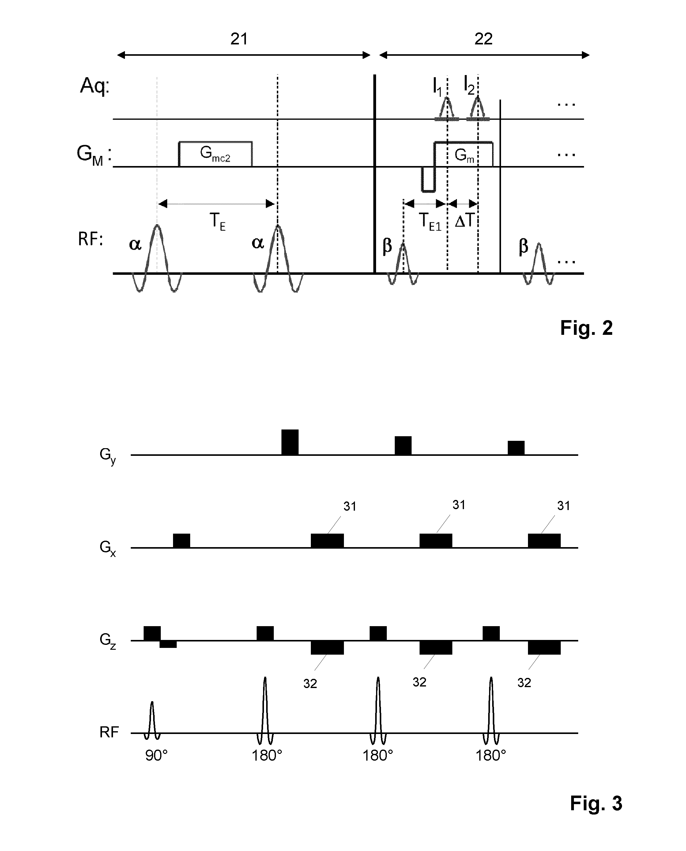
[0028]In a preferred embodiment of the invention, a stimulated echo sequence may be selected as a metal resistant SENSE reference scan. In general, a stimulated echo sequence comprises three (for example 60° or 90°) RF pulses, wherein the first two RF pulses are preparation pulses. The first preparation RF pulse excites magnetic resonance and transforms the longitudinal nuclear magnetization into transverse nuclear magnetization. The second preparation RF pulse stores a fraction of the de-phased transverse nuclear magnetization along the longitudinal axis. In case of 90° RF pulses this fraction is almost half of the de-phased transverse magnetization. The third RF pulse is applied during the acquisition period which is temporally subsequent to the preparation period. The third RF pulse (“reading RF pulse”) transforms the stored longitudinal nuclear magnetization into transverse nuclear magnetization again, thereby generating a so-called stimulated echo. Furthermore, it generates a FID signal from the remaining longitudinal magnetisation. Other RF refocused echoes could potentially be generated by this three RF pulse sequence, but those are not of interest here and may be suppressed by appropriate gradient switching regimes running in parallel to the RF irradiation. The stimulated echo MR signal together with the FID signal, which is also generated by the third RF pulse, is acquired according to the invention. MR imaging on the basis of the stimulated echoes can be accelerated by replacing the third RF pulse by a train of low-flip angle reading RF pulses, wherein each reading R

Problems solved by technology

MR imaging near metal is typically compromised by susceptibility issues degrading the magnetic fields used for image formation locally.
In diagnostic MR imaging scans, the susceptibility of the metal parts causes MR signal pile-up, signal voids and other geometric distortions.
When the susceptibility of metal parts compromises the quality of the SENSE reference scan, it may cause SENSE unfolding probl

Method used


Image

  • Metal resistant mr imaging
  • Metal resistant mr imaging
  • Metal resistant mr imaging

Examples


Embodiment Construction

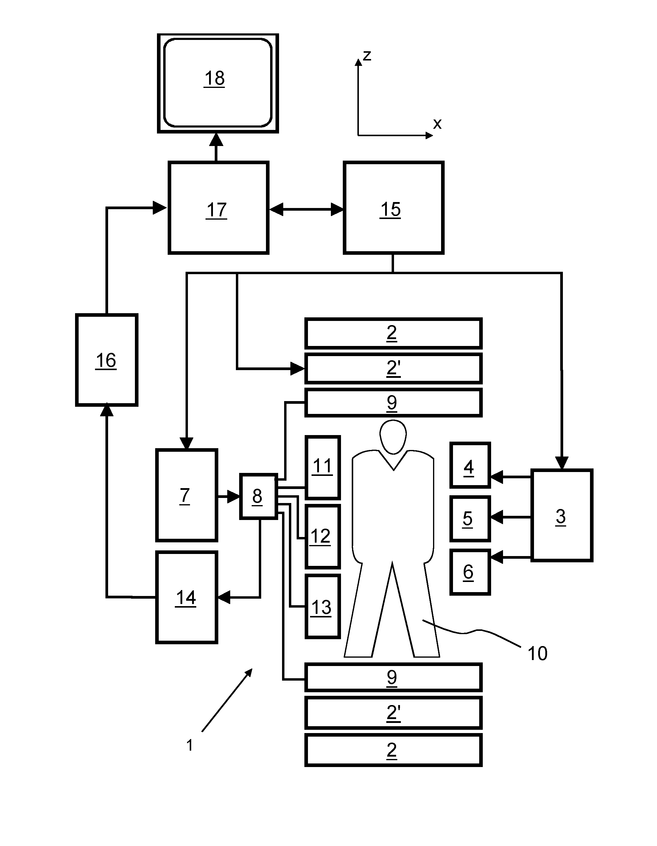
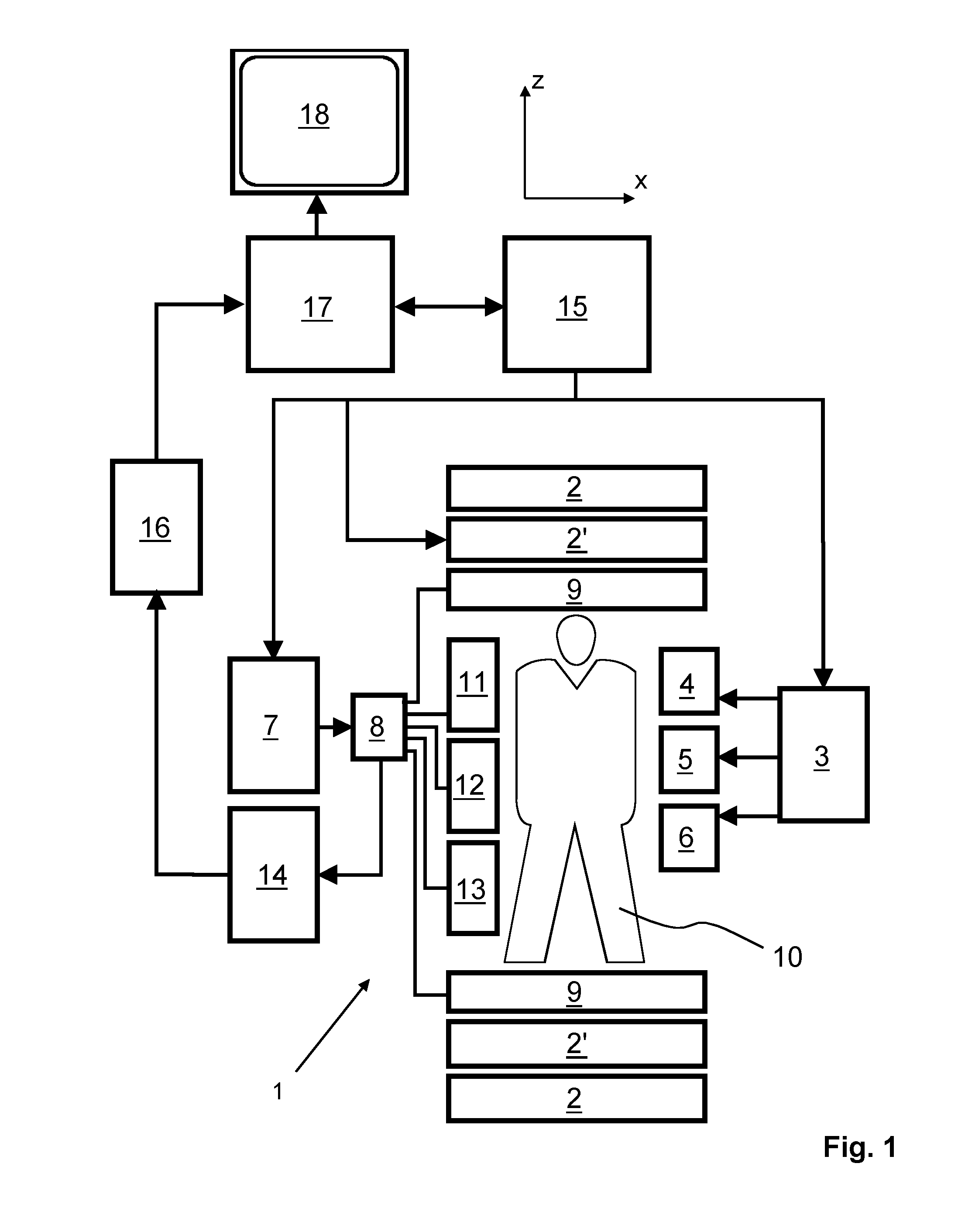
[0039]With reference to FIG. 1, a MR device 1 is shown. The device comprises superconducting or resistive main magnet coils 2 such that a substantially uniform, temporally constant main magnetic field B0 is created along a z-axis through an examination volume. The device further comprises a set of (1st, 2nd, and—where applicable—3rd order) shimming coils 2′, wherein the current flow through the individual shimming coils of the set 2′ is controllable for the purpose of minimizing B0 deviations within the examination volume.

[0040]A magnetic resonance generation and manipulation system applies a series of RF pulses and switched magnetic field gradients (also referred to as ‘gradient pulses’) to invert or excite nuclear magnetic spins, induce magnetic resonance, refocus magnetic resonance, manipulate magnetic resonance, spatially and otherwise encode the magnetic resonance, saturate spins, and the like to perform MR imaging.

[0041]More specifically, a gradient pulse amplifier 3 applies c...


PUM


Abstract

The invention relates to a method of parallel MR imaging which comprises the steps of: —subjecting a portion of a body (10) to a first imaging sequence (21,22) of RF pulses and switched magnetic field gradients, wherein first MR signals (11,12) are acquired via at least two RF coils having different spatial sensitivity profiles within the examination volume, —deriving the spatial sensitivity profiles of the at least two RF coils from the acquired first MR signals, —subjecting the portion of the body to a second imaging sequence of RF pulses and switched magnetic field gradients, wherein second MR signals are acquired by parallel acquisition via the at least two RF coils with sub-sampling of k-space, and —reconstructing a MR image from the acquired second MR signals and from the spatial sensitivity profiles of the at least two RF coils. According to the invention, the type and/or parameters of the first imaging sequence are selected automatically depending on the presence of a metal implant in the body. The selection of the type of the first imaging sequence is made between a gradient echo sequence, if no metal implants are present, and a spin echo sequence or a stimulated echo sequence, if a metal implant is present.

Description

FIELD OF THE INVENTION[0001]The invention relates to the field of magnetic resonance (MR) imaging. It concerns a method of MR imaging of at least a portion of a body. The invention also relates to a MR device and to a computer program to be run on a MR device.[0002]The international application WO2005 / 071429 addresses to problem of distortions in the calibration (reference) scan (for coil sensitivity estimation) due to rapid magnetic field changes. This known approach resolves this problem by applying a spin echo type of short echo time gradient echo type acquisition for the calibration scan. Further, the phase-encoding direction for the parallel imaging scan is matched to the phase encoding direction of the calibration scan.BACKGROUND OF THE INVENTION[0003]Image-forming MR methods which utilize the interaction between magnetic fields and nuclear spins in order to form two-dimensional or three-dimensional images are widely used nowadays, notably in the field of medical diagnostics, ...

Claims


Application Information

Patent Timeline
IPC IPC(8): G01R33/565G01R33/385G01R33/561
CPCG01R33/246G01R33/5611G01R33/5615G01R33/385G01R33/56536
Inventor DEN HARDER, JOHAN MICHIELVAN MEEL, MARIUS JOHANNES
Owner KONINKLJIJKE PHILIPS NV
Why Eureka
Try Eureka